

Necrosis avid near infrared fluorescent cyanines for imaging cell death and their use to monitor therapeutic efficacy in mouse tumor models
- PMID: 26472022
- PMCID: PMC4770755
- DOI: 10.18632/oncotarget.5498
Necrosis avid near infrared fluorescent cyanines for imaging cell death and their use to monitor therapeutic efficacy in mouse tumor models
Abstract
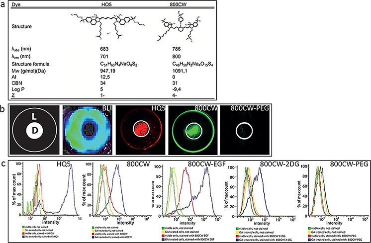
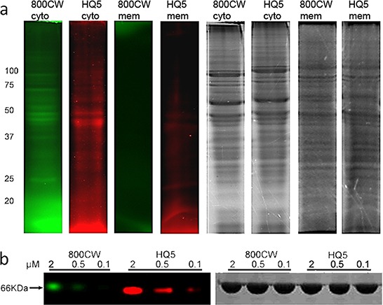
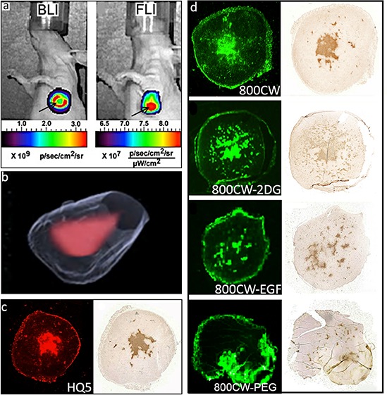
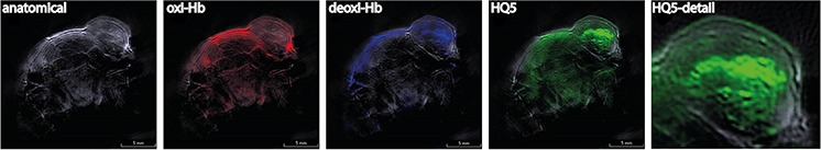
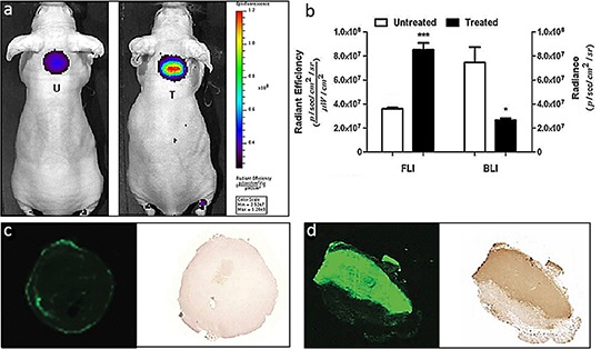
Quantification of tumor necrosis in cancer patients is of diagnostic value as the amount of necrosis is correlated with disease prognosis and it could also be used to predict early efficacy of anti-cancer treatments. In the present study, we identified two near infrared fluorescent (NIRF) carboxylated cyanines, HQ5 and IRDye 800CW (800CW), which possess strong necrosis avidity. In vitro studies showed that both dyes selectively bind to cytoplasmic proteins of dead cells that have lost membrane integrity. Affinity for cytoplasmic proteins was confirmed using quantitative structure activity relations modeling. In vivo results, using NIRF and optoacoustic imaging, confirmed the necrosis avid properties of HQ5 and 800CW in a mouse 4T1 breast cancer tumor model of spontaneous necrosis. Finally, in a mouse EL4 lymphoma tumor model, already 24 h post chemotherapy, a significant increase in 800CW fluorescence intensity was observed in treated compared to untreated tumors. In conclusion, we show, for the first time, that the NIRF carboxylated cyanines HQ5 and 800CW possess strong necrosis avid properties in vitro and in vivo. When translated to the clinic, these dyes may be used for diagnostic or prognostic purposes and for monitoring in vivo tumor response early after the start of treatment.
Keywords: cancer; cell death; cyanines; imaging; necrosis avid contrast agents.
Conflict of interest statement
The authors have no conflicts of interest to declare.
Figures

Similar articles
-
Pre-clinical Evaluation of a Cyanine-Based SPECT Probe for Multimodal Tumor Necrosis Imaging.Mol Imaging Biol. 2016 Dec;18(6):905-915. doi: 10.1007/s11307-016-0972-7. Mol Imaging Biol. 2016. PMID: 27277828 Free PMC article.
-
In Vivo Evaluation of Indium-111-Labeled 800CW as a Necrosis-Avid Contrast Agent.Mol Imaging Biol. 2020 Oct;22(5):1333-1341. doi: 10.1007/s11307-020-01511-x. Mol Imaging Biol. 2020. PMID: 32514888 Free PMC article.
-
Evaluation of four affibody-based near-infrared fluorescent probes for optical imaging of epidermal growth factor receptor positive tumors.Bioconjug Chem. 2012 Jun 20;23(6):1149-56. doi: 10.1021/bc200596a. Epub 2012 Jun 4. Bioconjug Chem. 2012. PMID: 22621238
-
Hows and Whys of Tumor-Seeking Dyes.Acc Chem Res. 2021 May 4;54(9):2121-2131. doi: 10.1021/acs.accounts.0c00733. Epub 2021 Apr 20. Acc Chem Res. 2021. PMID: 33877807 Review.
-
Discovery of necrosis avidity of rhein and its applications in necrosis imaging.J Drug Target. 2020 Nov;28(9):904-912. doi: 10.1080/1061186X.2020.1759079. Epub 2020 Apr 30. J Drug Target. 2020. PMID: 32314601 Review.
Cited by
-
Small Molecule Optoacoustic Contrast Agents: An Unexplored Avenue for Enhancing In Vivo Imaging.Molecules. 2018 Oct 25;23(11):2766. doi: 10.3390/molecules23112766. Molecules. 2018. PMID: 30366395 Free PMC article. Review.
-
Comparison of 5-aminolevulinic acid and MMP-14 targeted peptide probes in preclinical models of GBM.Theranostics. 2025 Feb 24;15(8):3517-3531. doi: 10.7150/thno.107210. eCollection 2025. Theranostics. 2025. PMID: 40093889 Free PMC article.
-
Necrosis binding of Ac-Lys0(IRDye800CW)-Tyr3-octreotate: a consequence from cyanine-labeling of small molecules.EJNMMI Res. 2021 May 10;11(1):47. doi: 10.1186/s13550-021-00789-4. EJNMMI Res. 2021. PMID: 33970376 Free PMC article.
-
The Necrosis-Avid Small Molecule HQ4-DTPA as a Multimodal Imaging Agent for Monitoring Radiation Therapy-Induced Tumor Cell Death.Front Oncol. 2016 Oct 21;6:221. doi: 10.3389/fonc.2016.00221. eCollection 2016. Front Oncol. 2016. PMID: 27818949 Free PMC article.
-
A novel multimodal nanoplatform for targeting tumor necrosis.RSC Adv. 2021 Sep 15;11(47):29486-29497. doi: 10.1039/d1ra05658a. eCollection 2021 Sep 1. RSC Adv. 2021. PMID: 35479549 Free PMC article.
References
-
- de Bruin EC, Medema JP. Apoptosis and non-apoptotic deaths in cancer development and treatment response. Cancer treatment reviews. 2008;34:737–749. - PubMed
-
- Venkatramani R, Wang L, Malvar J, Dias D, Sposto R, Malogolowkin MH, Mascarenhas L. Tumor necrosis predicts survival following neo-adjuvant chemotherapy for hepatoblastoma. Pediatric blood & cancer. 2012;59:493–498. - PubMed
-
- Hiraoka N, Ino Y, Sekine S, Tsuda H, Shimada K, Kosuge T, Zavada J, Yoshida M, Yamada K, Koyama T, Kanai Y. Tumour necrosis is a postoperative prognostic marker for pancreatic cancer patients with a high interobserver reproducibility in histological evaluation. British journal of cancer. 2010;103:1057–1065. - PMC - PubMed
-
- Kato T, Kameoka S, Kimura T, Tanaka S, Nishikawa T, Kobayashi M. p53, mitosis, apoptosis and necrosis as prognostic indicators of long-term survival in breast cancer. Anticancer research. 2002;22:1105–1112. - PubMed
-
- Maiorano E, Regan MM, Viale G, Mastropasqua MG, Colleoni M, Castiglione-Gertsch M, Price KN, Gelber RD, Goldhirsch A, Coates AS. Prognostic and predictive impact of central necrosis and fibrosis in early breast cancer: results from two International Breast Cancer Study Group randomized trials of chemoendocrine adjuvant therapy. Breast cancer research and treatment. 2010;121:211–218. - PMC - PubMed
Publication types
MeSH terms
Substances
LinkOut - more resources
Full Text Sources
Other Literature Sources
Medical
