. 2015 Oct 27;112(43):13255-60.
doi: 10.1073/pnas.1511209112. Epub 2015 Oct 15.

Myocardial NF-κB activation is essential for zebrafish heart regeneration

Affiliations

Myocardial NF-κB activation is essential for zebrafish heart regeneration

Ravi Karra et al. Proc Natl Acad Sci U S A. .

Abstract

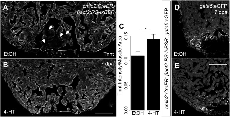
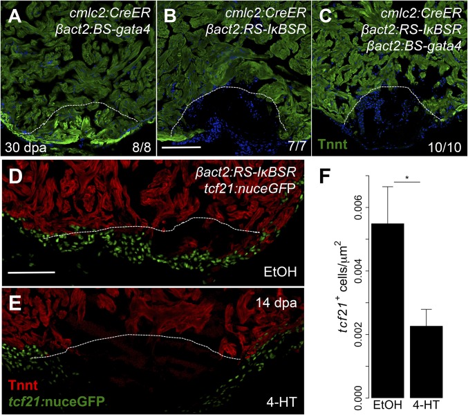
Heart regeneration offers a novel therapeutic strategy for heart failure. Unlike mammals, lower vertebrates such as zebrafish mount a strong regenerative response following cardiac injury. Heart regeneration in zebrafish occurs by cardiomyocyte proliferation and reactivation of a cardiac developmental program, as evidenced by induction of gata4 regulatory sequences in regenerating cardiomyocytes. Although many of the cellular determinants of heart regeneration have been elucidated, how injury triggers a regenerative program through dedifferentiation and epicardial activation is a critical outstanding question. Here, we show that NF-κB signaling is induced in cardiomyocytes following injury. Myocardial inhibition of NF-κB activity blocks heart regeneration with pleiotropic effects, decreasing both cardiomyocyte proliferation and epicardial responses. Activation of gata4 regulatory sequences is also prevented by NF-κB signaling antagonism, suggesting an underlying defect in cardiomyocyte dedifferentiation. Our results implicate NF-κB signaling as a key node between cardiac injury and tissue regeneration.

Keywords: NF-κB; cardiomyocyte; epicardium; heart regeneration; zebrafish.


Conflict of interest statement

The authors declare no conflict of interest.

Figures

Fig. 1.
Fig. 1.
NF-κB activity is induced by injury and during regeneration. (A) qPCR for NF-κB target genes in regenerating ventricles at 7 dpi in cmlc2:CreER; βact2:RS-DTA animals treated with tamoxifen (n = 6) compared with ventricles from cmlc2:CreER; βact2:RS-DTA animals treated with vehicle (n = 6). Error bars indicate SE. *P < 0.05, **P < 0.01, ***P < 0.001, Student’s t test, two-tailed. Each replicate consists of a pool of 2–3 ventricles. (BE) In situ hybridization for nfkbiaa or nfkbiab on sections from ventricles of cmlc2:CreER; βact2:RS-DTA animals treated with vehicle (B and D) or tamoxifen (C and E). Violet staining indicates expression. (FI) Time course of NF-κB:eGFP induction during regeneration in sections from an uninjured ventricle, a 1 dpa ventricle, a 7 dpa ventricle, and a 14 dpa ventricle. Ventricles are counterstained with an antibody against Tnnt (gray) to delineate cardiomyocytes. (Scale bars: 100 μm.)
Fig. S1.
Fig. S1.
NF-κB factors during heart regeneration. (AH) In situ hybridization for NF-κB transcription factors on sections from ventricles of cmlc2:CreER; βact2:RS-DTA animals treated with vehicle (AD) or tamoxifen (EH) to diffusely ablate cardiomyocytes. nfkb1 was the only gene from this group to show a detectable signal in cardiomyocytes. (IK) Staining of uninjured adult mouse hearts for NF-κB1 and Tnnt, indicating that NF-κB1 is present in murine cardiomyocytes (arrowheads). We did not detect an obvious difference in NF-κB1+ cardiomyocytes in compact versus trabecular muscle. (Scale bar: 100 μm.)
Fig. 2.
Fig. 2.
NF-κB is required for heart regeneration. (A) Schematic of βact2:RS-IκBSR transgene. (B and C) Section images of cmlc2:CreER; βact2:RS-IκBSR; NF-κB:eGFP hearts 60 d after treatment (dpt) with vehicle (n = 12) or 4-HT (n = 12). (D and E) Section images of cmlc2:CreER; βact2:RS-IκBSR ventricles at 30 d after amputation after treatment with vehicle or 4-HT. Images are stained for troponin to denote cardiac muscle. Dashed line indicates approximate resection plane. (F and G). Section images of cmlc2:CreER; βact2:RS-IκBSR ventricles at 30 d after amputation after treatment with vehicle or 4-HT. Images are stained with acid fuchsin orange G (AFOG) to identify scar (blue) and fibrin (red). (Scale bars: 100 μm.)
Fig. 3.
Fig. 3.
NF-κB modulates cardiomyocyte proliferation and dedifferentiation. (A and B) Section images from cmlc2:CreER; βact2:RS-IκBSR hearts treated with vehicle or 4-HT at 7 dpa. Sections are stained for Mef2 (red) and PCNA (green). Arrowheads indicate Mef2+/PCNA+ nuclei. Dashed line indicates approximate resection plane. (C) Quantification of cardiomyocyte proliferation in cmlc2:CreER; βact2:RS-IκBSR hearts treated with vehicle (n = 15) or 4-HT (n = 17) at 7 dpa. Error bars indicate SEM. *P = 0.004, Mann–Whitney test. (DI) Sections from NF-κB:eGFP; gata4:DsRed2 hearts at 14 and 30 dpa. (JQ) Section images from cmlc2:CreER; βact2:RS-IκBSR; gata4:eGFP hearts treated with vehicle (n = 9) or 4-HT (n = 10) at 7 dpa. Inset shows unique high-magnification images for Tnnt, eGFP, and a merge of both channels. (R) Plot for density of putative NFκB1 sites in the gata4 promoter. seqLogo is for the consensus NFκB1 binding site used for analysis. Arrow indicates the region of the promoter with highest density of NFκB1 sites. (S) ChIP-PCR for the region of the gata4 promoter highlighted by arrowhead in R after immunoprecipitation for NFκB1 from cardiac chromatin extracts of cmlc2:CreER; βact2:RS-IκBSR fish treated with vehicle or 4-HT at 7 dpa. (Scale bars: 100 μm.)
Fig. S2.
Fig. S2.
Cardiomyocyte apoptosis assays during NF-κB inhibition. TUNEL staining on sections of 7 dpa hearts from cmlc2:CreER; βact2:RS-IκBSR fish treated with vehicle (A) or 4-HT (B). No gross differences in TUNEL staining were noted during NF-κB inhibition. Arrowheads indicate TUNEL + nuclei. (Scale bar: 100 μm.)
Fig. S3.
Fig. S3.
NF-κB signaling contributes to cardiomyocyte dedifferentiation. (A and B) Representative images of 7 dpa ventricles from cmlc2:CreER; βact2:RS-IκBSR treated with vehicle (n = 6) or 4-HT (n = 7) stained for Tnnt (gray). Arrowheads show diminished Tnnt staining in cardiomyocytes adjacent to the wound in control animals, suggestive of sarcomere disassembly. (C) Quantification of intensity of Tnnt intensity per muscle area suggests decreased sarcomere disassembly in ventricles with defective NF-κB signaling (n = 13). (D and E) Section images of 7 dpa ventricles from cmlc2:CreER; βact2:RS-IκBSR; gata5:eGFP fish treated with vehicle (n = 5) or 4-HT (n = 5). eGFP fluorescence is shown in grayscale. *P < 0.05, Student’s t test, two-tailed. (Scale bars: 100 μm.)
Fig. 4.
Fig. 4.
NF-κB has pleiotropic inhibitory effects on heart regeneration. (AC) Section images of cmlc2:CreER; βact2:BS-gata4, cmlc2:CreER; βact2:RS-IκBSR, and cmlc2:CreER; βact2:BS-gata4; βact2:RS-IκBSR ventricles at 30 dpa after treatment with 4-HT. Images are stained for Tnnt to demarcate cardiac muscle. Dashed line indicates approximate resection plane. (D and E) Tilescan images of cmlc2:CreER; βact2:RS-IκBSR; tcf21:nuceGFP ventricles at 14 dpa after treatment with vehicle or 4-HT. Images are stained for Tnnt to denote cardiac muscle. (F) Quantification of tcf21+ cell responses in cmlc2:CreER; βact2:RS-IκBSR hearts treated with vehicle (n = 10) or 4-HT (n = 12) at 14 dpa. Error bars indicate SE. *P < 0.05, Student’s t test, two-tailed. (Scale bars: 100 μm.)
Fig. S4.
Fig. S4.
gata4 overexpression in cardiomyocytes. (A) Schematic of βact2:BS-gata4 transgene. (B and C) Section images of cmlc2:CreER; βact2:BS-gata4 ventricles after treatment with vehicle or 4-HT. Images are stained with an anti-FLAG antibody to identify overexpressed Gata4. (Scale bar: 100 μm.)
Fig. S5.
Fig. S5.
gata4 overexpression does not alter cardiomyocyte proliferation defects caused by NF-κB depletion. (AC) Section images of 7 dpa ventricles from cmlc2:CreER; βact2:RS-IκBSR; βact2:BS-gata4 fish treated with 4-HT. Sections are stained for Mef2 (red) and PCNA (green). Arrowheads indicate Mef2+/PCNA+ nuclei. Dashed line indicates approximate resection plane. (D) Quantification of cardiomyocyte proliferation from 7 dpa cmlc2:CreER; βact2:BS-gata4 (n = 7), cmlc2:CreER; βact2:RS-IκBSR (n = 5), and cmlc2:CreER; βact2:RS-IκBSR; βact2:BS-gata4 (n = 8) ventricles treated. Error bars indicate SEM. *P < 0.05, Mann–Whitney test.

Similar articles

Cited by

References

    1. Mozaffarian D, et al. American Heart Association Statistics Committee and Stroke Statistics Subcommittee Heart disease and stroke statistics--2015 update: A report from the American Heart Association. Circulation. 2015;131(4):e29–e322. - PubMed
    1. Bergmann O, et al. Evidence for cardiomyocyte renewal in humans. Science. 2009;324(5923):98–102. - PMC - PubMed
    1. Senyo SE, et al. Mammalian heart renewal by pre-existing cardiomyocytes. Nature. 2013;493(7432):433–436. - PMC - PubMed
    1. Poss KD, Wilson LG, Keating MT. Heart regeneration in zebrafish. Science. 2002;298(5601):2188–2190. - PubMed
    1. Kikuchi K, et al. Primary contribution to zebrafish heart regeneration by gata4(+) cardiomyocytes. Nature. 2010;464(7288):601–605. - PMC - PubMed

Publication types

LinkOut - more resources