. 2016 Jan;245(1):87-95.
doi: 10.1002/dvdy.24359. Epub 2015 Nov 3.

Expression of the Drosophila homeobox gene, Distal-less, supports an ancestral role in neural development

Affiliations

Expression of the Drosophila homeobox gene, Distal-less, supports an ancestral role in neural development

Jessica S Plavicki et al. Dev Dyn. 2016 Jan.

Abstract

Background: Distal-less (Dll) encodes a homeodomain transcription factor expressed in developing appendages of organisms throughout metazoan phylogeny. Based on earlier observations in the limbless nematode Caenorhabditis elegans and the primitive chordate amphioxus, it was proposed that Dll had an ancestral function in nervous system development. Consistent with this hypothesis, Dll is necessary for the development of both peripheral and central components of the Drosophila olfactory system. Furthermore, vertebrate homologs of Dll, the Dlx genes, play critical roles in mammalian brain development.

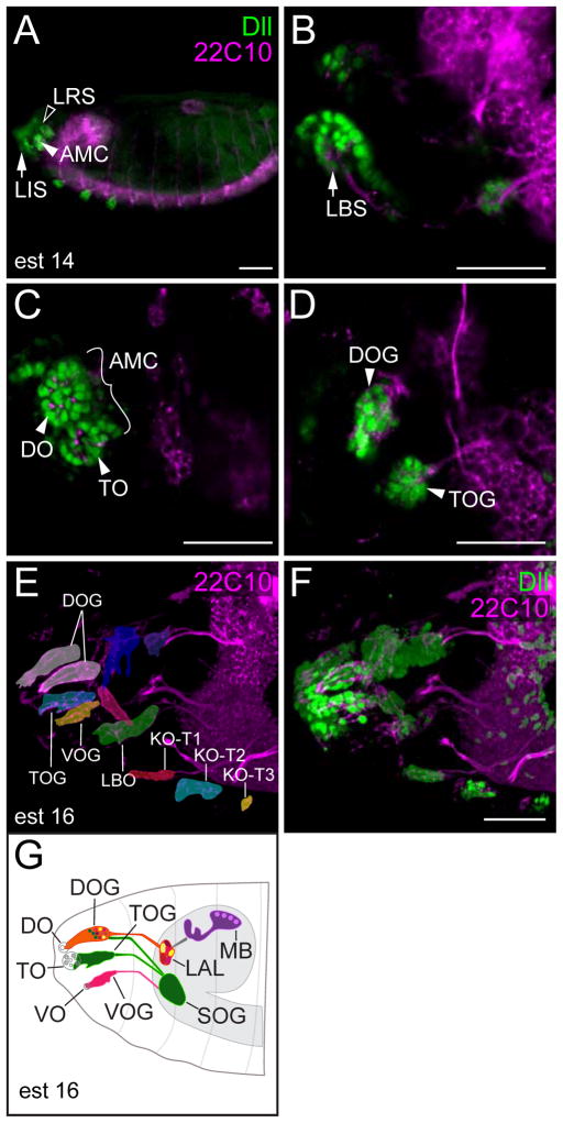
Results: Using fluorescent immunohistochemistry of fixed samples and multiphoton microscopy of living Drosophila embryos, we show that Dll is expressed in the embryonic, larval and adult central nervous system and peripheral nervous system (PNS) in embryonic and larval neurons, brain and ventral nerve cord glia, as well as in PNS structures associated with chemosensation. In adult flies, Dll expression is expressed in the optic lobes, central brain regions and the antennal lobes.

Conclusions: Characterization of Dll expression in the developing nervous system supports a role of Dll in neural development and function and establishes an important basis for determining the specific functional roles of Dll in Drosophila development and for comparative studies of Drosophila Dll functions with those of its vertebrate counterparts.

Keywords: Dll; Dlx; nervous system; olfaction; sensory system.


Figures

Figure 1
Figure 1. In vivo multiphoton imaging of Dll expressing cells during embryogenesis
A–J: Still shots from a multiphoton movie spanning late germ-band elongation (stage 10; A) to head involution (stage 17; J) of an embryo of genotype: w1118; P{UAS-GFP.nls}14 P{GawB}Dllmd23/CyO in which nuclear GFP expression is driven by a Dll-Gal4 enhancer trap. Dll expression is detected in the antennal and maxillary head segments (asterisks in A–C) that give rise to the olfactory and gustatory organs (asterisks in D–J) as well as presumptive (arrowheads in B–E) and delaminated cells of the brain (arrowheads in F–J). Still images are taken from Supplemental Movie 1 which was initiated at ~6 hours after egg laying and continued until ~14 hours after egg laying. Minutes indicated are relative to start of imaging. Scale bar = 50 μm.
Figure 2
Figure 2. Colocalization of GFP expression regulated by P{GawB}Dllmd23 driver and Dll protein
A-B″: Dorsal views of stage 16 embryos. A: Still image from Supplemental Movie 2 (frame #97). B: w1118; P{UAS-GFP.nls}14 P{GawB}Dllmd23/CyO embryo stained for Dll (red) and 22C10 (blue). Monoclonal antibody 22C10 recognizes the microtubule associated protein Futsch and is used to delineate neuronal morphology (Hummel et al., 2000). Dll protein, which is nuclear, is contained within a broader domain of GFP expression. This is expected due to both the perdurance of GFP and GFP localizing to cytoplasm as well as nucleus. We note that while there does not appear to be GFP outside the Dll expression domains, there is some Dll protein expression where we do not detect GFP (e.g. red alone in panel B). GFP and Dll are expressed in the dorsal organ ganglion (arrows), the terminal organ ganglion (arrowheads), and the neurons in the labral sensory organ complex (asterisks). B′: GFP expression relative to 22C10. B″: Dll protein expression relative to 22C10. A–B″: 40× confocal images. Scale bar = 50 μm.
Figure 3
Figure 3. Dll expression in sensory organ precursors
A-G: Lateral views of a stage 13 embryo stained for Dll (green), Senseless (Sens; red), and Prospero (Pros; blue). A, E–G″: Single focal planes. B–D: Brightest point projections of a z-series from the boxed region in (A). C: Dll is coexpressed with a subset of Sens-expressing cells in the antennal (an), maxillary (mx), and labial (lb) head segments and the thoracic segments (T1–3). D: Dll is coexpressed with a subset of Pros-expressing cells in the an, mx, and lb head segments and the thoracic segments. E: Boxes 1 and 2 mark the segments shown at higher magnification in F–F, and G–G, respectively. In the mx segment, there are many Dll-expressing cells that lack both Pros and Sens, while in the T1 segment almost all of the Dll-expressing cells have Pros and/or Sens expression. F: Dll, Sens and Pros expression in the mx segment. F′: Dll and Sens. F″: Dll and Pros. G: Dll, Sens and Pros expression in T1. G′: Dll and Sens. G″: Dll and Pros. A: 20× confocal image. B–G″: 40× confocal images. Scale bars = 50 μm.
Figure 4
Figure 4. Dll is expressed in subsets of glia in the ventral nerve cord (VNC) and in both neurons and glia in the supraesophageal ganglion
Lateral (A, B), ventral (C, D), and dorsal (E, F) views of wild type stage 14 embryos stained for Dll (green), the glial marker Repo (red), and the neuronal morphology marker 22C10 (blue). Yellow indicates areas of overlapping Dll and Repo expression. A, B: Dll expression in cell body associated glia (arrows in B) and dorsal longitudinal glia (arrowheads in B). Boxed region in (A) is shown at higher magnification in (B). C, D: Dll expression in dorsal longitudinal glia (arrowheads in D). Boxed region in (C) is shown at higher magnification in (D). E, F: Dll expression in neurons (asterisks in F) and glia (arrows in F) in the supraesophageal ganglion. Boxed region in (E) is shown at higher magnification in (F). G, H, I: Dorsal views of a w1118; P{UAS-GFP.nls}14 P{GawB}Dllmd23/CyO third instar larval brain stained with anti-Repo (red) and anti-horseradish peroxidase (anti-HRP; blue). The region marked by boxes 1 & 2 are shown at higher magnification in (H) and (I), respectively. Dll continues to be expressed in brain (H) and VNC glia (I) during larval stages. J, K: X-gal staining of an early third instar larval central nervous system from an animal carrying a Dll-lacZ enhancer trap (Dll01092; Goto and Hayashi, 1997). Boxed region in (J) is shown at higher magnification in (K). A, C, E, G: 10× confocal images. B, D, F, H, I: 40× confocal images. J: 10× image. K: 40× image. A, C, E: Scale bars = 100 μm. B, D, F, H, I, K: Scale bars = 25 μm. G, J: Scale bar = 50 μm.
Figure 5
Figure 5. Dll expression in anterior sense organs of the embryonic peripheral nervous system (PNS)
A–D: Lateral views of a wild type stage 14 embryo stained for Dll (green) and 22C10 (purple). A: Dll is expressed in the labial (LIS; arrow), labral (LRS; open arrowhead) and antennomaxillary sensory complexes (AMC: arrowhead). B: Dll expression in the labial sensory complex. C: Dll expression in the developing cuticular components of the DO and TO. D: Dll expression in the dorsal organ ganglion (DOG) neurons that innervate the dorsal organ (DO in panel C) and in the neurons of the terminal organ ganglion (TOG) that innervate the terminal organ (TO in panel C). E, F: Lateral view of a stage 16 embryo stained for Dll (green) and 22C10 (purple). Dll is expressed in both neurons and supporting cells of multiple sense organs in the head, including the DOG, TOG, ventral organ ganglion (VOG), labial organ (LBO). Dll also is expressed in the mechanosensory Keilin’s organs (KO) of the three thoracic segments (T1–T3). The Dll-expressing sense organs in (F) are highlighted in (E). G: Schematic of the large, Dll-expressing chemosensory organs in (E) and (F). DO, TO, and VO are the dorsal organ, terminal organ, and ventral organ, respectively. These organs lie in the larval epidermis and are innervated by neurons with cell bodies in their respective ganglia (DOG, TOG and VOG). From the ganglia, axons project to targets within the larval brain as. Olfactory DOG axons project to the larval antennal lobe (LAL). The LAL relays olfactory information to the mushroom body (MB) and lateral horn (not shown). Gustatory DOG axons, along with gustatory TOG and VOG axons project to the subesophageal ganglion (SOG). A: 10× confocal image. B, C, D, E, F: 40× confocal images. A: Scale bar = 100 μm. B, C, D, F: Scale bars = 50 μm.
Figure 6
Figure 6. Dll is expressed in larval and adult chemosensory neurons
A: Dorsal view of a w1118; P{UAS-GFP.nls}14 P{GawB}Dllmd23/CyO (green) third instar larval brain stained with anti-horseradish peroxidase (HRP; purple). Anti-HRP recognizes Nervana proteins in neuronal membranes and is used to visualize the neuropil (Jan and Jan, 1982). Dll expression is seen in central brain regions. Dll-expressing olfactory receptor neurons (ORNs) in the dorsal organ ganglion (DOG) project to the larval antennal lobe (LAL) whereas Dll-expressing gustatory receptor neurons in the DOG, terminal organ ganglion (TOG) and the ventral organ (VOG) project to the subesophageal ganglion (SOG). B: Co-localization of Dll antibody staining with the neuronal marker Elav (O’Neill et al., 1994) and with GFP driven by P{GawB}Dllmd23 (Calleja et al., 1996). Dll is expressed in TOG neurons. C: Higher magnification view of the LAL boxed in panel A. D: Anterior view of the adult antennal lobes (AL). Both larval and adult Dll expressing ORNs project to their respective antennal lobes. E: Schematic of an adult brain from a frontal view. The mushroom body (MB), lateral horn (LH), and antennal lobe (AL) are indicated. The inset is a schematic of an adult antenna with the olfactory receptor neuron (ORN) cell bodies indicated in red, orange, and yellow. ORNs expressing the same odorant receptor project to a single glomerulus in the AL. A: 10× confocal images. B, C, D: 40× confocal images. A, B, D: Scale bars = 50 μm. C: Scale bar = 12.5 μm.
Figure 7
Figure 7. Dll is expressed in the larval and adult optic lobes
A: Lateral view of a w1118; P{UAS-GFP.nls}14 P{GawB}Dllmd23/CyO (green) third instar larval optic lobe stained for Dac (blue) and Elav (red). Dac is an early marker of differentiated laminal neurons, Elav marks laminal neurons in later stages of optic lobe development (Morante and Desplan, 2008; Bertet et al., 2014). B: Higher magnification view of optic lobe shown in (A). C: Dorsal view of a larval third instar brain stained for Dll (green) and Fas2 (red). Fas2 marks a subset of neuronal membranes. Dll is expressed in laminal neurons (box in C) and the medulla (arrows in C). D: High magnification view of boxed area from (C). E: Anterior view of an adult brain stained for Dll (green), Repo (red) and anti-HRP (blue). Dll is expressed in neurons in central regions of the adult brain (asterisks) and in the optic lobes (arrow and arrowhead). Arrow indicates Dll-expressing cells in the medulla, while arrowhead indicates Dll-expressing cells in the lamina. F: High magnification view of boxed area in (E). Dll is expressed in adult optic lobe neurons, but not glia. (G) X-gal staining of an adult brain from an animal carrying a reporter insertion into the Dll locus (Dll01092; Goto and Hayashi, 1997). This reporter faithfully recapitulates Dll expression in both the central brain (asterisks) as well as the medulla (arrow) and lamina (arrowhead) of the optic lobes. A, C: 20× confocal images. E: 10× confocal image. B, D, F: 40× confocal images. A, C, E, F, G: Scale bars = 50 μm. B, D: Scale bars = 25 μm.

Similar articles

Cited by

References

    1. Alfonso T, Jones B. gcm2 promotes glial cell differentiation and is required with glial cells missing for macrophage development in Drosophila. Dev Biol. 2002;248:369–383. - PubMed
    1. Arendt D, Nubler-Jung K. Comparison of early nerve cord development in insects and vertebrates. Development. 1999;126:2309–2325. - PubMed
    1. Bertet C, Li X, Erclik T, Cavey M, Wells B, Desplan C. Temporal patterning of neuroblasts controls Notch-mediated cell survival through regulation of Hid or Reaper. Cell. 2014;158:1173–1186. - PMC - PubMed
    1. Calleja M, Moreno E, Pelaz S, Morata G. Visualization of gene expression in living adult Drosophila. Science. 1996;274(5285):252–255. - PubMed
    1. Campbell G, Goring H, Lin T, Spana E, Andersson S, Doe CQ, Tomlinson A. RK2, a glial-specific homeodomain protein required for embryonic nerve cord condensation and viability in Drosophila. Development. 1994;120:2957–2966. - PubMed

Publication types

MeSH terms

LinkOut - more resources