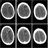
Multiple calcified brain metastases in a man with invasive ductal breast cancer
- PMID: 26472289
- PMCID: PMC4612730
- DOI: 10.1136/bcr-2015-211777
Multiple calcified brain metastases in a man with invasive ductal breast cancer
Abstract
We report a case of a 52-year-old Caucasian man with invasive ductal carcinoma of the breast. One year after initial diagnosis, he developed a generalised epileptic seizure and neuroimaging showed multiple, calcified intracerebral lesions. Owing to these atypical cerebral imaging findings, comprehensive serological and cerebrospinal fluid analysis was conducted and a latent toxoplasmosis was suspected. In order to distinguish between metastases and an infectious disease, a cerebral biopsy was performed, which verified brain metastases. The patient received whole-brain radiotherapy. The last cerebral CT scan, 18 months later showed stable disease. Calcification of brain metastases in patients with breast cancer is very rare. Owing to their non-characteristic radiological appearance with a lack of contrast enhancement, diagnosis of metastases can be difficult. Infectious diseases should be considered within the diagnostic work up. Owing to possible pitfalls, we recommend a widespread differential diagnostic work up in similar cases, and even in cases with a confirmed primary tumour.
2015 BMJ Publishing Group Ltd.
Figures


Similar articles
-
[Late metastases from breast cancer--report of two cases].Khirurgiia (Sofiia). 2010;(1):62-6. Khirurgiia (Sofiia). 2010. PMID: 21972709 Bulgarian.
-
Cortical blindness and choroidal metastases secondary to metastatic breast carcinoma in a male patient.Ophthalmic Surg Lasers Imaging Retina. 2013 Jan-Feb;44(1):81-4. doi: 10.3928/23258160-20121221-18. Ophthalmic Surg Lasers Imaging Retina. 2013. PMID: 23410813
-
A case report of advanced male breast cancer with an objective response to tamoxifen treatment.Breast Cancer. 2000;7(3):256-60. doi: 10.1007/BF02967470. Breast Cancer. 2000. PMID: 11029808
-
[Pineal metastasis of breast cancer: case report and review].Cancer Radiother. 2014 Mar;18(2):139-41. doi: 10.1016/j.canrad.2013.10.012. Epub 2014 Jan 10. Cancer Radiother. 2014. PMID: 24418000 Review. French.
-
[Calcified cerebral metastases. Study of two cases and review of literature].Neurologia. 2000 Mar;15(3):136-9. Neurologia. 2000. PMID: 10846876 Review. Spanish.
Cited by
-
A cerebellopontine angle metastatis of a male breast cancer: Case report.Ann Med Surg (Lond). 2022 Mar 1;75:103421. doi: 10.1016/j.amsu.2022.103421. eCollection 2022 Mar. Ann Med Surg (Lond). 2022. PMID: 35386782 Free PMC article.
-
Unveiling the Hidden Burden: A Systematic Review on the Prevalence and Clinical Implications of Calcified Brain Metastases.Biomolecules. 2024 Dec 11;14(12):1585. doi: 10.3390/biom14121585. Biomolecules. 2024. PMID: 39766292 Free PMC article.
References
-
- Fatehi F, Basiri K, Mehr GK. Calcified brain metastasis of breast cancer. Acta Neurol Belg 2010;110:124–5. - PubMed
Publication types
MeSH terms
LinkOut - more resources
Full Text Sources
Other Literature Sources
Medical
