Review
. 2016 Jan;73(1):119-27.
doi: 10.1007/s00018-015-2058-0.

Gated entry into the ciliary compartment

Review

Gated entry into the ciliary compartment

Daisuke Takao et al. Cell Mol Life Sci. 2016 Jan.

Abstract

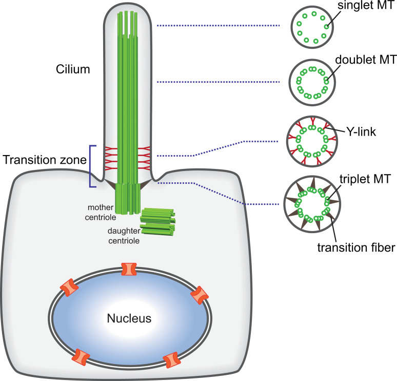
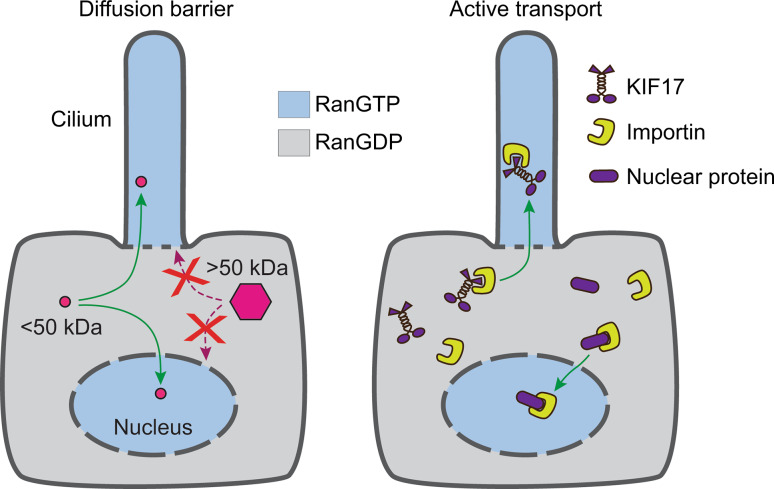
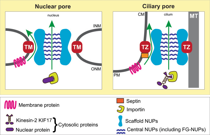
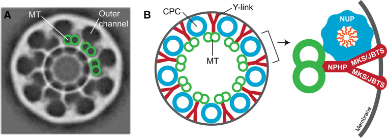
Cilia and flagella play important roles in cell motility and cell signaling. These functions require that the cilium establishes and maintains a unique lipid and protein composition. Recent work indicates that a specialized region at the base of the cilium, the transition zone, serves as both a barrier to entry and a gate for passage of select components. For at least some cytosolic proteins, the barrier and gate functions are provided by a ciliary pore complex (CPC) that shares molecular and mechanistic properties with nuclear gating. Specifically, nucleoporins of the CPC limit the diffusional entry of cytosolic proteins in a size-dependent manner and enable the active transport of large molecules and complexes via targeting signals, importins, and the small G protein Ran. For membrane proteins, the septin protein SEPT2 is part of the barrier to entry whereas the gating function is carried out and/or regulated by proteins associated with ciliary diseases (ciliopathies) such as nephronophthisis, Meckel–Gruber syndrome and Joubert syndrome. Here, we discuss the evidence behind these models of ciliary gating as well as the similarities to and differences from nuclear gating.


Figures

Fig. 1
Fig. 1
General structure of the primary cilium. The mother centriole contains nine triplet microtubules (MT) and is anchored to the periciliary membrane by transition fibers. Nine doublet microtubules protrude from the triplet microtubules and form the axoneme. The proximal end of the cilium is termed the transition zone and is the site for gated entry into the ciliary compartment. Here, Y-shaped structures (the Y-links) connect the doublet microtubules to the ciliary membrane. At the distal end of the cilium in some cells and species, the doublet microtubules convert to singlet microtubules
Fig. 2
Fig. 2
Gated entry of cytosolic molecules into the ciliary and nuclear compartments. Entry of cytosolic molecules is characterized by a size-exclusion barrier (left) and active transport across the barrier (right). The size-exclusion barrier limits the diffusional entry of soluble molecules larger than ~50 kDa. Large molecules gain entry via the active transport process in which importins bind to nuclear or ciliary proteins and mediate their transport across the barriers. The directionality of transport is specified by a RanGTP–RanGDP gradient across the ciliary-cytoplasmic and nuclear-cytoplasmic barriers
Fig. 3
Fig. 3
Models for nuclear and ciliary gating. Both nuclear and ciliary pores contain a central channel for the gated entry of cytosolic proteins with central pore NUPs (e.g., the FG-NUP NUP62) providing this sieve-like barrier. Gated entry of membrane proteins utilizes peripheral channels of the NPC for nuclear gating, while transition zone (TZ) proteins may form a peripheral channel for gated entry of ciliary membrane proteins. Both nuclear and ciliary pores contain scaffold NUPs (e.g., NUP93, NUP35). For the NPC, the scaffold is anchored by transmembrane (TM) NUPs whereas in the CPC, the scaffold may be anchored by transition zone proteins. TM transmembrane NUP, TZ transition zone protein, MT microtubule
Fig. 4
Fig. 4
Possible structural relationship between the ciliary pore and the transition zone. a Electron cryotomography reveals nine pores (outer channels) in isolated basal bodies from Tetrahymena. Each outer channel is located adjacent to a doublet microtubule (MT) and may be a pore for the entry of ciliary components. Reprinted from Ounjai et al. [91], with permission from Elsevier. b Schematic drawings of the putative arrangement of the CPC and Y-link structures. The CPCs are comprised of scaffold and central NUPs and form a barrier for entry of cytosolic proteins, whereas the Y-links are comprised of NPHP, MKS, and JBTS proteins and form a barrier for entry of membrane proteins. NPHP nephronophthisis, MKS Meckel–Gruber syndrome, JBTS Joubert syndrome, MT microtubule

References

    1. Basten SG, Giles RH. Functional aspects of primary cilia in signaling, cell cycle and tumorigenesis. Cilia. 2013;2:6. doi: 10.1186/2046-2530-2-6. - DOI - PMC - PubMed
    1. Goetz SC, Anderson KV. The primary cilium: a signalling centre during vertebrate development. Nat Rev Genet. 2010;11:331–344. doi: 10.1038/nrg2774. - DOI - PMC - PubMed
    1. Silverman MA, Leroux MR. Intraflagellar transport and the generation of dynamic, structurally and functionally diverse cilia. Trends Cell Biol. 2009;19:306–316. doi: 10.1016/j.tcb.2009.04.002. - DOI - PubMed
    1. Sung CH, Leroux MR. The roles of evolutionarily conserved functional modules in cilia-related trafficking. Nat Cell Biol. 2013;15:1387–1397. doi: 10.1038/ncb2888. - DOI - PMC - PubMed
    1. Wei Q, Ling K, Hu J. The essential roles of transition fibers in the context of cilia. Curr Opin Cell Biol. 2015;35:98–105. doi: 10.1016/j.ceb.2015.04.015. - DOI - PMC - PubMed

Publication types

Substances

LinkOut - more resources