Review
. 2015:124:3-19.
doi: 10.1016/bs.irn.2015.07.001. Epub 2015 Aug 21.

Neuronal Nicotinic Acetylcholine Receptor Structure and Function and Response to Nicotine

Affiliations
Review

Neuronal Nicotinic Acetylcholine Receptor Structure and Function and Response to Nicotine

John A Dani. Int Rev Neurobiol. 2015.

Abstract

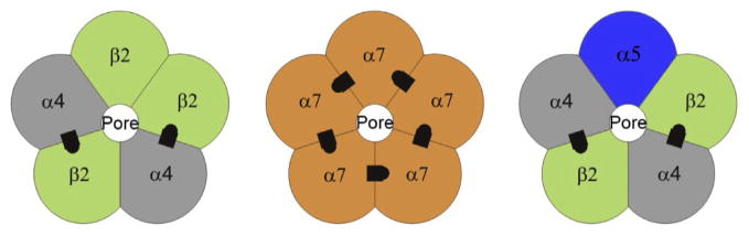
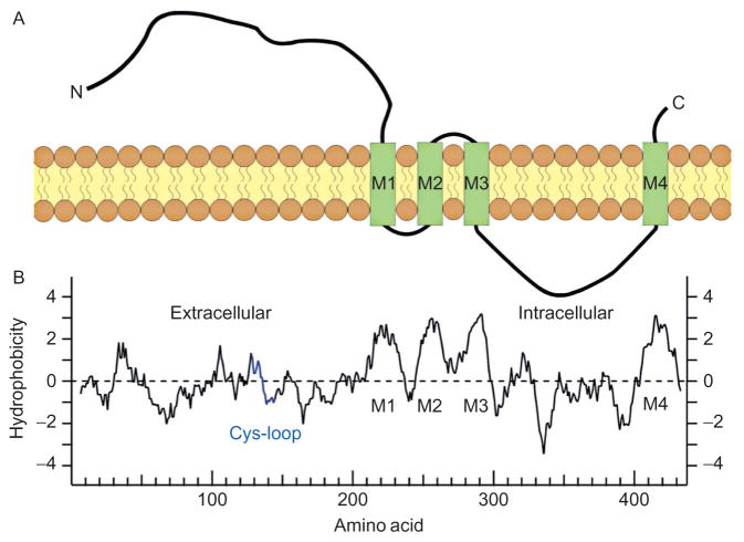
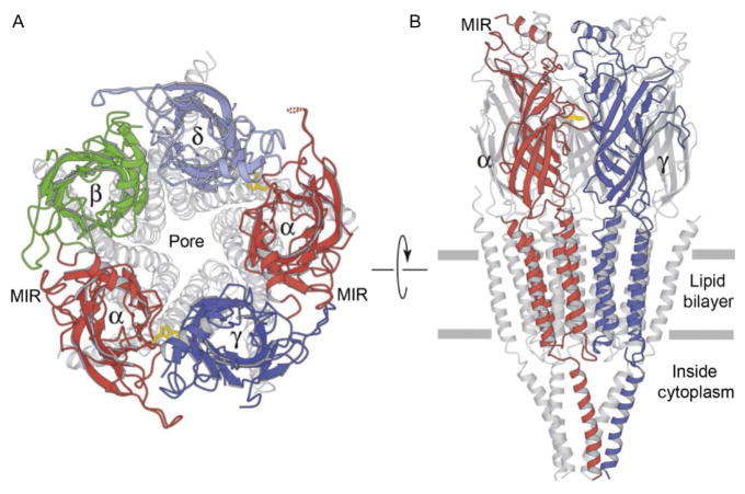
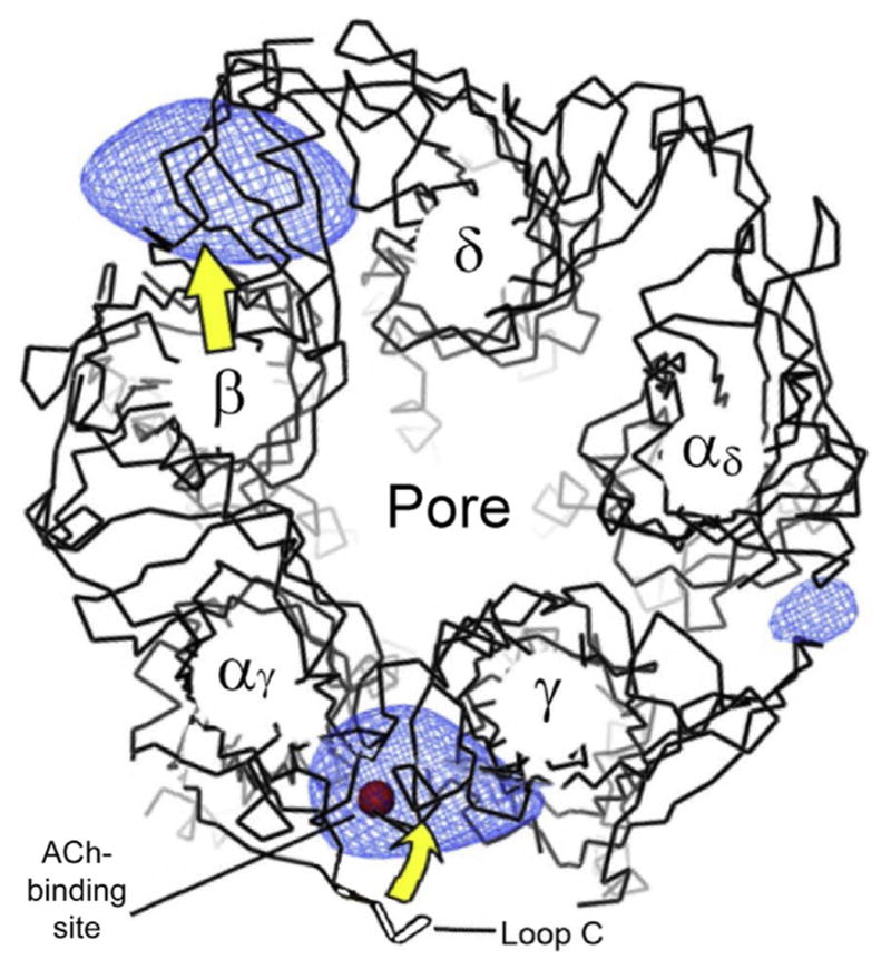
Nicotinic acetylcholine receptors (nAChRs) belong to the "Cys-loop" superfamily of ligand-gated ion channels that includes GABAA, glycine, and serotonin (5-HT3) receptors. There are 16 homologous mammalian nAChR subunits encoded by a multigene family. These subunits combine to form many different nAChR subtypes with various expression patterns, diverse functional properties, and differing pharmacological characteristics. Because cholinergic innervation is pervasive and nAChR expression is extremely broad, practically every area of the brain is impinged upon by nicotinic mechanisms. This review briefly examines the structural and functional properties of the receptor/channel complex itself. The review also summarizes activation and desensitization of nAChRs by the low nicotine concentrations obtained from tobacco. Knowledge of the three-dimensional structure and the structural characteristics of channel gating has reached an advanced stage. Likewise, the basic functional properties of the channel also are reasonably well understood. It is these receptor/channel properties that underlie the participation of nAChRs in nearly every anatomical region of the mammalian brain.

Keywords: Calcium permeability; Gating; Nicotine; Permeation; Presynaptic; Synaptic; nAChR.


Figures

Figure 1
Figure 1
Didactic illustration of the nAChR subunits arranged as pentamers around the water-filled cation-permeable pore. The most common nAChRs in the brain are hetero-oligomeric α4β2 nAChRs and homo-oligomeric α7 nAChRs. The recognized ACh-binding sites are indicated by black asymmetric designs located between adjacent subunits. Adapted from Fig. 1B of McKay, Placzek, and Dani (2007).
Figure 2
Figure 2
Transmembrane topology of a nAChR subunit. (A) A didactic illustration of the linear structure of the nAChR subunit with four transmembrane domains (M1–M4) passing through the lipid bilayer member. (B) A plot of the hydrophobicity profile of a human α1 subunit. The profile is aligned with the linear representation of the subunit just above. Panel (A): Adapted from Fig. 1A of McKay et al. (2007). Panel (B): Adapted from Fig. 1A of Papke (2014).
Figure 3
Figure 3
Illustrations of ribbon diagrams of the nAChR. (A) View from the top into the pore with only the upper most portion highlighted in colors (gray shades in the print version). (B) View from the side with only the front two subunits highlighted in colors (gray shades in the print version). The plane of the membrane is indicated by the two horizontal gray lines. Adapted from Fig. 3 of Unwin (2005).
Figure 4
Figure 4
Illustration of the structural changes induced by ACh (red (dark gray in the print version) sphere) binding into the pocket formed by the closing of loop C, as viewed from the top into the pore. The blue (light gray in the print version) shaded regions represent the most significant increases in density of the open channel relative to the closed channel. The yellow (light gray in the print version) arrows indicate the general structural displacement caused by opening. Adapted from Fig. 2 of Unwin and Fujiyoshi (2012).
Figure 5
Figure 5
Representation of the structural change of the nAChR pore when transitioning from closed (dashed black) to open (red (dark gray in the print version)). The pore is represented to run vertically with the gray horizontal lines delimiting the position of the lipid bilayer membrane. Upon opening, the diameter of the pore increases in the constricting hydrophobic region near the middle of the membrane (at Z =0), and the narrowest region shifts near to the intracellular membrane surface where the pore is lined by polar residues (near Z =−14). Adapted from Fig. 10 of Unwin and Fujiyoshi (2012).

References

    1. Albuquerque EX, Pereira EF, Alkondon M, Rogers SW. Mammalian nicotinic acetylcholine receptors: From structure to function. Physiological Reviews. 2009;89:73–120. - PMC - PubMed
    1. Amador M, Dani JA. Mechanism for modulation of nicotinic acetylcholine receptors that can influence synaptic transmission. The Journal of Neuroscience: The Official Journal of the Society for Neuroscience. 1995;15:4525–4532. - PMC - PubMed
    1. Auerbach A. Agonist activation of a nicotinic acetylcholine receptor. Neuropharmacology. 2014;96:150–156. - PMC - PubMed
    1. Bertrand D, Galzi JL, Devillers-Thiery A, Bertrand S, Changeux JP. Mutations at two distinct sites within the channel domain M2 alter calcium permeability of neuronal alpha 7 nicotinic receptor. Proceedings of the National Academy of Sciences of the United States of America. 1993b;90:6971–6975. - PMC - PubMed
    1. Bertrand D, Galzi JL, Devillers-Thiery A, Bertrand S, Changeux JP. Stratification of the channel domain in neurotransmitter receptors. Current Opinion in Cell Biology. 1993a;5:688–693. - PubMed

Publication types

LinkOut - more resources