Comparative Study
. 2016 Mar;32(3):451-9.
doi: 10.1007/s10554-015-0787-7. Epub 2015 Oct 16.

Increased native T1-values at the interventricular insertion regions in precapillary pulmonary hypertension

Affiliations
Comparative Study

Increased native T1-values at the interventricular insertion regions in precapillary pulmonary hypertension

Onno A Spruijt et al. Int J Cardiovasc Imaging. 2016 Mar.

Abstract

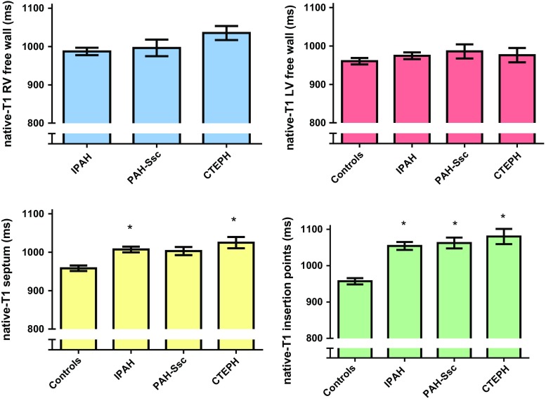
Cardiac magnetic resonance imaging of the pressure overloaded right ventricle (RV) of precapillary pulmonary hypertension (PH) patients, exhibits late gadolinium enhancement at the interventricular insertion regions, a phenomenon which has been linked to focal fibrosis. Native T1-mapping is an alternative technique to characterize myocardium and has the advantage of not requiring the use of contrast agents. The aim of this study was to characterize the myocardium of idiopathic pulmonary arterial hypertension (IPAH), systemic scleroderma related PH (PAH-Ssc) and chronic thromboembolic PH (CTEPH) patients using native T1-mapping and to see whether native T1-values were related to disease severity. Furthermore, we compared native T1-values between the different precapillary PH categories. Native T1-mapping was performed in 46 IPAH, 14 PAH-SSc and 10 CTEPH patients and 10 control subjects. Native T1-values were assessed using regions of interest at the RV and LV free wall, interventricular septum and interventricular insertion regions. In PH patients, native T1-values of the interventricular insertion regions were significantly higher than the native T1-values of the RV free wall, LV free wall and interventricular septum. Native T1-values at the insertion regions were significantly related to disease severity. Native T1-values were not different between IPAH, PAH-Ssc and CTEPH patients. Native T1-values of the interventricular insertion regions are significantly increased in precapillary PH and are related to disease severity. Native T1-mapping can be developed as an alternative technique for the characterization of the interventricular insertion regions and has the advantage of not requiring the use of contrast agents.

Keywords: Myocardium; Non-contrast T1; Pulmonary hypertension; T1-mapping.


Figures

Fig. 1
Fig. 1
Schematic illustration of ROIs. Schematic illustration of a mid-ventricular short-axis image. ROIs of the different regions are marked with colors. ROIs of the LV free wall (red) covered the total LV free wall. ROIs of the RV free wall (blue), were only analyzed in the inferior part of the RV free wall. Attempts to cover the total RV free wall failed because in a majority of the patients the free wall was too thin, resulting in unreliable native T1-values due to partial volume effects. ROIs of the interventricular septum are marked in yellow. ROIs of the interventricular insertion regions are marked in green
Fig. 2
Fig. 2
Native T1-values (ms) of the RV free wall, LV free wall, interventricular septum and the interventricular insertion regions. Data is presented as mean and standard error of the mean. a Regional differences in the myocardium of control subjects. No regional differences were found in control subjects. b Regional differences in the myocardium of PH patients. T1-values of the RV free wall of PH patients were not different from T1-values of the LV free wall and interventricular septum. *T1-values of the interventricular septum of PH patients were significantly higher compared to native T1-values of the LV free wall. #Native T1-values of the interventricular insertion regions of PH patients were significantly higher compared to native T1-values of the RV free wall, LV free wall and interventricular septum
Fig. 3
Fig. 3
Native T1-maps showing increased native T1-values at the interventricular insertion regions. Two examples of native T1-maps of a control subject (a) and an IPAH (b) patient. The white arrows indicate the increased native T1-values at the interventricular insertion regions
Fig. 4
Fig. 4
Correlations between native T1-values of the interventricular insertion regions and RVEDVI, RVESVI, RVEF and RAP in PH patients. Native T1-values of the interventricular insertion regions of PH patients were significantly related to RVEDVI, RVESVI, RVEF and RAP. RVEDVI right ventricular end-diastolic volume index, RVESVI right ventricular end-systolic volume index, RVEF right ventricular ejection fraction, RAP right atrial pressure
Fig. 5
Fig. 5
Comparison of native T1-values between IPAH, PAH-SSc and CTEPH patients and control subjects. Data is presented as mean and standard error of the mean. The Y-axis Native T1-values of the RV free wall (blue), LV free wall (red), interventricular septum (yellow) and interventricular insertion regions (green) were not significantly different between IPAH, PAH-SSc and CTEPH patients. Native T1-values of the LV free wall was not significantly different between control subjects and PH patients. Native T1-values of the interventricular septum was significantly higher in IPAH and CTEPH patients compared to control subjects. Native T1-values of the interventricular insertion regions were significantly increased in all PH categories compared to control subjects

References

    1. van de Veerdonk MC, Kind T, Marcus JT, Mauritz GJ, Heymans MW, Bogaard HJ, Boonstra A, Marques KM, Westerhof N, Vonk-Noordegraaf A. Progressive right ventricular dysfunction in patients with pulmonary arterial hypertension responding to therapy. J Am Coll Cardiol. 2011;58:2511–2519. doi: 10.1016/j.jacc.2011.06.068. - DOI - PubMed
    1. Blyth KG, Groenning BA, Martin TN, Foster JE, Mark PB, Dargie HJ, Peacock AJ. Contrast enhanced-cardiovascular magnetic resonance imaging in patients with pulmonary hypertension. Eur Heart J. 2005;26:1993–1999. doi: 10.1093/eurheartj/ehi328. - DOI - PubMed
    1. McCann GP, Beek AM, Vonk-Noordegraaf A, van Rossum AC. Delayed contrast-enhanced magnetic resonance imaging in pulmonary arterial hypertension. Circulation. 2005;112:e268. doi: 10.1161/CIRCULATIONAHA.104.512848. - DOI - PubMed
    1. McCann GP, Gan CT, Beek AM, Niessen HW, Vonk Noordegraaf A, van Rossum AC. Extent of MRI delayed enhancement of myocardial mass is related to right ventricular dysfunction in pulmonary artery hypertension. Am J Roentgenol. 2007;188:349–355. doi: 10.2214/AJR.05.1259. - DOI - PubMed
    1. Sanz J, Dellegrottaglie S, Kariisa M, Sulica R, Poon M, O’Donnell TP, Mehta D, Fuster V, Rajagopalan S. Prevalence and correlates of septal delayed contrast enhancement in patients with pulmonary hypertension. Am J Cardiol. 2007;100:731–735. doi: 10.1016/j.amjcard.2007.03.094. - DOI - PubMed

Publication types

MeSH terms

LinkOut - more resources