. 2015 Oct 16;350(6258):aac4383.
doi: 10.1126/science.aac4383. Epub 2015 Oct 15.

Structural basis of histone H3K27 trimethylation by an active polycomb repressive complex 2

Affiliations

Structural basis of histone H3K27 trimethylation by an active polycomb repressive complex 2

Lianying Jiao et al. Science. .

Abstract

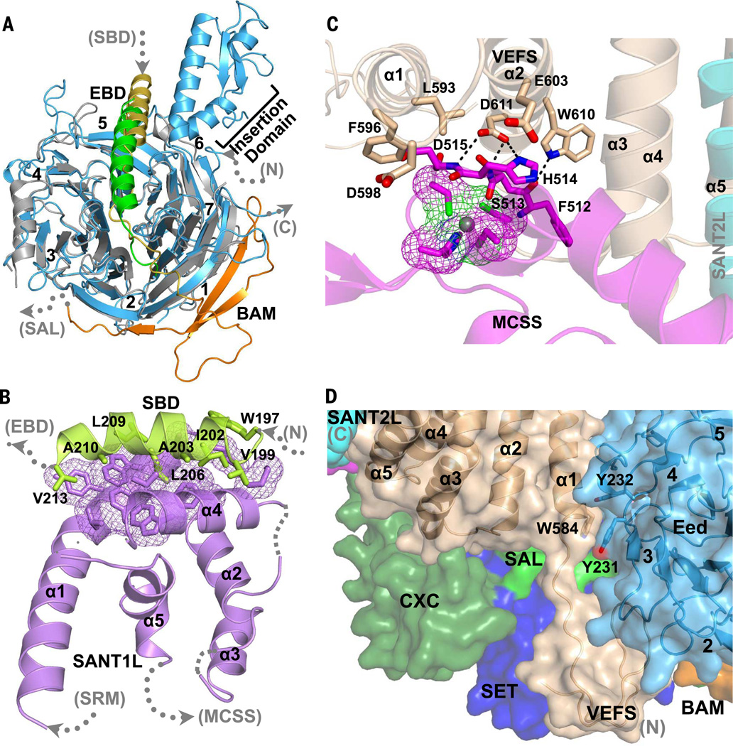
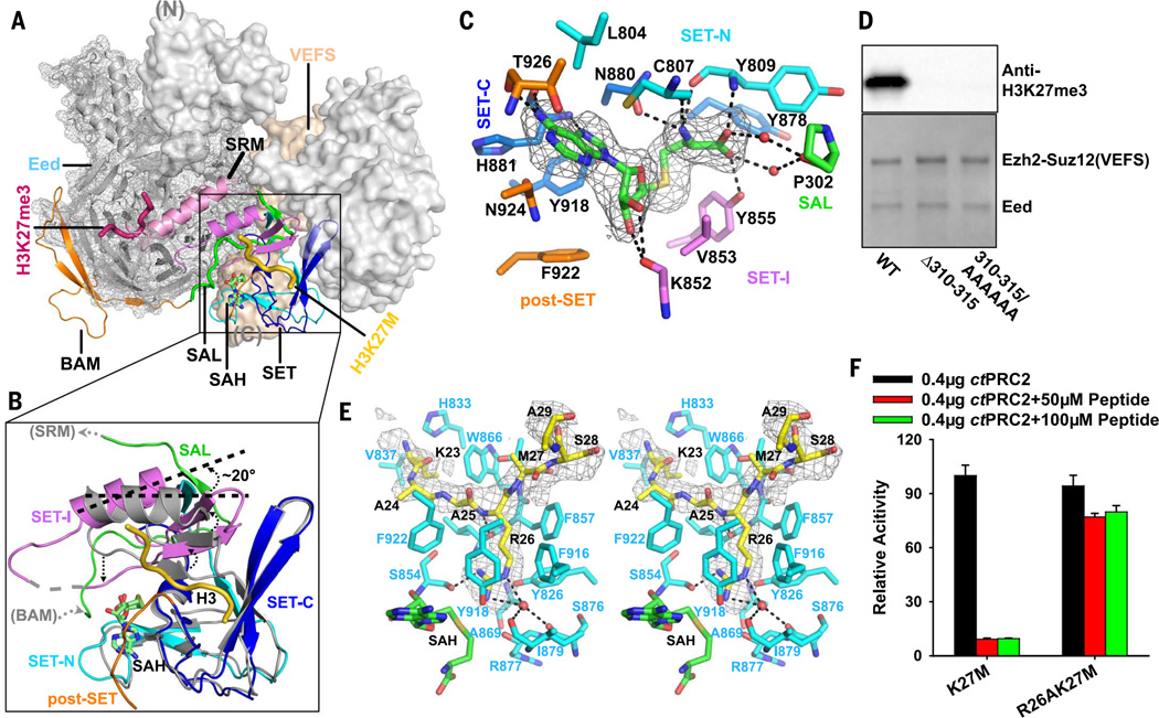
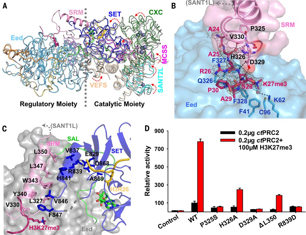
Polycomb repressive complex 2 (PRC2) catalyzes histone H3K27 trimethylation (H3K27me3), a hallmark of gene silencing. Here we report the crystal structures of an active PRC2 complex of 170 kilodaltons from the yeast Chaetomium thermophilum in both basal and stimulated states, which contain Ezh2, Eed, and the VEFS domain of Suz12 and are bound to a cancer-associated inhibiting H3K27M peptide and a S-adenosyl-l-homocysteine cofactor. The stimulated complex also contains an additional stimulating H3K27me3 peptide. Eed is engulfed by a belt-like structure of Ezh2, and Suz12(VEFS) contacts both of these two subunits to confer an unusual split active SET domain for catalysis. Comparison of PRC2 in the basal and stimulated states reveals a mobile Ezh2 motif that responds to stimulation to allosterically regulate the active site.


Figures

Fig. 1
Fig. 1. Overall structure of the active Ezh2-Eed-Suz12 (VEFS) ternary complex
(A) Schematic domain structures of Ezh2, Eed, and Suz12 (VEFS) are shown. Individual functional domains of Ezh2 are labeled, and locations of zinc-coordinating structures are indicated. Seven blades of the β-propeller structure of the Eed WD40 domain are numbered, and an insertion domain specific to C. thermophilum Eed is indicated. Peptides included for crystallization are also shown, with modified or mutated residues colored in red. (B) The overall structure of the active ternary complex bound to the stimulating H3K27me3 peptide, inhibiting H3K27M cancer mutant peptide, and cofactor SAH is shown in cartoon representation. Functional domains are colored as in (A). Zn2+ ions and SAH are shown as spheres, and the two peptide ligands are shown as thick ribbons. The regulatory and catalytic moieties of the complex are also indicated. (C) Ezh2 and Suz12(VEFS) subunits of the ternary complex are shown in surface representation, and Eed is in mesh. Eed is engulfed by the belt-like structure of Ezh2 within the regulatory moiety.
Fig. 2
Fig. 2. Close-up views of the intra- and intermolecular interactions in the active ternary complex
(A) The crystal structure of the stimulated ternary complex is superimposed with the mouse Eed-EBD binary complex (PDB: 2QXV). Mouse Eed and EBD are shown in gray and green, respectively. The BAM of Ezh2 in orange is also shown interacting with the WD40 domain of Eed. To indicate the close-up views in the context of the overall structure, Ezh2 domains preceding and following the shown ones are hereafter denoted by the dotted gray arrows. A map of the close-up views is also provided in fig. S6 for the same purpose. (B) Intramolecular hydrophobic interactions between the SBD and SANT1L regions of Ezh2 are shown, with involved residues highlighted as sticks. (C) Interactions between the MCSS region of Ezh2 and Suz12(VEFS) are shown, with the disease mutation residues of Suz12(VEFS) and their interacting residues in the MCSS highlighted as sticks. The zinc-coordinating motif of the MCSS is shown as mesh. (D) Suz12(VEFS) sits in the interface of the regulatory and catalytic moieties on the back of the SET domain. Conserved interacting residues of Suz12(VEFS) and Eed are highlighted as sticks.
Fig. 3
Fig. 3. Conformation of the active catalytic SET domain of Ezh2
(A) The trajectory of the SAL of Ezh2 is highlighted as a green ribbon in the context of the overall structure. The catalytic SET domain of Ezh2 is zoomed in and shown in Fig. 3B. (B) The catalytic SET domain from the current structure is superimposed with the isolated inactive SET domain of human Ezh2 (PDB:4MI0, shown in gray). SET-I swings about 20° counterclockwise relative to the inactive conformation. The SAL region of Ezh2 required to maintain the active conformation is also shown as a green ribbon. (C) Close-up view of Ezh2 residues involved in SAH interaction. The Fo − Fc omit electron density map for SAH contoured to 2.2σ is also shown as a gray mesh. (D) Deletion or mutation of residues 310 to 315 in the SAL of Ezh2 abolishes enzyme activity. Purified wild-type and mutant ternary complexes are shown below the result of the Western blot–based activity assay. (E) The lysine access channel of the active SET domain is shown as a stereo pair. The Fo − Fc omit electron density map contoured at 2.5σ that corresponds to the inhibiting H3K27M peptide is indicated by a gray mesh. Residue H3R26 occupying the lysine channel is unambiguously defined by the electron density map. (F) Inhibition of PRC2 enzyme activity by the H3K27M and H3R26AK27M mutant histone peptides. Relative enzyme activities from the ELISA-based assays are shown. Data are mean ± SD based on three trials.
Fig. 4
Fig. 4. Allosteric regulation of the PRC2 enzyme activity
(A) The crystal structures of the ternary complexes in the basal (gray ribbons) and stimulated (colored ribbons) states are aligned on the basis of Eed. The catalytic moiety is rotated counterclockwise toward the SRM from the basal to the stimulated state. The SRM of Ezh2, which experiences disorder-to-order conformational transition, is highlighted in cartoon representation. (B) Close-up view of interactions between the H3K27me3 peptide (red), the SRM (pink), and Eed (light blue). Interacting residues are shown as sticks. The Fo − Fc omit electron density map corresponding to the H3K27me3 peptide is contoured at 2.5σ and is shown as a gray mesh. (C) Close-up view of interactions between the SRM (pink) and the SET domain (deep blue) of Ezh2. (D) Enzyme stimulation assay of the wild-type and mutant ternary complexes. Relative enzyme activities from the triplicated ELISA-based assays are shown. Data are mean ± SD based on three trials.

Comment in

Similar articles

Cited by

References

    1. Cao R, Zhang Y. The functions of E(Z)/EZH2-mediated methylation of lysine 27 in histone H3. Curr. Opin. Genet. Dev. 2004;14:155–164. pmid: 15196462. - PubMed
    1. Simon JA, Kingston RE. Mechanisms of polycomb gene silencing: Knowns and unknowns. Nat. Rev. Mol. Cell Biol. 2009;10:697–708. pmid: 19738629. - PubMed
    1. Margueron R, Reinberg D. The Polycomb complex PRC2 and its mark in life. Nature. 2011;469:343–349. pmid: 21248841. - PMC - PubMed
    1. Delest A, Sexton T, Cavalli G. Polycomb: A paradigm for genome organization from one to three dimensions. Curr. Opin. Cell Biol. 2012;24:405–414. pmid: 22336329. - PubMed
    1. Di Croce L, Helin K. Transcriptional regulation by Polycomb group proteins. Nat. Struct. Mol. Biol. 2013;20:1147–1155. pmid: 24096405. - PubMed

Publication types

MeSH terms