Cellular Effect of Curcumin and Citral Combination on Breast Cancer Cells: Induction of Apoptosis and Cell Cycle Arrest
- PMID: 26472972
- PMCID: PMC4600686
- DOI: 10.4048/jbc.2015.18.3.225
Cellular Effect of Curcumin and Citral Combination on Breast Cancer Cells: Induction of Apoptosis and Cell Cycle Arrest
Abstract
Purpose: The unmanageable side effects caused by current chemotherapy regimens to treat cancer are an unresolved problem. Although many phytonutrients are useful as chemoprevention without side effects, their effects are slower and smaller than conventional chemotherapy. In the present work, we examined the cumulative effect of two phytonutrients, curcumin and citral, on breast cancer cell lines and compared their effect with the known chemotherapy regimen of cyclophosphamide, methotrexate, and 5-fluorouracil.
Methods: Using cultured breast cancer and normal epithelial cells, the cytotoxic and apoptotic effect of curcumin and citral was evaluated in vitro. The synergistic effect of curcumin and citral was calculated by a combination index study using the method by Chou and Talalay. Cell death pathways and mechanisms were analyzed by measuring intracellular reactive oxygen species (ROS) and apoptotic protein levels.
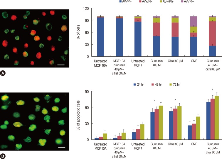
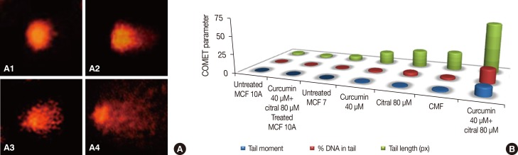
Results: Curcumin and citral caused dose and time dependent cell death and showed a synergistic effect at effective concentration EC50 and above concentrations in breast cancer cells without disturbing normal breast epithelial cells. With combination curcumin and citral treatment, apoptosis induction and cell cycle arrest at G0/G1 phase in breast cancer cells were observed. Curcumin and citral generated ROS and activated p53 and poly (ADP-ribose) polymerase-1 mediated apoptotic pathways.
Conclusion: The results of this study suggest that curcumin and citral in combination may be a useful therapeutic intervention for breast cancer.
Keywords: Apoptosis; Breast cancer cell line; Cell cycle checkpoints; Citral; Curcumin.
Conflict of interest statement
Figures


Similar articles
-
Curcumin induces apoptosis and cell cycle arrest via the activation of reactive oxygen species-independent mitochondrial apoptotic pathway in Smad4 and p53 mutated colon adenocarcinoma HT29 cells.Nutr Res. 2018 Mar;51:67-81. doi: 10.1016/j.nutres.2017.12.011. Epub 2018 Jan 6. Nutr Res. 2018. PMID: 29673545
-
Curcumin enhances the anticancer effects of trichostatin a in breast cancer cells.Mol Carcinog. 2013 May;52(5):404-11. doi: 10.1002/mc.21875. Epub 2012 Jan 30. Mol Carcinog. 2013. PMID: 22290509
-
Photodynamic treatment with anionic nanoclays containing curcumin on human triple-negative breast cancer cells: Cellular and biochemical studies.J Cell Biochem. 2019 Apr;120(4):4998-5009. doi: 10.1002/jcb.27775. Epub 2018 Oct 9. J Cell Biochem. 2019. PMID: 30302810
-
Methylglyoxal in combination with 5-Fluorouracil elicits improved chemosensitivity in breast cancer through apoptosis and cell cycle inhibition.Biomed Pharmacother. 2019 Jun;114:108855. doi: 10.1016/j.biopha.2019.108855. Epub 2019 Apr 16. Biomed Pharmacother. 2019. PMID: 31003140
-
Curcumin and paclitaxel induce cell death in breast cancer cell lines.Oncol Rep. 2018 Oct;40(4):2381-2388. doi: 10.3892/or.2018.6603. Epub 2018 Jul 26. Oncol Rep. 2018. PMID: 30066930
Cited by
-
Lemongrass Essential Oil Components with Antimicrobial and Anticancer Activities.Antioxidants (Basel). 2021 Dec 22;11(1):20. doi: 10.3390/antiox11010020. Antioxidants (Basel). 2021. PMID: 35052524 Free PMC article. Review.
-
Prominent Naturally Derived Oxidative-Stress-Targeting Drugs and Their Applications in Cancer Treatment.Antioxidants (Basel). 2025 Jan 3;14(1):49. doi: 10.3390/antiox14010049. Antioxidants (Basel). 2025. PMID: 39857383 Free PMC article. Review.
-
Effect of Heat Pasteurization and Sterilization on Milk Safety, Composition, Sensory Properties, and Nutritional Quality.Foods. 2025 Apr 14;14(8):1342. doi: 10.3390/foods14081342. Foods. 2025. PMID: 40282744 Free PMC article. Review.
-
Hematopoietic Effects of Angelica gigas Nakai Extract on Cyclophosphamide-Induced Myelosuppression.Plants (Basel). 2022 Dec 12;11(24):3476. doi: 10.3390/plants11243476. Plants (Basel). 2022. PMID: 36559587 Free PMC article.
-
Natural Compounds as Modulators of Cell Cycle Arrest: Application for Anticancer Chemotherapies.Curr Genomics. 2017 Apr;18(2):106-131. doi: 10.2174/1389202917666160808125645. Curr Genomics. 2017. PMID: 28367072 Free PMC article. Review.
References
-
- Pan MH, Ho CT. Chemopreventive effects of natural dietary compounds on cancer development. Chem Soc Rev. 2008;37:2558–2574. - PubMed
-
- Ramachandran C, You W. Differential sensitivity of human mammary epithelial and breast carcinoma cell lines to curcumin. Breast Cancer Res Treat. 1999;54:269–278. - PubMed
LinkOut - more resources
Full Text Sources
Other Literature Sources
Research Materials
Miscellaneous