. 2016 May;54(5):718-27.
doi: 10.1165/rcmb.2015-0180OC.

Peripheral Airway Smooth Muscle, but Not the Trachealis, Is Hypercontractile in an Equine Model of Asthma

Affiliations

Peripheral Airway Smooth Muscle, but Not the Trachealis, Is Hypercontractile in an Equine Model of Asthma

Oleg S Matusovsky et al. Am J Respir Cell Mol Biol. 2016 May.

Abstract

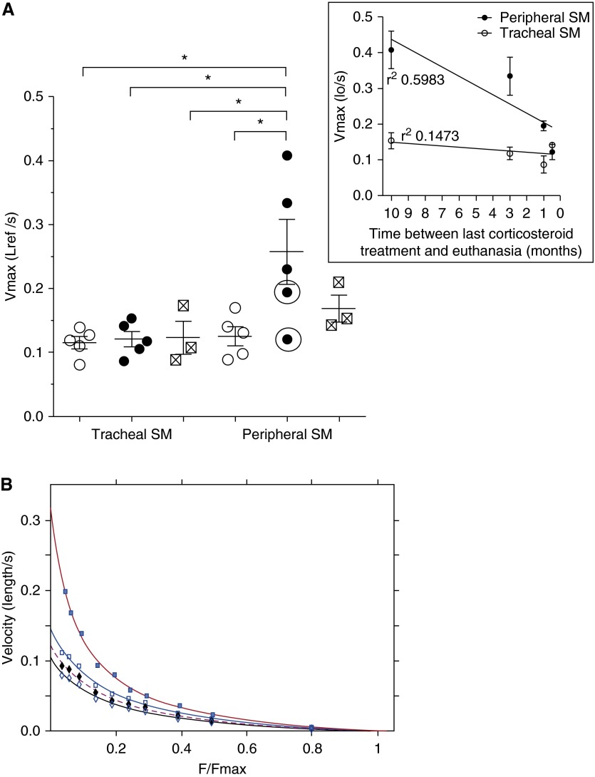
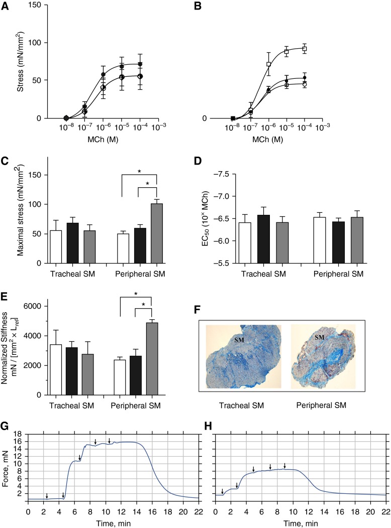
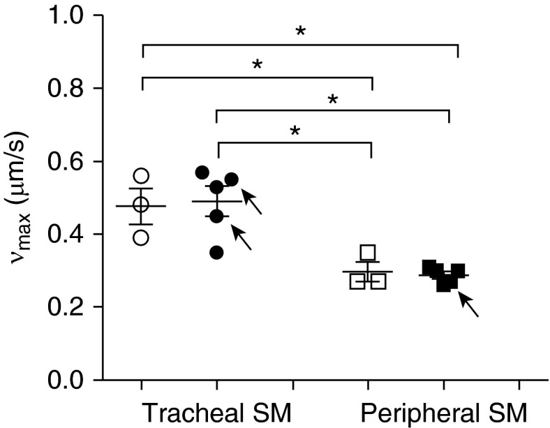
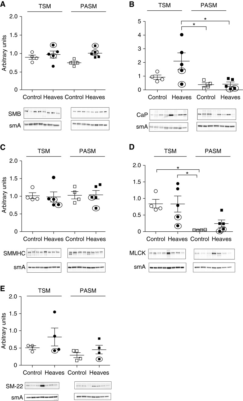
Heaves is a naturally occurring equine disease that shares many similarities with human asthma, including reversible antigen-induced bronchoconstriction, airway inflammation, and remodeling. The purpose of this study was to determine whether the trachealis muscle is mechanically representative of the peripheral airway smooth muscle (ASM) in an equine model of asthma. Tracheal and peripheral ASM of heaves-affected horses under exacerbation, or under clinical remission of the disease, and control horses were dissected and freed of epithelium to measure unloaded shortening velocity (Vmax), stress (force/cross-sectional area), methacholine effective concentration at which 50% of the maximum response is obtained, and stiffness. Myofibrillar Mg(2+)-ATPase activity, actomyosin in vitro motility, and contractile protein expression were also measured. Horses with heaves had significantly greater Vmax and Mg(2+)-ATPase activity in peripheral airway but not in tracheal smooth muscle. In addition, a significant correlation was found between Vmax and the time elapsed since the end of the corticosteroid treatment for the peripheral airways in horses with heaves. Maximal stress and stiffness were greater in the peripheral airways of the horses under remission compared with controls and the horses under exacerbation, potentially due to remodeling. Actomyosin in vitro motility was not different between controls and horses with heaves. These data demonstrate that peripheral ASM is mechanically and biochemically altered in heaves, whereas the trachealis behaves as in control horses. It is therefore conceivable that the trachealis muscle may not be representative of the peripheral ASM in human asthma either, but this will require further investigation.

Keywords: airway hyperresponsiveness; airway smooth muscle; asthma; smooth muscle mechanics.


Figures

Figure 1.
Figure 1.
(A) Unloaded shortening velocity (Vmax) of tracheal and peripheral airway smooth muscle (ASM) from controls (open circles; n = 5 horses), horses with heaves during exacerbation (solid circles; n = 5 horses), and horses experiencing remission (crossed squares; n = 3 horses) of the disease. The two circled values are from horses affected with heaves but recently treated with corticosteroids (horses 8 and 9 in Table 1). *P< 0.05. Inset, Vmax of tracheal (open circles) and peripheral (solid circles) ASM as a function of time between the last corticosteroid treatments and killing, measured in the horses with heaves. Lo and Lref, reference length. (B) Representative force–velocity curves for tracheal control (open diamonds) or peripheral control (open squares) smooth muscle (SM) and tracheal heaves–affected (solid diamonds) or peripheral heaves–affected (solid squares) SM. F/Fmax, force normalized to maximal force. The force–velocity relationships were accurately fitted by the Hill hyperbolic model (r2 > 0.98). Data are presented as mean (±SEM).
Figure 2.
Figure 2.
Mean cumulative methacholine (MCh) dose–response curves expressed as SM stress for (A) tracheal SM and (B) peripheral ASM from control horses (open circles; n = 4 horses), heaves-affected horses (solid circles; n = 5 horses), and horses under remission (open squares; n = 3 horses). (C) Maximal stress generated by tracheal and peripheral ASM at 10−4 M MCh (maximal force normalized to SM cross-sectional area [CSA]) in control (white bars), heaves (black bars), and heaves under remission (gray bars). *P < 0.05. (D) Effective concentration at which 50% of the maximum stress (EC50) is generated for the three groups of animals. (E) Active stiffness (force normalized to SM CSA and divided by Lref) measured in tracheal and peripheral ASM of control horses (white bars), horses with heaves (black bars), and horses under clinical remission (gray bars). *P < 0.05. (F) Cross-section of tracheal and peripheral ASM strips used for CSA quantification. (G and H) Representative cumulative MCh dose–response traces of tracheal (G) and peripheral airway (H) SM. Arrows denote time points of MCh injection. Data are presented as mean (±SEM).
Figure 3.
Figure 3.
Mg2+-ATPase activity of myofibrils isolated from tracheal and peripheral ASM of control (open circles and open squares, respectively; n = 3 horses for both) and heaves-affected horses during exacerbation (solid circles and solid squares; n = 3 horses for both). *P< 0.05. Data are presented as mean (±SEM).
Figure 4.
Figure 4.
Velocity of actin filament (νmax) when propelled by thiophosphorylated myosin purified from tracheal and peripheral ASM of controls (open circles and open squares, respectively; n = 3 horses for both) or heaves-affected horses (solid circles and solid squares; n = 5 horses for both), as measured in the in vitro motility assay. Arrows show νmax for myosin purified from tracheal or peripheral ASM of horses affected by heaves recently treated with corticosteroids. *P< 0.0001. Data are presented as mean (±SEM).
Figure 5.
Figure 5.
Western blot analysis: (A) (+) insert SM myosin heavy chain (SMMHC) isoform B (SM-B); (B) calponin (CaP); (C) total SMMHC; (D) myosin light chain kinase (MLCK); and (E) transgelin (SM22) of tracheal SM (TSM) of control (open circles) or heaves-affected horses (solid circles) and peripheral ASM (PASM) of control (open squares) or heaves-affected horses (solid squares); n = 4 for TSM and PASM of control horses (except for SM22, where n = 3 horses); n = 5 for TSM and PASM of heaves-affected horses (except for SM22, where n = 4 horses). Smooth muscle actin (smA) was used as a loading control. The circled values are from horses affected with heaves that were recently treated with corticosteroids (horses 8 and 9 in Table 1). Representative Western blots are shown. *P< 0.05. Data are presented as mean (±SEM).

Similar articles

Cited by

References

    1. Bateman ED, Hurd SS, Barnes PJ, Bousquet J, Drazen JM, FitzGerald M, Gibson P, Ohta K, O’Byrne P, Pedersen SE, et al. Global strategy for asthma management and prevention: GINA executive summary. Eur Respir J. 2008;31:143–178. - PubMed
    1. Antonissen LA, Mitchell RW, Kroeger EA, Kepron W, Tse KS, Stephens NL. Mechanical alterations of airway smooth muscle in a canine asthmatic model. J Appl Physiol. 1979;46:681–687. - PubMed
    1. Jiang H, Rao K, Halayko AJ, Kepron W, Stephens NL. Bronchial smooth muscle mechanics of a canine model of allergic airway hyperresponsiveness. J Appl Physiol (1985) 1992;72:39–45. - PubMed
    1. Fan T, Yang M, Halayko A, Mohapatra SS, Stephens NL. Airway responsiveness in two inbred strains of mouse disparate in IgE and IL-4 production. Am J Respir Cell Mol Biol. 1997;17:156–163. - PubMed
    1. Duguet A, Biyah K, Minshall E, Gomes R, Wang CG, Taoudi-Benchekroun M, Bates JH, Eidelman DH. Bronchial responsiveness among inbred mouse strains: role of airway smooth-muscle shortening velocity. Am J Respir Crit Care Med. 2000;161:839–848. - PubMed

Publication types