

Review
doi: 10.1002/art.39458.
Review: Enthesitis: New Insights Into Pathogenesis, Diagnostic Modalities, and Treatment
Affiliations
- PMID: 26473401
- PMCID: PMC5195265
- DOI: 10.1002/art.39458
Item in Clipboard
Review
Review: Enthesitis: New Insights Into Pathogenesis, Diagnostic Modalities, and Treatment
Arthritis Rheumatol.
2016 Feb.
No abstract available
Figures
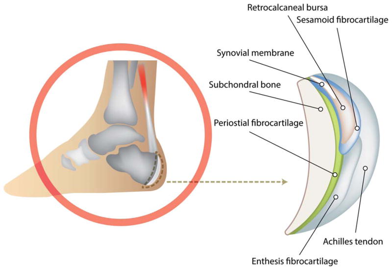
Illustration of the Achilles enthesis organ.
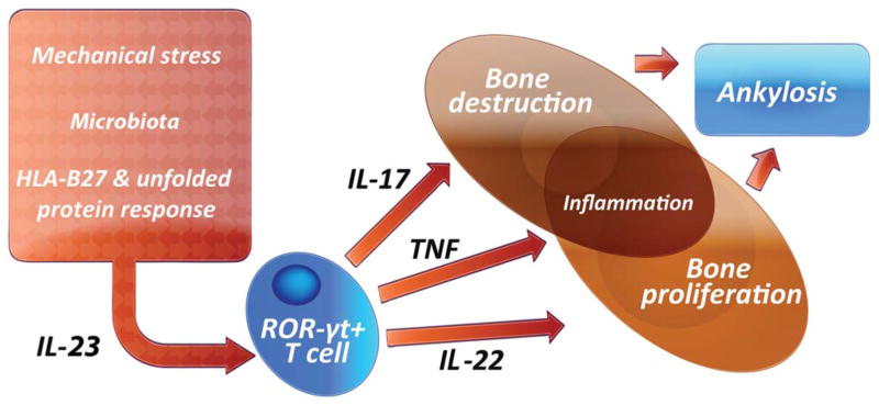
Interleukin-23 (IL-23) is activated via a variety of pathways including the HLA–B27 unfolded protein response. IL-23 then activates resident T cells within the enthesis, which then promotes inflammation and bone remodeling, with inflammation mediated by IL-17 and osteoproliferation mediated by IL-22. The net result is bone ankylosis in the spine. RORγt = retinoic acid receptor–related orphan nuclear receptor γt; TNF = tumor necrosis factor. Color figure can be viewed in the online issue, which is available at http://onlinelibrary.wiley.com/journal/doi/10.1002/art.39458/abstract
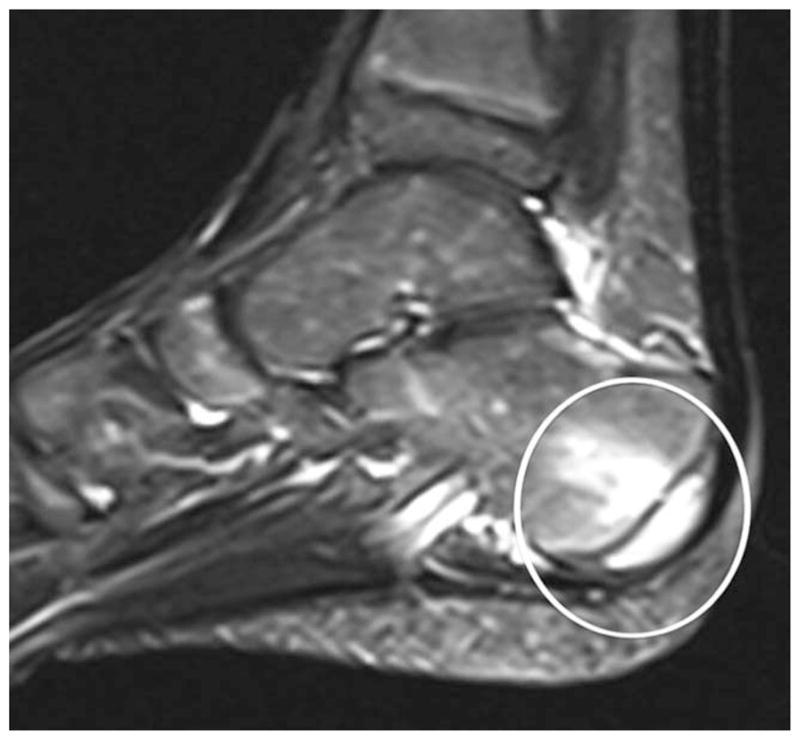
Achilles tendon insertion into the calcaneus. An abnormal signal is seen at the posterior calcaneus at the site of the Achilles entheseal insertion (encircled area) on magnetic resonance imaging using STIR sequences. Reproduced from ref. .
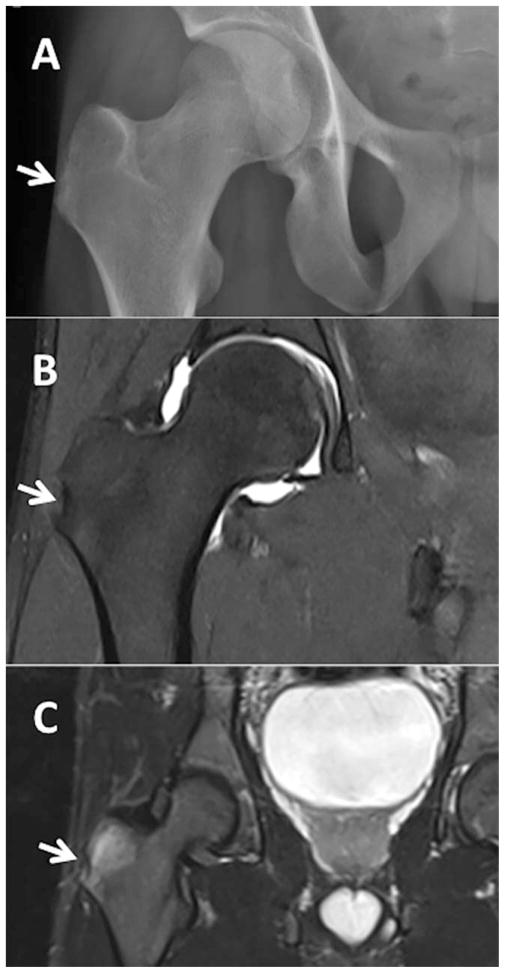
Radiographic findings of enthesitis in a 21-year-old man with ankylosing spondylitis. A, Reference plain radiograph. B, Magnetic resonance image with T1 sequence showing a small erosion at the right greater trochanter. C, Axial T2 fat-saturated sequence of the right hip showing edema of the right gluteus minimus tendon at its insertion, consistent with enthesitis. Arrows indicate the region of the right greater trochanter. Images courtesy of Dr. Joseph Robinson (Cedars-Sinai Medical Center).
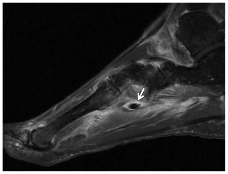
Fat-suppressed T1-weighted sequence magnetic resonance image (MRI) of the right midfoot of a 58-year-old woman with HLA–B27–positive peripheral spondyloarthropathy. The MRI demonstrates extensive enthesitis, synovitis, and tenosynovitis of the peroneus longus, peroneus brevis, tibialis posterior, flexor digitorum longus, and extensor digitorum tendons. This is characterized by excess fluid and enhancement in and around the tendon sheaths. Arrow indicates extensive edema and enhancement at the plantar aspect of the midfoot, involving insertions of intrinsic musculature and capsular ligaments consistent with enthesitis. Image courtesy of Dr. Joseph Robinson (Cedars-Sinai Medical Center).

A, Ultrasound image of a patient with psoriatic arthritis with enthesitis (long-axis view of the lateral epicondyle [high-frequency, 18-MHz probe]). Precise delineation of blood flow seen at the cortical interface on B-flow imaging is shown. Calcification is seen adjacent to the epicondyle. B and C, 3T magnetic resonance image of the same patient, in the same orientation as the ultrasound in A, showing proton density (B) and fat presaturation (C). Boxed areas show the region of the lateral epicondyle. Images courtesy of Dr. Ralph Thiele (University of Rochester, Rochester, NY). Color figure can be viewed in the online issue, which is available at http://onlinelibrary.wiley.com/journal/doi/10.1002/art.39458/abstract
References
-
- Lampman JH. Origin of enthesopathy. J Rheumatol. 1985;12:1030–1. - PubMed
-
- Francois RJ, Braun J, Khan MA. Entheses and enthesitis: a histopathologic review and relevance to spondyloarthritides. Curr Opin Rheumatol. 2001;13:255–64. - PubMed
-
- Benjamin M, Moriggl B, Brenner E, Emery P, McGonagle D, Redman S. The “enthesis organ” concept: why enthesopathies may not present as focal insertional disorders. Arthritis Rheum. 2004;50:3306–13. - PubMed
-
- McGonagle D, Stockwin L, Isaacs J, Emery P. An enthesitis based model for the pathogenesis of spondyloarthropathy: additive effects of microbial adjuvant and biomechanical factors at disease sites. J Rheumatol. 2001;28:2155–9. - PubMed
Publication types
MeSH terms
Substances
Grants and funding
LinkOut - more resources
Full Text Sources
Other Literature Sources
Medical
