. 2015 Oct 16;11(10):e1005201.
doi: 10.1371/journal.ppat.1005201. eCollection 2015 Oct.

Peripheral Vγ9Vδ2 T Cells Are a Novel Reservoir of Latent HIV Infection

Affiliations

Peripheral Vγ9Vδ2 T Cells Are a Novel Reservoir of Latent HIV Infection

Natalia Soriano-Sarabia et al. PLoS Pathog. .

Abstract

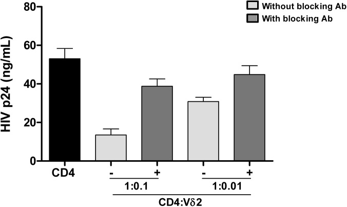
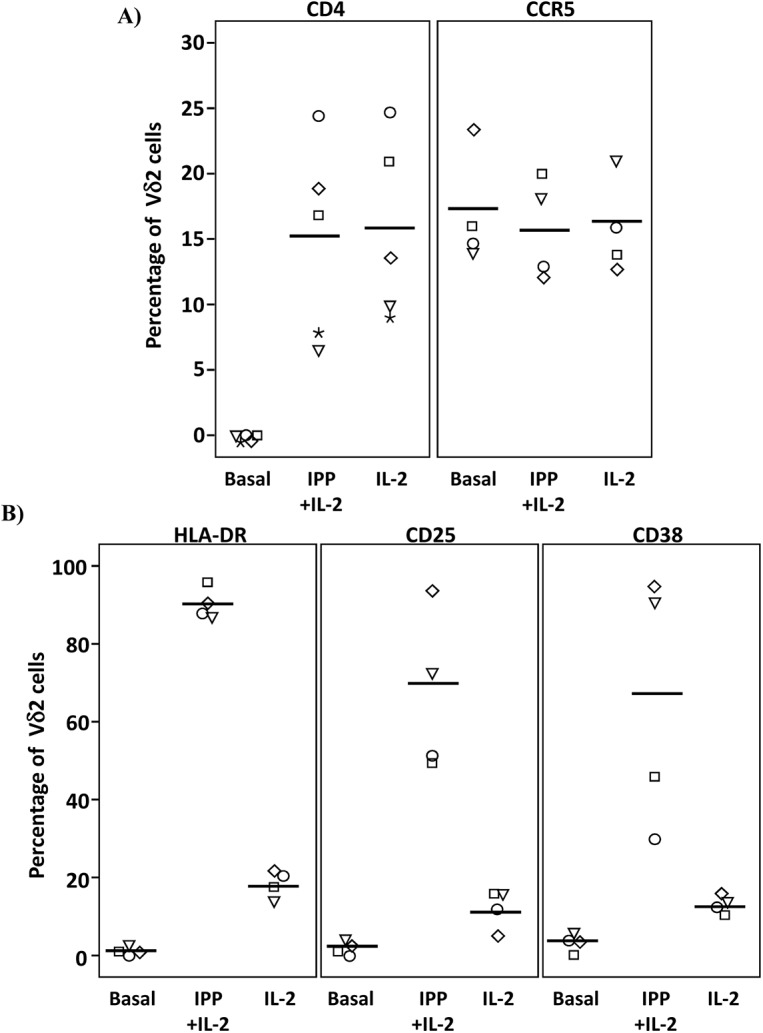
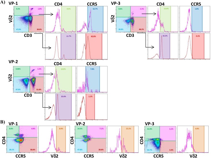
Eradication of HIV infection will require the identification of all cellular reservoirs that harbor latent infection. Despite low or lack of CD4 receptor expression on Vδ2 T cells, infection of these cells has previously been reported. We found that upregulation of the CD4 receptor may render primary Vδ2 cells target for HIV infection in vitro and we propose that HIV-induced immune activation may allow infection of γδ T cells in vivo. We assessed the presence of latent HIV infection by measurements of DNA and outgrowth assays within Vδ2 cells in 18 aviremic patients on long-standing antiretroviral therapy. In 14 patients we recovered latent but replication-competent HIV from highly purified Vδ2 cells demonstrating that peripheral Vδ2 T cells are a previously unrecognized reservoir in which latent HIV infection is unexpectedly frequent.


Conflict of interest statement

The authors have declared that no competing interests exist.

Figures

Fig 1
Fig 1. Vδ2 T cell sorting strategy and purity.
A) An example of the gating strategy to sort pre-enriched Vδ2 cells (panel 1) using a 2-step doublet discrimination (panels 2 and 3), showing that CD3+ Vδ2 cells (panel 4) lack expression of HLA-DR (panel 5). B) Dot plot examples showing lack of αβ-TCR (panel 6) and CD4 (panel 7) expression in the presort sample. C) Purity of the sorted cell population. After the sort an aliquot of sorted cells was acquired and analyzed. Plots show high purity of CD3+ Vδ2 cells (panel 8) and lack of HLA-DR expression (panel 9). Post-sort analysis shows lack of αβ-TCR and CD4 (panels 10 and 11).
Fig 2
Fig 2. Quantification of total HIV DNA levels.
A) Total pol HIV copies were quantified by ddPCR within Vδ2 cells (n = 12), total resting CD4+ T cells (r-CD4) (n = 8) and unfractionated PBMC (n = 12) from HIV-1 suppressed patients treated in the acute HIV infection (AHI) or in the chronic HIV infection (CHI). Limit of quantitation (LOQ) was 50.6 copies/ 106 Vδ2 cells and 5.1 copies/ 106 r-CD4 cells and PBMC, and is depicted with a dotted line. Each color represents one patient. B) Pie charts reflecting the contribution of Vδ2 cells (purple) and r-CD4 cells (red) to the total HIV DNA+ PBMC in CHI patients (left pie) and AHI patients (right pie).
Fig 3
Fig 3. Recovery of replication-competent virus from isolated Vδ2 cells.
Representation of the culture conditions in isolated peripheral resting Vδ2 T cells in patients treated in acute HIV infection (AHI) and in chronic HIV infection (CHI). Each square represents one culture replicate that is gray when replication-competent HIV was recovered, and is open when culture replicates were negative. Vδ2 T cells were recovered at a higher frequency in patients treated in AHI than in patients treated in CHI, allowing more replicates for the outgrowth assays. However, HIV was more frequently recovered from CHI patients than from AHI patients.
Fig 4
Fig 4. Frequency of replication-competent HIV expressed as infectious units per million (IUPM) cells.
A) Comparison between the frequency of infection within isolated Vδ2 cells between patients treated in the acute HIV infection (AHI) and patients treated in the chronic HIV infection (CHI). B) Point estimate (95% CI) of the frequency of infection in isolated Vδ2 cells (circles) and resting CD4+ T (r-CD4) cells (triangles) in individual patients treated in AHI (left panel) or treated in CHI (right panel). Open symbols means HIVp24 was below the limit of detection, that was <10.67 for Vδ2 cells in patient A6, <0.1 for r-CD4 cells in patient A7, and <31.9 for Vδ2 cells in patient C3. C) Comparison of the point estimate of the frequency of infection between Vδ2 cells and r-CD4 cells in patients treated in AHI (left panel) and patients treated in CHI (right panel). p> 0.05, Wilcoxon signed-ranked test.
Fig 5
Fig 5. Vδ2 cells exhibit potent anti-HIV activity in vitro.
Isolated Vδ2 cells from healthy donors were co-cultured with autologous in vitro infected HIV- CD4+ T cells at 1:0.1 or 1:0.01 ratios (CD4:Vδ2) for seven days. HIV p24 production was inhibited in a cell-dose-dependent manner. When a cocktail of blocking antibodies against CD8, CD16 and NKG2D were added, cytotoxic function of Vδ2 cells was partially abrogated. Mean±SE of three different donors is represented.
Fig 6
Fig 6. Expression of CD4, CCR5 and activation markers on Vδ2 cells.
PBMC from HIV-uninfected donors were cultured and treated with IL-2 alone or IPP and IL-2 for six days. The surface expression of CD4, CCR5, and activation markers (HLA-DR, CD38 and CD25) on Vδ2 cells were analyzed by flow cytometry on days 0 and 6. A) Majority of peripheral Vδ2 cells do not express the CD4 receptor but CD4 is significantly upregulated after six days in culture with IL-2 or with IPP and IL-2 (*p<0.01). However, CCR5 is expressed on the surface of Vδ2 cells at baseline without a significant induction after treatments. B) Analysis of activation markers (HLA-DR, CD25 and CD38) showed peak activation after six days of culture with IPP and IL-2 with moderate activation by IL-2 alone (*p<0.05).
Fig 7
Fig 7. Vδ2 cells from untreated recently HIV-infected patients express CD4 and CCR5.
A) Dot plots from three recently infected patient (VP1, VP2 and VP3) showing surface expression of CD4 and CCR5 on CD3+Vδ2+ cells, and on CD3+Vδ2- cells for comparison. B) Density plots showing the percentage of Vδ2 cells coexpressing CD4 and CCR5 in the same three patients.

Similar articles

Cited by

References

    1. Stevenson M, Stanwick TL, Dempsey MP, Lamonica CA. HIV-1 replication is controlled at the level of T cell activation and proviral integration. EMBO J. 1990; 9: 1551–1560. - PMC - PubMed
    1. Chun TW, Carruth L, Finzi D, Shen X, DiGiuseppe JA, Taylor H, et al. Quantification of latent tissue reservoirs and total body viral load in HIV-1 infection. Nature 1997; 387: 183–188. - PubMed
    1. Finzi D, Blankson J, Siliciano JD, Margolisk JD, Chadwick K, Pierson T, et al. Latent infection of CD4+ T cells provides a mechanism for lifelong persistence of HIV-1, even in patients on effective combination therapy. Nat Med. 1999; 5: 512–517. - PubMed
    1. Wong J, Hezareh M, Günthard HF, Havlir DV, Ignacio CC, Richman DD. Recovery of replication-competent HIV despite prolonged suppression of plasma viremia Science 1997; 278: 1291–1294. - PubMed
    1. Siliciano JD, Kajdas J, Finzi D, Quinn TC, Margolick JB, Kovacs C, et al. Longterm follow-up studies confirm the stability of the latent reservoir for HIV-1 in resting CD4+ T cells. Nat Med. 2003; 9: 727–728. - PubMed

Publication types

Substances