

Multiparametric MRI of Epiphyseal Cartilage Necrosis (Osteochondrosis) with Histological Validation in a Goat Model
- PMID: 26473611
- PMCID: PMC4608749
- DOI: 10.1371/journal.pone.0140400
Multiparametric MRI of Epiphyseal Cartilage Necrosis (Osteochondrosis) with Histological Validation in a Goat Model
Abstract
Purpose: To evaluate multiple MRI parameters in a surgical model of osteochondrosis (OC) in goats.
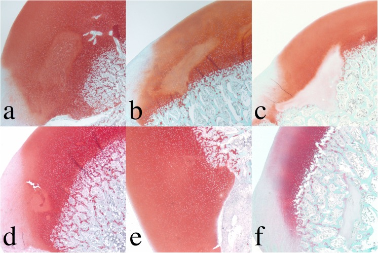
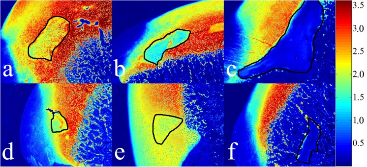
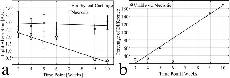
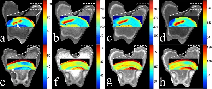
Methods: Focal ischemic lesions of two different sizes were induced in the epiphyseal cartilage of the medial femoral condyles of goats at 4 days of age by surgical transection of cartilage canal blood vessels. Goats were euthanized and specimens harvested 3, 4, 5, 6, 9 and 10 weeks post-op. Ex vivo MRI scans were conducted at 9.4 Tesla for mapping the T1, T2, T1ρ, adiabatic T1ρ and TRAFF relaxation times of articular cartilage, unaffected epiphyseal cartilage, and epiphyseal cartilage within the area of the induced lesion. After MRI scans, safranin O staining was conducted to validate areas of ischemic necrosis induced in the medial femoral condyles of six goats, and to allow comparison of MRI findings with the semi-quantitative proteoglycan assessment in corresponding safranin O-stained histological sections.
Results: All relaxation time constants differentiated normal epiphyseal cartilage from lesions of ischemic cartilage necrosis, and the histological staining results confirmed the proteoglycan (PG) loss in the areas of ischemia. In the scanned specimens, all of the measured relaxation time constants were higher in the articular than in the normal epiphyseal cartilage, consistently allowing differentiation between these two tissues.
Conclusions: Multiparametric MRI provided a sensitive approach to discriminate between necrotic and viable epiphyseal cartilage and between articular and epiphyseal cartilage, which may be useful for diagnosing and monitoring OC lesions and, potentially, for assessing effectiveness of treatment interventions.
Conflict of interest statement
Figures
Similar articles
-
In vivo visualization using MRI T2 mapping of induced osteochondrosis and osteochondritis dissecans lesions in goats undergoing controlled exercise.J Orthop Res. 2017 Apr;35(4):868-875. doi: 10.1002/jor.23332. Epub 2016 Jun 19. J Orthop Res. 2017. PMID: 27283998 Free PMC article.
-
Quantitative susceptibility mapping detects abnormalities in cartilage canals in a goat model of preclinical osteochondritis dissecans.Magn Reson Med. 2017 Mar;77(3):1276-1283. doi: 10.1002/mrm.26214. Epub 2016 Mar 28. Magn Reson Med. 2017. PMID: 27018370 Free PMC article.
-
Surgical induction, histological evaluation, and MRI identification of cartilage necrosis in the distal femur in goats to model early lesions of osteochondrosis.Osteoarthritis Cartilage. 2015 Feb;23(2):300-7. doi: 10.1016/j.joca.2014.11.009. Epub 2014 Nov 15. Osteoarthritis Cartilage. 2015. PMID: 25463443 Free PMC article.
-
Pathogenesis of epiphyseal osteochondrosis.Vet J. 2013 Jul;197(1):3-12. doi: 10.1016/j.tvjl.2013.03.035. Epub 2013 May 4. Vet J. 2013. PMID: 23647656 Review.
-
An Update on the Pathogenesis of Osteochondrosis.Vet Pathol. 2015 Sep;52(5):785-802. doi: 10.1177/0300985815588778. Epub 2015 Jun 16. Vet Pathol. 2015. PMID: 26080832 Review.
Cited by
-
In vivo visualization using MRI T2 mapping of induced osteochondrosis and osteochondritis dissecans lesions in goats undergoing controlled exercise.J Orthop Res. 2017 Apr;35(4):868-875. doi: 10.1002/jor.23332. Epub 2016 Jun 19. J Orthop Res. 2017. PMID: 27283998 Free PMC article.
-
Relationship between the extent of vascular injury and the evolution of surgically induced osteochondrosis lesions in a piglet model.PLoS One. 2024 Aug 8;19(8):e0308641. doi: 10.1371/journal.pone.0308641. eCollection 2024. PLoS One. 2024. PMID: 39116161 Free PMC article.
-
Identification of Areas of Epiphyseal Cartilage Necrosis at Predilection Sites of Juvenile Osteochondritis Dissecans in Pediatric Cadavers.J Bone Joint Surg Am. 2018 Dec 19;100(24):2132-2139. doi: 10.2106/JBJS.18.00464. J Bone Joint Surg Am. 2018. PMID: 30562294 Free PMC article.
-
Quantitative susceptibility mapping detects abnormalities in cartilage canals in a goat model of preclinical osteochondritis dissecans.Magn Reson Med. 2017 Mar;77(3):1276-1283. doi: 10.1002/mrm.26214. Epub 2016 Mar 28. Magn Reson Med. 2017. PMID: 27018370 Free PMC article.
-
Longitudinal 3T MRI T2 * mapping of Juvenile osteochondritis dissecans (JOCD) lesions differentiates operative from non-operative patients-Pilot study.J Orthop Res. 2023 Jan;41(1):150-160. doi: 10.1002/jor.25343. Epub 2022 Apr 30. J Orthop Res. 2023. PMID: 35430743 Free PMC article.
References
Publication types
MeSH terms
Grants and funding
LinkOut - more resources
Full Text Sources
Other Literature Sources
Medical
Miscellaneous
