

Somatic IDH1 mutation in a pituitary adenoma of a patient with Maffucci syndrome
- PMID: 26473790
- PMCID: PMC5121660
- DOI: 10.3171/2015.4.JNS15191
Somatic IDH1 mutation in a pituitary adenoma of a patient with Maffucci syndrome
Abstract
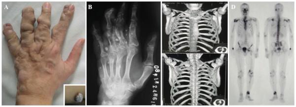
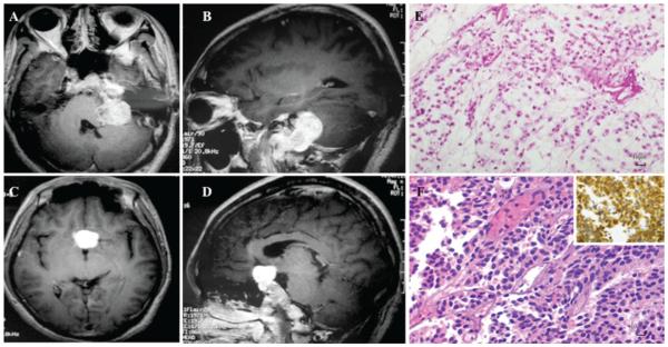
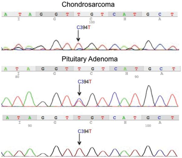
Maffucci syndrome is a rare disease characterized by multiple enchondromas and soft-tissue hemangiomas. Additionally, neuroendocrine tumors including pituitary adenomas have been described in these patients. The underlying genetic etiology lies in somatic mosaicism of mutations in isocitrate dehydrogenase 1 (IDH1) or isocitrate dehydrogenase 2 (IDH2). This report describes a patient with Maffucci syndrome who presented with intracranial tumors of the skull base and suprasellar region. The patient underwent resection of both intracranial tumors, revealing histopathological diagnoses of chondrosarcoma and pituitary adenoma. DNA sequencing of the tumors was performed to identify common IDH1/2 mutations. Clinical, radiological, and biochemical assessments were performed. Genotypic studies used standard Sanger sequencing in conjunction with a target-specific peptide nucleic acid to detect IDH1 mutations in tumor tissues. DNA sequencing demonstrated identical IDH1 mutations (c.394C > T) in both tumors. To the authors' knowledge, this report provides the first genetic evidence for the inclusion of pituitary adenomas among tumors characterizing Maffucci syndrome. In patients who are newly diagnosed with Maffucci syndrome, it is appropriate to monitor for development of pituitary pathology and neuroendocrine dysfunction.
Keywords: ACTH = adrenocorticotropic hormone; FSH = follicle-stimulating hormone; GH = growth hormone; IDH1 = isocitrate dehydrogenase 1; IDH2 = isocitrate dehydrogenase 2; LH = luteinizing hormone; MEN1 = multiple endocrine neoplasia 1; Maffucci syndrome; PCR = polymerase chain reaction; PNA = peptide nucleic acid; PRL = prolactin; RET = rearranged during transfection; SDH = succinate dehydrogenase; TSH = thyroid-stimulating hormone; isocitrate dehydrogenase; oncology; pituitary adenoma; somatic mosaicism.
Figures
References
-
- Amary MF, Damato S, Halai D, Eskandarpour M, Berisha F, Bonar F, et al. Ollier disease and Maffucci syndrome are caused by somatic mosaic mutations of IDH1 and IDH2. Nat Genet. 2011;43:1262–1265. - PubMed
-
- Auyeung J, Mohanty K, Tayton K. Maffucci lymphangioma syndrome: an unusual variant of Ollier’s disease, a case report and a review of the literature. J Pediatr Orthop B. 2003;12:147–150. - PubMed
-
- Balcer LJ, Galetta SL, Cornblath WT, Liu GT. Neuroophthalmologic manifestations of Maffucci’s syndrome and Ollier’s disease. J Neuroophthalmol. 1999;19:62–66. - PubMed
-
- Balss J, Meyer J, Mueller W, Korshunov A, Hartmann C, von Deimling A. Analysis of the IDH1 codon 132 mutation in brain tumors. Acta Neuropathol. 2008;116:597–602. - PubMed
-
- Baradnay G, Hoffmann J, Okros J. [Dyschondroplasia and hemangiomatosis (Maffuci’s syndrome)] Orv Hetil. 1960;101:1753–1755. [Hungarian] - PubMed
Publication types
MeSH terms
Substances
Grants and funding
LinkOut - more resources
Full Text Sources
Other Literature Sources
Medical
Miscellaneous
