. 2016 Mar;68(3):730-739.
doi: 10.1002/art.39453.

ApoE Receptor 2 Mediation of Trophoblast Dysfunction and Pregnancy Complications Induced by Antiphospholipid Antibodies in Mice

Affiliations

ApoE Receptor 2 Mediation of Trophoblast Dysfunction and Pregnancy Complications Induced by Antiphospholipid Antibodies in Mice

Victoria Ulrich et al. Arthritis Rheumatol. 2016 Mar.

Abstract

Objective: Pregnancies in women with the antiphospholipid syndrome (APS) are frequently complicated by fetal loss and intrauterine growth restriction (IUGR). How circulating antiphospholipid antibodies (aPL) cause pregnancy complications in APS is poorly understood. We sought to determine whether the low-density lipoprotein receptor family member apolipoprotein E receptor 2 (ApoER2) mediates trophoblast dysfunction and pregnancy complications induced by aPL.

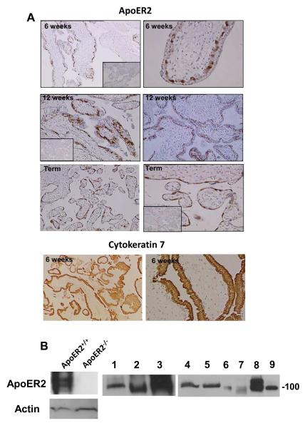
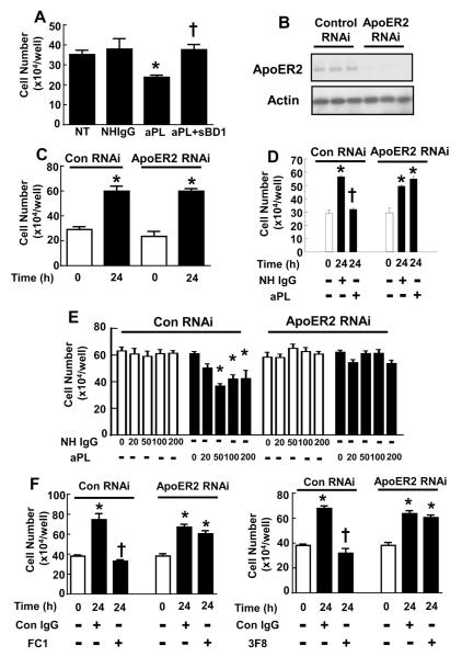
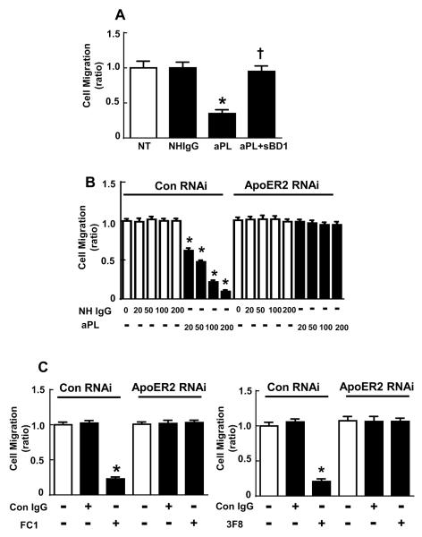
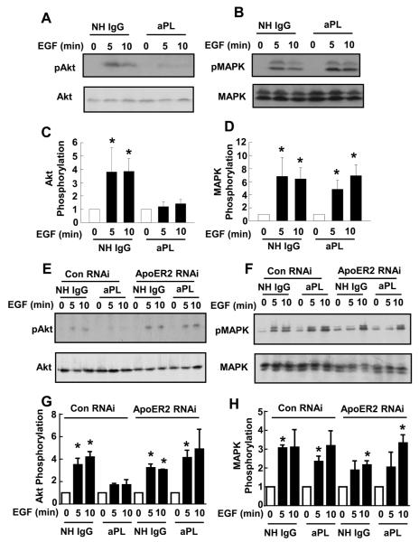
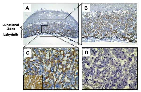
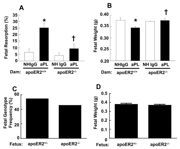
Methods: Placental and trophoblast ApoER2 expression was evaluated by immunohistochemistry and immunoblotting. Normal human IgG and aPL were purified from healthy individuals and APS patients, respectively. The role of ApoER2 in aPL-induced changes in trophoblast proliferation and migration and in kinase activation was assessed using RNA interference in HTR-8/SVneo cells. The participation of ApoER2 in aPL-induced pregnancy loss and IUGR was evaluated in pregnant ApoER2(+/+) and ApoER2(-/-) mice injected with aPL or normal human IgG.

Results: We found that ApoER2 is abundant in human and mouse placental trophoblasts and in multiple trophoblast-derived cell lines, including HTR-8/SVneo cells. ApoER2 and its interaction with the cell surface protein β2 -glycoprotein I were required for aPL-induced inhibition of cultured trophoblast proliferation and migration. In parallel, aPL antagonism of Akt kinase activation by epidermal growth factor in trophoblasts was mediated by ApoER2. Furthermore, in a murine passive-transfer model of pregnancy complications of APS, ApoER2(-/-) mice were protected from both aPL-induced fetal loss and aPL-induced IUGR.

Conclusion: ApoER2 plays a major role in the attenuation of trophoblast function by aPL, and the receptor mediates aPL-induced pregnancy complications in vivo in mice. ApoER2-directed interventions can now potentially be developed to combat the pregnancy complications associated with APS.


Figures

Figure 1
Figure 1
ApoER2 is expressed in human and mouse placental trophoblasts. A. Human placental villous tissue from first trimester (6-12 weeks gestation) and normal term deliveries (37-41 weeks gestation) was immunostained for ApoER2 or cytokeratin 7. Inserts show negative immunostaining using a rabbit IgG control. Images are at 10X (left panels) and at 40X magnification (right panels). B. ApoER2 expression in human placental tissue, primary trophoblasts and trophoblast cell lines was evaluated by immunoblot analysis. Brain lysates from apoER2−/− and apoER2+/+ mice (5 μg total protein loaded) were used as controls. Lanes: 1; mouse placenta (25 μg), 2; mouse SM9-1 cells (mouse trophoblast cell line, 25 μg), 3; BeWo cells (human choriocarcinoma cell line, 25 μg), 4,5; human primary trophoblasts (term delivery, 25 μg), 6,7; human primary trophoblasts (first trimester) (6 μg), 8; Sw.71 cells (first trimester trophoblast cell line, 20 μg), 9; HTR-8/SVneo cells (first trimester trophoblast cell line, 20 μg).
Figure 2
Figure 2
ApoER2 and β2GPI-apoER2 interaction are required for the suppression of trophoblast proliferation by aPL. A. HTR-8/SVneo cells were plated at 10 × 104cells/well and incubated with PBS (NT), NHIgG or aPL (100 μg/ml) in the presence or absence of sBD1 (50 μg/ml). Cell number was counted after 24h incubation (N=9, mean±SEM, *p<0.05 vs. NT, †p<0.05 vs. aPL alone). B. HTR-8/SVneo cells were transfected with control siRNA or double-stranded siRNA targeting apoER2, and whole cell lysates were immunoblotted for apoER2 and actin. Results for 3 samples for each condition are shown. C. HTR-8/SVneo cells were transfected with control siRNA or double-stranded siRNA targeting apoER2, and following transfection the increase in cell number over 24 h was evaluated. N=3, Mean±SEM, *p<0.05 vs. 0h. D, E. Following transfection with control siRNA or siRNA targeting apoER2, the increase in cell number over 24 h was evaluated in cells treated with various concentrations of NHIgG or aPL. (0-200 μg/ml, N=3, Mean±SEM, *p<0.05 vs. 0h, †p<0.05 vs. NHIgG). F. Following transfection with control siRNA or siRNA targeting apoER2, the increase in cell number over 24 h was evaluated in cells treated with anti-β2GPI monoclonal antibodies (FC1 or 3F8) or isotype-matched control IgG (Con IgG). (N=3, Mean±SEM, *p<0.05 vs. 0h, †p<0.05 vs. Con IgG).
Figure 3
Figure 3
ApoER2 and β2GPI-apoER2 interaction are required for the attenuation of trophoblast migration by aPL. A. HTR-8/SVneo cells were treated with PBS (NT), NHIgG (100 μg/ml), aPL (100 μg/ml), or aPL+sBD1 (50 μg/ml) for 24 h during scratch assays. Migration was expressed relative to migration with NT (N=4, Mean±SEM, *p<0.05 vs. NT, †p<0.05 vs. aPL alone). B. Scratch assays were performed in HTR-8/SVneo cells transfected with control siRNA or apoER2 siRNA in the presence of various concentrations of NHIgG of aPL (0-200 μg/ml) for 24 h. Migration was expressed relative to migration of NHIgG-treated cells (N=10, Mean±SEM, *p<0.05 vs. NHIgG). C. Scratch assays were performed in HTR-8/SVneo cells transfected with control siRNA or apoER2 siRNA in the presence of control IgG (Con IgG) or anti-β2 GPI monoclonal antibodies (FC1 or 3F8) Migration was expressed relative to migration of Control IgG-treated cells (N=10, Mean±SEM, *p<0.05 vs. NHIgG).
Figure 4
Figure 4
APL inhibit EGF-induced Akt activation via apoER2. A-D. HTR-8/SVneo cells were incubated with NHIgG or aPL (100 μg/ml) for 16 h, then treated with EGF (100 ng/ml) for 0, 5, or 10 min. Cell lysates were immunoblotted for phospho-Akt (S473), total Akt, phospho-MAPK, and total MAPK A, B. Representative immunoblots are shown. C, D. Summary data for 3 experiments, mean±SEM, *p<0.05 vs. time 0. E-H. HTR-8/SVneo cells were transfected with control siRNA or double-stranded siRNA targeting apoER2. Twenty-four hours following transfection, the cells were treated and immunoblot analyses were done as in A-D. E, F. Representative immunoblots are shown. G, H. Summary data for 3 experiments, mean±SEM, *p<0.05 vs.time 0.
Figure 5
Figure 5
ApoER2 is expressed in mouse placental trophoblasts in vivo. A-C. Immunostaining of ApoER2 was performed in wild-type mouse placenta (E11.5). ApoER2 protein was detected in trophoblasts in the labyrinth. Images are at 4X (A), 10X (B) and 40X magnification (C). Inset in C shows cytokeratin 7 staining in an adjacent placenta section. D. Control staining without primary antibody.
Figure 6
Figure 6
ApoER2−/− mice are protected from aPL-induced fetal resorption and IUGR. Following mating of apoER2+/+ or apoER2+/− males with apoER2+/+ or apoER2−/− females respectively, the females were injected with NHIgG or aPL (10 mg/mouse IP) on day 8 and 12 of pregnancy. Mice were euthanized on day 15 of pregnancy, uteri were dissected, embryos were weighed, and fetal resorption rates were calculated (number of resorptions/number of live fetuses + number of resorptions). A. Fetal resorption frequency. N=7814, Mean±SEM. *p<0.05 vs. NHIgG, p<0.05 vs. apoER2+/+. B. Fetal weights. N=55896, Mean±SEM. *p<0.05 vs. NHIgG, p<0.05 vs. apoER2+/+. C, D. Surviving fetuses from apoER2−/− pregnant mice treated with aPL were genotyped for apoER2 (C) and fetal weights were determined (D). In a total of 66 fetuses, 36 were apoER2+/− and 30 were apoER2−/−.

Comment in

References

    1. Empson M, Lassere M, Craig J, Scott J. Prevention of recurrent miscarriage for women with antiphospholipid antibody or lupus anticoagulant. Cochrane Database Syst Rev. 2005;(2):CD002859. - PMC - PubMed
    1. Meroni PL, Borghi MO, Raschi E, Tedesco F. Pathogenesis of antiphospholipid syndrome: understanding the antibodies. Nat Rev Rheumatol. 2011;7(6):330–9. - PubMed
    1. Ruiz-Irastorza G, Crowther M, Branch W, Khamashta MA. Antiphospholipid syndrome. Lancet. 2010;376(9751):1498–509. - PubMed
    1. Miyakis S, Lockshin MD, Atsumi T, Branch DW, Brey RL, Cervera R, et al. International consensus statement on an update of the classification criteria for definite antiphospholipid syndrome (APS) J Thromb Haemost. 2006;4(2):295–306. - PubMed
    1. Levine JS, Branch DW, Rauch J. The antiphospholipid syndrome. N Engl J Med. 2002;346(10):752–63. - PubMed

Publication types

Substances