. 2015 Oct 16:6:199.
doi: 10.1186/s13287-015-0192-0.

Combination therapy of menstrual derived mesenchymal stem cells and antibiotics ameliorates survival in sepsis

Affiliations

Combination therapy of menstrual derived mesenchymal stem cells and antibiotics ameliorates survival in sepsis

Francisca Alcayaga-Miranda et al. Stem Cell Res Ther. .

Abstract

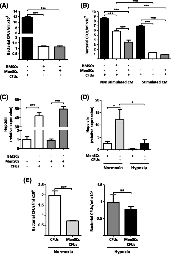
Introduction: Sepsis is a clinical syndrome associated with a severe systemic inflammation induced by infection. Although different anti-microbial drugs have been used as treatments, morbidity and mortality rates remain high. Mesenchymal stem cells (MSCs) derived from the bone marrow have demonstrated a partial protective effect in sepsis. Menstrual derived MSCs (MenSCs) emerge as an attractive candidate because they present important advantages over other sources, including improved proliferation rates and paracrine response under specific stress conditions. Here, we evaluate their therapeutic effect in a polymicrobial severe sepsis model.

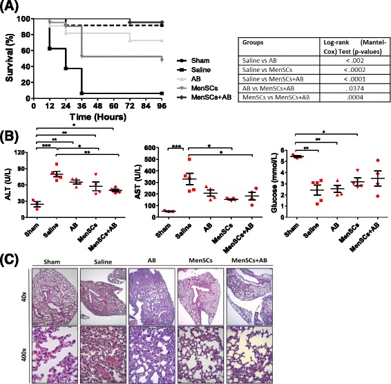
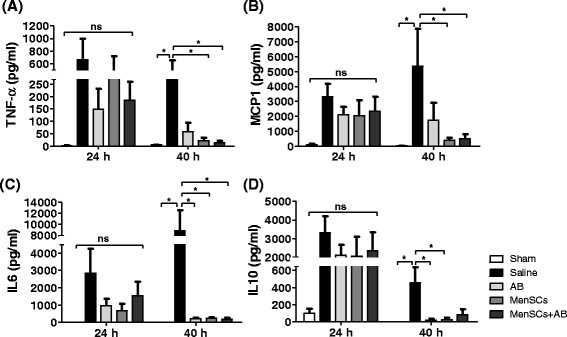
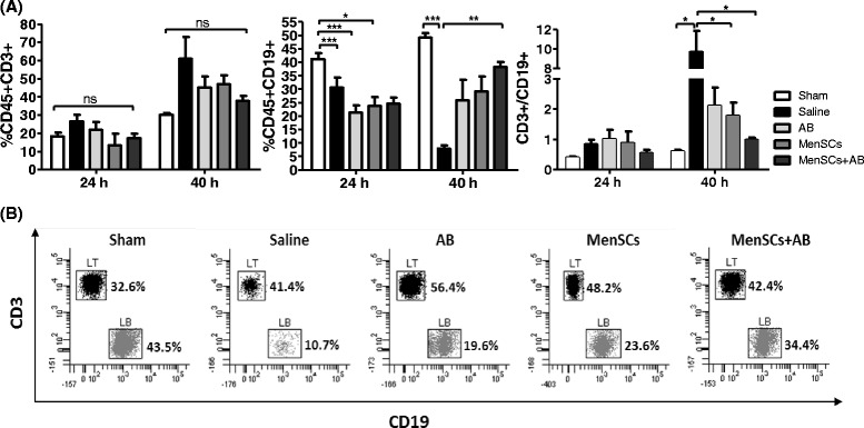
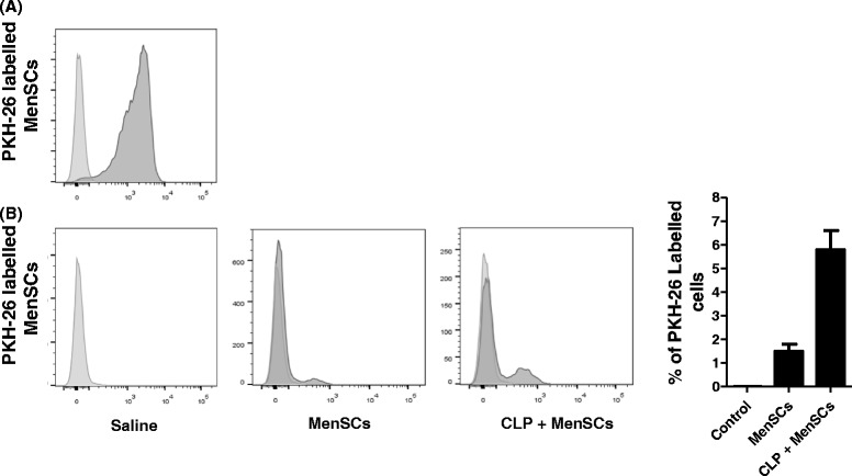
Methods: The antimicrobial activity of MenSCs was determined in vitro through direct and indirect bacterial growth assays and the measurement of the expression levels of different antimicrobial peptides (AMPs) by quantitative reverse transcription-polymerase chain reaction. The therapeutic effect of MenSCs was determined in the cecal ligation and puncture (CLP) mouse model. Mice were then treated with antibiotics (AB) or MenSCs alone or in combination. The survival rates and histological and biochemical parameters were evaluated, and the systemic levels of pro- and anti-inflammatory cytokines as well as the response of specific lymphocyte subsets were determined by flow cytometry.

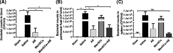
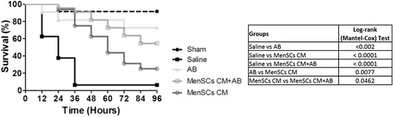
Results: MenSCs exerted an important antimicrobial effect in vitro, mediated by a higher expression of the AMP-hepcidin. In the CLP mouse model, MenSCs in synergy with AB (a) improved the survival rate (95 %) in comparison with saline (6 %), AB (73 %), and MenSCs alone (48 %) groups; (b) enhanced bacterial clearance in the peritoneal fluids and blood; (c) reduced organ injuries evaluated by lower concentrations of the liver enzymes alanine aminotransferase and aspartate aminotransferase; and (d) modulated the inflammatory response through reduction of pro- and anti-inflammatory cytokines without significant loss of T and B lymphocytes.

Conclusions: We conclude that MenSCs in combination with AB enhance survival in CLP-induced sepsis by acting on multiples targets. MenSCs thus constitute a feasible approach for the future clinical treatment of sepsis.


Figures

Fig. 1
Fig. 1
MenSCs exert an important in vitro anti-microbial effect, which is associated in part with the increased expression of hepcidin. MenSCs and BMSCs were evaluated in their capacity to inhibit bacterial proliferation. a Direct inhibition of the bacterial proliferation. Cells were cultured in direct contact with a bacterial mixture. After 6 hours, the CM was collected and plated on blood-agar plates. Values are expressed as CFUs per milliliter. b Indirect inhibition of bacterial proliferation. CM of cells cultured with or without bacterial stimulation was collected and their antimicrobial activity was tested by the incubation with a bacterial mixture. Values are expressed as CFUs per milliliter. c Relative expression of hepcidin. The mRNA expression levels of hepcidin under both basal and stimulated conditions are determined by qRT-PCR. d Inhibition of hepcidin by hypoxia. The mRNA expression levels of hepcidin under normoxic and hypoxic culture conditions in basal and stimulated MenSCs are determined by qRT-PCR. e Direct inhibition of the bacterial proliferation under hypoxic conditions. MenSCs were cultured in direct contact with a bacterial mixture under normoxic and hypoxic conditions for 24 hours. After 24 hours, the CM was collected and plated on blood-agar plates. Values are expressed as CFUs per milliliter. Data are presented as the mean ± standard error. BMSCs bone marrow-derived mesenchymal stem cells, CFU colony-forming unit, CM conditioned medium, MenSCs menstrual derived mesenchymal stem cells, ns non-significant, qRT-PCR quantitative reverse transcription-polymerase chain reaction
Fig. 2
Fig. 2
Treatment with MenSCs alone or in combination with antibiotics improves survival and protects against multiorgan dysfunction in CLP-induced sepsis. a Survival curves in mice with polymicrobial sepsis and different treatments. C57BL6/J mice subjected to CLP-induced sepsis were randomized into five groups: sham (n = 12), saline (n = 16), antibiotics (AB) (n = 22), MenSCs (n = 21), and MenSCs + AB (n = 22). Three hours post-surgery, mice were injected with saline (vehicle), AB and/or MenSCs. Survival percentages of untreated and treated mice after CLP are presented as a Kaplan-Meier survival curve. b Serum concentration of alanine aminotransferase (ALT), aspartate aminotransferase (AST), and glucose after CLP-induced sepsis. Serum was isolated 24 hours post-surgery and treatment administration (sham, n = 3; saline, n = 5; AB, n = 4; MenSCs, n = 4; MenSCs + AB, n = 4), and the concentrations of biomarkers of liver function and total glucose were determined. Dot plots represent individual values, horizontal bars represent mean values, and vertical bars represent standard error values. c Lung histology after CLP-induced sepsis. At 40 hours post-surgery and treatment infusion, lungs were collected, fixed, embedded in paraffin, and stained with hematoxylin-eosin. Pictures are representative images of lungs from the different experimental groups. AB antibiotics, CLP cecal ligation puncture, MenSCs menstrual derived mesenchymal stem cells
Fig. 3
Fig. 3
MenSCs modulate the response of the host immune system to sepsis. At 24 and 40 hours after CLP-induced sepsis and treatment administration, blood samples were obtained to determine the serum concentration of the inflammatory cytokines (a) TNF-α, (b) MCP-1, (c) IL-6, and (d) IL-10 using a Cytometric Bead Array and analyzed by flow cytometry (sham, n = 6; saline, n = 3–11; AB, n = 4–6; MenSCs, n = 5–6; MenSCs + AB, n = 6). Histograms represent the mean ± standard error. AB antibiotics, CLP cecal ligation and puncture, IL interleukin, MCP-1 monocyte chemoattractant protein-1, MenSC menstrual derived mesenchymal stem cells, ns non-significant, TNF-α tumor necrosis factor-alpha
Fig. 4
Fig. 4
MenSCs in combination with antibiotics prevent the decrease of CD45+ CD3+ and CD45+ CD19+ lymphocyte levels after CLP-induced sepsis. Blood samples were obtained 24 and 40 hours after administration of various treatments post-CLP, and specific lymphocyte subsets were determined by fluorescence-activated cell sorting. a Graphs represent the percentage of CD45+ CD3+ and CD45+ CD19+ lymphocytes and the ratio CD45+ CD3+/CD45+ CD19 (sham, n = 3–6; saline, n = 3–10; AB, n = 3–6; MenSCs, n = 3–6; MenSCs + AB, n = 3–6). Histograms represent the mean ± standard error. b Representative dot plots of specific lymphocyte subsets described in (a) at 40 hours post-induction of sepsis and administration of different treatments. AB antibiotics, CLP cecal ligation and puncture, LB B lymphocytes, LT T lymphocytes, MenSCs menstrual derived mesenchymal stem cells, ns non-significant
Fig. 5
Fig. 5
MenSCs alone or in combination with antibiotic treatment promote bacterial clearance in CLP-induced sepsis in mice. Blood, peritoneal fluid, and spleen were obtained 24 hours after administration of the different treatments (sham, n = 6; saline, n = 11; AB, n = 6; MenSCs, n = 5; MenSCs + AB, n = 6). Bacterial loads were determined after incubating at 37 °C overnight and are expressed as CFUs per milliliter. a Bacterial load in blood. b Bacterial load in peritoneal fluid. c Bacterial load in spleen. Data are presented as mean ± standard error. AB antibiotics, CFU colony-forming unit, CLP cecal ligation and puncture, MenSCs menstrual derived mesenchymal stem cells, ns non-significant
Fig. 6
Fig. 6
Septic mice show higher retention of the injected MenSCs in the peritoneal cavity in comparison with healthy control mice. MenSCs were labelled with PKH-26 and analyzed by flow cytometry. a Fluorescence-activated cell sorting plot showing a 95 % labelling efficiency. Around 2 × 106 cells were injected intraperitoneally in septic or healthy mice. b Histogram and representative flow cytometry plots showing the percentage of remaining cells in peritoneal lavage at 24 hours post-injection. CLP cecal ligation puncture, MenSCs menstrual derived mesenchymal stem cells
Fig. 7
Fig. 7
MenSCs CM improves the survival in CLP-induced sepsis in mice. C57BL6/J mice with CLP-induced sepsis were randomized in five groups: sham (n = 12), saline (n = 16), antibiotics (AB) (n = 22), MenSCs CM (n = 16), and MenSC CM + AB (n = 22). After 3 hours post-sepsis induction, mice were injected with saline (vehicle), AB and/or MenSC CM. Survival percentages of untreated and treated mice are represented as a Kaplan-Meier survival curve. AB antibiotics, CFU colony-forming unit, CLP cecal ligation and puncture, CM conditioned medium, MenSCs menstrual derived mesenchymal stem cells

References

    1. Hall SR, Tsoyi K, Ith B, Padera RF, Lederer JA, Wang Z, et al. Mesenchymal stromal cells improve survival during sepsis in the absence of heme oxygenase-1: The importance of neutrophils. Stem Cells. 2013;31:397–407. doi: 10.1002/stem.1270. - DOI - PMC - PubMed
    1. Martin GS, Mannino DM, Eaton S, Moss M. The epidemiology of sepsis in the United States from 1979 through 2000. N Engl J Med. 2003;348:1546–1554. doi: 10.1056/NEJMoa022139. - DOI - PubMed
    1. Németh K, Leelahavanichkul A, Yuen PS, Mayer B, Parmelee A, Doi K, et al. Bone marrow stromal cells attenuate sepsis via prostaglandin E(2)-dependent reprogramming of host macrophages to increase their interleukin-10 production. Nat Med. 2009;15:42–49. doi: 10.1038/nm.1905. - DOI - PMC - PubMed
    1. Cohen J. The immunopathogenesis of sepsis. Nature. 2002;420:885–891. doi: 10.1038/nature01326. - DOI - PubMed
    1. Mei SH, Haitsma JJ, Dos Santos CC, Deng Y, Lai PF, Slutsky AS, et al. Mesenchymal stem cells reduce inflammation while enhancing bacterial clearance and improving survival in sepsis. Am J Respir Crit Care Med. 2010;182:1047–1057. doi: 10.1164/rccm.201001-0010OC. - DOI - PubMed

Publication types

MeSH terms