Review
. 2015 Nov;16(11):1439-53.
doi: 10.15252/embr.201540749. Epub 2015 Oct 15.

The H1 linker histones: multifunctional proteins beyond the nucleosomal core particle

Affiliations
Review

The H1 linker histones: multifunctional proteins beyond the nucleosomal core particle

Sonja P Hergeth et al. EMBO Rep. 2015 Nov.

Abstract

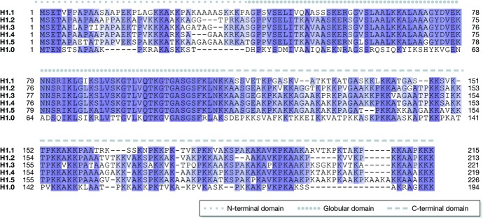
The linker histone H1 family members are a key component of chromatin and bind to the nucleosomal core particle around the DNA entry and exit sites. H1 can stabilize both nucleosome structure and higher-order chromatin architecture. In general, H1 molecules consist of a central globular domain with more flexible tail regions at both their N- and C-terminal ends. The existence of multiple H1 subtypes and a large variety of posttranslational modifications brings about a considerable degree of complexity and makes studying this protein family challenging. Here, we review recent progress in understanding the function of linker histones and their subtypes beyond their role as merely structural chromatin components. We summarize current findings on the role of H1 in heterochromatin formation, transcriptional regulation and embryogenesis with a focus on H1 subtypes and their specific modifications.

Keywords: chromatin; epigenetics; linker histone; modifications; subtypes.


Figures

Figure 1
Figure 1. Alignment of the somatic human H1 isotypes and H1.0
The globular domain (solid line) is highly conserved, whereas the N- and C-terminal domains (dotted and dashed lines, respectively) are more variable . Conserved residues are highlighted by shades of blue. Darker colour represents higher conservation.
Figure 2
Figure 2. Cell cycle-dependent phosphorylation of H1.4
In interphase (top panel), two phosphorylated serine residues have been detected in the C-terminal tail, S172 and S187 ,. Both residues are part of a CDK consensus sequence (S/T-P-X-K) and are likely to be phosphorylated by Cdk2 ,,. S172p and S187p have been linked to active transcription by RNAP I and II . Phosphorylation levels are thought to increase during S phase and are highest in mitosis (bottom panel), where up to six phosphorylation sites have been identified in both the N- and C-terminal tails. Threonine residues are phosphorylated mainly during mitosis . Ser/Thr residues located in CDK consensus sites are thought to be targeted by the mitotic kinase CDK1/CycB . H1.4S27 is phosphorylated by Aurora B, a member of the chromosomal passenger complex . Full phosphorylation during mitosis may allow for a structural rearrangement inducing chromatin compaction . p: phosphorylation, S/T-P-X-K: CDK consensus site.
Figure 3
Figure 3. H1.4 modifications and their cellular functions
Among the various modifications of linker histones, only few were characterized with site-specific antibodies. Mostly modifications on the subtype H1.4 have been characterized. H1.4 is methylated at K26, which is catalysed by G9a/GLP1 and potentially also by EZH2 ,. This methylation provides a binding platform for HP1 and is thus linked to transcriptional repression and heterochromatin formation. Phosphorylation of S27 inhibits binding of HP1 to K26me . The C-terminal phosphorylations S172p and S187p are present on H1.4 also in interphase and have been linked to active transcription . H1.4K34ac is set by the acetyltransferase GCN5 and is enriched at active transcription start sites. It can positively regulate transcription by (i) recruiting the bromodomain-containing TAF1 subunit of the TFIID transcription complex and (ii) increasing H1 mobility . The conversion of R54 to citrulline by PADI4 has been shown to occur on several mouse H1 subtypes, among them H1.4. This modification in a DNA-binding site results in eviction of H1 and global chromatin decondensation in pluripotent cells. p: phosphorylation, ac: acetylation, me: methylation, cit: citrullination, violet: enzymes, orange: readers.
Figure 4
Figure 4. Model of H1 modes of action
Based on current knowledge, we propose a dual model of H1 function: (i) H1 is a key structural component of chromatin. It can stabilize nucleosome structure and influence nucleosome spacing, and it is required for chromatin compaction (left panel). These functions often seem to be redundant between different subtypes. (ii) H1 also functions through interaction with other proteins that will in turn modify chromatin or take part in DNA-based processes (right panel). In Drosophila melanogaster, H1 recruits S(uv)39 to chromatin, which is required for heterochromatin formation . These functions are often subtype specific, as, for example, the recruitment of Cul4A and PAF1 by H1.2 to support target gene transcription . Often, H1 modifications come into play, as was demonstrated for HP1 binding to H1.4K26me or TAFII binding to H1.4K34ac ,. Of course, these two modes of action are not always separable. For example, interaction with other proteins or H1 modifications can be required for H1 eviction from chromatin (f), so that it will no longer exert its stabilizing function, resulting in chromatin opening for DNA-based processes like transcription, replication and repair.

Similar articles

Cited by

References

    1. Luger K, Mäder AW, Richmond RK, Sargent DF, Richmond TJ. Crystal structure of the nucleosome core particle at 2.8 A resolution. Nature. 1997;389:251–260. - PubMed
    1. Woodcock CL, Skoultchi AI, Fan Y. Role of linker histone in chromatin structure and function: H1 stoichiometry and nucleosome repeat length. Chromosome Res. 2006;14:17–25. - PubMed
    1. Robinson PJ, Rhodes D. Structure of the “30 nm” chromatin fibre: a key role for the linker histone. Curr Opin Struct Biol. 2006;16:336–343. - PubMed
    1. Thoma F, Koller T, Klug A. Involvement of histone H1 in the organization of the nucleosome and of the salt-dependent superstructures of chromatin. J Cell Biol. 1979;83:403–427. - PMC - PubMed
    1. Schlissel MS, Brown DD. The transcriptional regulation of Xenopus 5s RNA genes in chromatin: the roles of active stable transcription complexes and histone H1. Cell. 1984;37:903–913. - PubMed

Publication types