. 2015 Dec;15(23-24):3969-79.
doi: 10.1002/pmic.201500256. Epub 2015 Nov 23.

Expression and distribution of neuropeptides in the nervous system of the crab Carcinus maenas and their roles in environmental stress

Affiliations

Expression and distribution of neuropeptides in the nervous system of the crab Carcinus maenas and their roles in environmental stress

Yuzhuo Zhang et al. Proteomics. 2015 Dec.

Abstract

Environmental fluctuations, such as salinity, impose serious challenges to marine animal survival. Neuropeptides, signaling molecules involved in the regulation process, and the dynamic changes of their full complement in the stress response have yet to be investigated. Here, a MALDI-MS-based stable isotope labeling quantitation strategy was used to investigate the relationship between neuropeptide expression and adaptability of Carcinus maenas to various salinity levels, including high (60 parts per thousand [p.p.t.]) and low (0 p.p.t.) salinity, in both the crustacean pericardial organ (PO) and brain. Moreover, a high salinity stress time course study was conducted. MS imaging (MSI) of neuropeptide localization in C. maenas PO was also performed. As a result of salinity stress, multiple neuropeptide families exhibited changes in their relative abundances, including RFamides (e.g. APQGNFLRFamide), RYamides (e.g. SSFRVGGSRYamide), B-type allatostatins (AST-B; e.g. VPNDWAHFRGSWamide), and orcokinins (e.g. NFDEIDRSSFGFV). The MSI data revealed distribution differences in several neuropeptides (e.g. SGFYANRYamide) between color morphs, but salinity stress appeared to not have a major effect on the localization of the neuropeptides.

Keywords: Crustacean; Isotopic labeling; Mass spectrometry imaging; Neuropeptides; Salinity stress.


Conflict of interest statement

The authors have declared no conflict of interest.

Figures
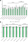
Figure 1
Figure 1
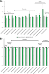
Content changes of neuropeptides of interest in Carcinus maenas (A) PO (n=6) and (B) brain (n=4) between a control and 5h of high salinity stress. For each pair, the bars on the left refer to changes in the green morph, while the bars on the right correspond to the red morph. Each group contained two POs or one brain. A ratio equal to one denotes no change between stress and control conditions, marked by a dotted line. The error bars depict the SEM of the measured ratio. Asterisks indicate significance levels (student’s t test): *, p<0.05, **, p<0.005.
Figure 2
Figure 2
Content changes of neuropeptides of interest in Carcinus maenas (A) PO (n=5) and (B) brain (n=5) between a control and 5h of low salinity stress. For each pair, the bars on the left refer to changes in the green morph, while the bars on the right correspond to the red morph. Each group contained two POs or one brain. A ratio equal to one denotes no change between stress and control conditions, marked by a dotted line. The error bars depict the SEM of the measured ratio. Asterisks indicate significance levels (student’s t test): *, p<0.05, **, p<0.005.
Figure 3
Figure 3
Time course study of Carcinus maenas neuropeptide content changes due to high salinity stress for the (A) PO and (B) brain for the green color morph. 6 groups for 5h, 10 groups for 12 h, 6 groups for 24h, 6 groups for 48h (each group has two POs or one brain). A ratio equal to one denotes no change between stress and control conditions, marked by a dotted line. The error bars depict the SEM of the measured ratio. Asterisks indicated significance levels (student’s t test): *, p<0.05, **, p<0.005. ANOVA tests where the peptide changes were found to be significant are indicated by a pound sign (#) by the peptide name.
Figure 4
Figure 4
MALDI-MSI analysis of several neuropeptides in the C. maenas PO. (A) Optical image of a PO on a MALDI plate. The main body of the decapod crustacean PO consists of anterior region, posterior region, dorsal trunk, and ventral trunk. Neuropeptide localization in (B) green shell and (C) red shell C. maenas PO for both control (left) and stressed (right) animal. Visualized neuropeptides pertain to three families, including RYamides ((i) FVGGSRYa (m/z 784.5), (ii) SGFYANRYa (m/z 976.5), and (iii) pEGFYSQRYa (m/z 1030.5)), RFamides ((iv) NRNFLRFa (m/z 965.5), (v) GNRNFLRFa (m/z 1022.6), and (vi) DGNRNFLRFa (m/z 1137.6)), and AST-Bs ((vii) QWSSMRGAWa (m/z 1107.6), (viii) STNWSSLRSAWa (m/z 1293.6), and (ix) VPNDWAHFRGSWa (m/z 1470.7)).

References

    1. Chen Y, Bender RA, Brunson KL, Pomper JK, et al. Modulation of dendritic differentiation by corticotropin-releasing factor in the developing hippocampus. Proc Natl Acad Sci U S A. 2004;101:15782–15787. - PMC - PubMed
    1. Hiremagalur B, Kvetnansky R, Nankova B, Fleischer J, et al. Stress elicits trans-synaptic activation of adrenal neuropeptide Y gene expression. Brain Res Mol Brain Res. 1994;27:138–144. - PubMed
    1. Delorenzi A, Dimant B, Frenkel L, Nahmod VE, et al. High environmental salinity induces memory enhancement and increases levels of brain angiotensin-like peptides in the crab Chasmagnathus granulatus. J Exp Biol. 2000;203:3369–3379. - PubMed
    1. Chung JS, Zmora N. Functional studies of crustacean hyperglycemic hormones (CHHs) of the blue crab, Callinectes sapidus - the expression and release of CHH in eyestalk and pericardial organ in response to environmental stress. Febs Journal. 2008;275:693–704. - PubMed
    1. McCormick DA, Nusbaum MP. Editorial overview: Neuromodulation: Tuning the properties of neurons, networks and behavior. Current Opinion in Neurobiology. 2014;29:IV–VII. - PMC - PubMed

Publication types

LinkOut - more resources