

Clinical and dermoscopic features of atypical Spitz tumors: A multicenter, retrospective, case-control study
- PMID: 26475536
- PMCID: PMC4806681
- DOI: 10.1016/j.jaad.2015.08.018
Clinical and dermoscopic features of atypical Spitz tumors: A multicenter, retrospective, case-control study
Abstract
Background: Few studies have described the clinical and dermoscopic features of atypical Spitz tumors.
Objective: We sought to describe the clinical and dermoscopic features of a series of atypical Spitz tumors as compared with those of conventional Spitz nevi.
Methods: This was a multicenter, retrospective, case-control study, analyzing the clinical and dermoscopic characteristics of 55 atypical Spitz tumors and 110 Spitz nevi that were excised and diagnosed histopathologically.
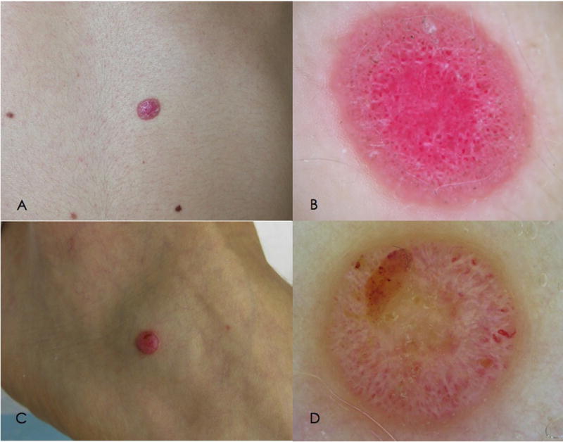
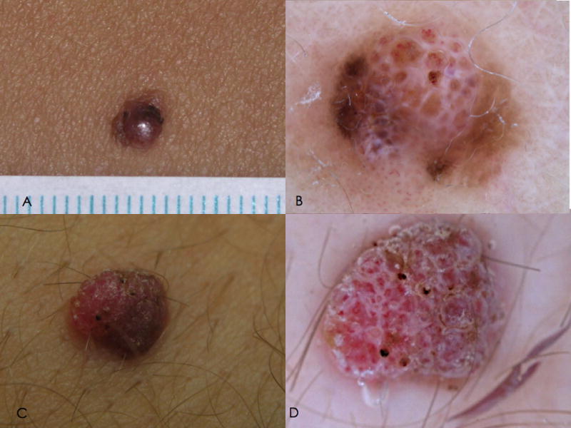
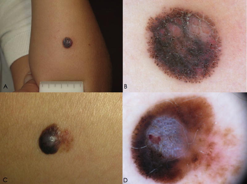
Results: The majority of atypical Spitz tumors presented clinically as a plaque or nodule, dermoscopically typified by a multicomponent or nonspecific pattern. A proportion of lesions (16.4%) exhibited the typical nonpigmented Spitzoid pattern of dotted vessels and white lines under dermoscopy. Nodularity, ulceration, linear vessels, polymorphic vessels, white lines, and blue-white veil were associated with atypical Spitz tumors by univariate analysis, but only nodularity and white lines remained significant after multivariate analysis. In contrast, a pigmented typical Spitzoid pattern was a potent predictor of Spitz nevi, associated with 6.5-fold increased probability.
Limitations: Differentiation from Spitzoid melanoma and other nonmelanocytic lesions was not investigated.
Conclusion: Atypical Spitz tumors are polymorphic melanocytic proliferations with a nodular clinical appearance. Dermoscopically they demonstrate a multicomponent and nonspecific pattern. A typical nonpigmented Spitzoid pattern on dermoscopy (with dotted vessels and white lines) does not exclude atypical Spitz tumors.
Keywords: Spitz nevus; atypical Spitz tumor; dermoscopy; histopathology; melanoma; skin cancer.
Copyright © 2015 American Academy of Dermatology, Inc. Published by Elsevier Inc. All rights reserved.
Figures
References
-
- Kernen JA, Ackerman LV. Spindle cell nevi and epithelioid cell nevi (so-called juvenile melanomas) in children and adults: a clinicopathologic study of 27 cases. Cancer. 1960;13:612–625. - PubMed
-
- Smith KJ, Barrett TL, Skelton HG, 3rd, et al. Spindle cell and epithelioid cell nevi with atypia and metastasis (malignant Spitz nevus) Am J Surg Pathol. 1989;13:931–939. - PubMed
-
- Barnhill RL, Flotte TJ, Fleischli M, et al. Cutaneous melanoma and atypical Spitz tumors in children. Cancer. 1995;76:1833–1845. - PubMed
-
- Barnhill RL, Argenyi ZB, From L, et al. Atypical Spitz nevi/tumor: lack of consensus for diagnosis, discrimination from melanoma, and prediction of outcome. Hum Pathol. 1999;30:513–520. - PubMed
-
- Mones JM, Ackerman AB. “Atypical”tSpitz’s nevus, “Malignant”aSpitz’s nevus, and “Metastasizing”eSpitz’s nevus: a critique in historical perspective of three concepts flawed fatally. Am J Dermatopathol. 2004;26:310–333. - PubMed
Publication types
MeSH terms
Grants and funding
LinkOut - more resources
Full Text Sources
Other Literature Sources
Medical
