. 2015 Dec;6(6):729-40.
doi: 10.1007/s13244-015-0441-x. Epub 2015 Oct 16.

Imaging characteristics of pleural tumours

Affiliations

Imaging characteristics of pleural tumours

Luca De Paoli et al. Insights Imaging. 2015 Dec.

Abstract

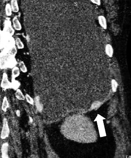
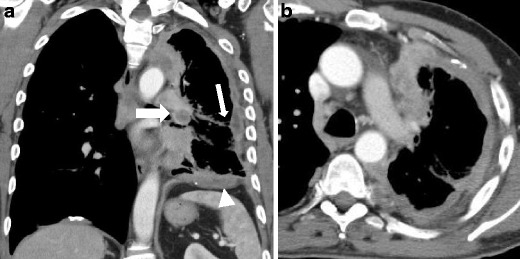
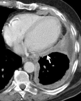
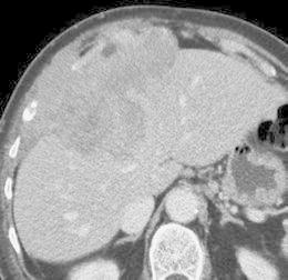
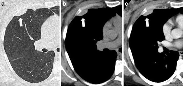
Malignant mesothelioma is doubtless the more known pleural tumour. However, according to the morphology code of the International Classification of Diseases for Oncology (ICD-O), there are several histological types of pleural neoplasms, divided into mesothelial, mesenchymal and lymphoproliferative tumours, that may be misdiagnosed. In this paper we summarise and illustrate the incidence aspects and the clinical, pathological and radiological features of these neoplasms.

Teaching points: • According to the ICD-O, there are 11 different histological types of pleural neoplasm. • Imaging, clinical and histopathological aspects of these neoplasms may be overlapping. • Knowledge of different pleural tumours plays an important role for diagnosis orientation.

Keywords: Malignant mesothelioma; Pleural neoplasm; Primary effusion lymphoma; Solitary fibrous tumour; Synovial sarcoma.


Figures

Fig. 1
Fig. 1
Schematic diagram of the pleurae and upper airways
Fig. 2
Fig. 2
Sarcomatoid mesothelioma; axial view contrast-enhanced CT scan: soft tissue (a) and bones (b) window images showing a large thoracic wall mass with inhomogeneous enhancement, involving the lateral arch of a rib, that appears eroded (white arrow), a typical infiltration sign
Fig. 3
Fig. 3
Biphasic mesothelioma; chest X-ray film (a) showing a right basal and lateral pleural thickening with fissure involvement (thick arrow). Lung MR confirm these findings, both on fat saturation T1-weighted image (b) and spin echo T2-weighted sequence (c) that better demonstrates a mild basal anterior pleural effusion (arrowhead)
Fig. 4
Fig. 4
Stage I of MM; contrast-enhanced CT scan: coronal (a) and axial (b) view. Diffuse circumferential thickening of the thoracic, mediastinal and diaphragmatic pleura involving even the fissure. No extra-pleural infiltration or significant lymphadenopaties are observed (only two small lymph nodes are reported—white arrows)
Fig. 5
Fig. 5
Stage II of MM; contrast-enhanced CT scan: coronal view. Focal pleural enhancing thickening that infiltrates the left hemidiaphragm (thick arrow)
Fig. 6
Fig. 6
Stage III of MM. Contrast-enhanced CT scan: coronal (a) and axial (b) view show a severe left lung hypoexpansion, with circumferential irregular pleural thickening and fissure thickening (white arrow); left hilar inhomogeneous pathological lymph node may be seen (thick arrow), and infiltration of the left hemidiaphragm is reported (arrowhead); this MM (sarcomatoid mesothelioma variant) is classified as T2, N1, M0
Fig. 7
Fig. 7
Stage III of MM; contrast-enhanced CT scan: axial view showing a circumferential pleural thickening, involving the mediastinal pleura and the pericardium (white arrow), classified as T3, N0, M0
Fig. 8
Fig. 8
Stage IV of MM; abdominal contrast-enhanced CT scan. Aggressive MM grown through the diaphragm. An axial view demonstrates the infiltration of the peritoneum and the liver (classified as T4)
Fig. 9
Fig. 9
Large left thoracic wall sarcoma; contrast-enhanced CT scan: coronal (a) and axial (b) view. Severe left hemithorax hypoexpansion, homolateral hemidiaphragm superelevation and presence of a large polylobulated mass, with faint enhancement and extraparietal extension. No precise fat plane may be seen between the mediastinal pleura and the pericardium (white arrow), a finding that is highly suspicious for mediastinal infiltration
Fig. 10
Fig. 10
An accidental case of solitary fibrous tumour in a 56-year-old man: standard chest radiography (a, b) demonstrates a well-defined, ovalar shaped chest wall mass. Contrast-enhanced CT, axial plane before (c) and after (d) contrast media administration showing a bulky, homogeneus and non-enhancing mass of the left posterior chest wall
Fig. 11
Fig. 11
Unusual case of giant solitary fibrous tumour; contrast-enhanced CT, axial (a) and coronal plane (b) showing a bulky and inhomogeneous contrast-enhancing mass of the right hemithorax. The mediastinal structures are compressed and contralaterally migrated. The hypodense areas (white arrows) may represent the presence of necrosis, haemorrhage or myxoid tissue
Fig. 12
Fig. 12
Primary effusion lymphoma; axial view contrast-enhanced CT scan: soft tissue (a) and lung (b) window images. A mild pleural effusion may be seen (arrowhead) with slightly hyperdensity, associated with a small lung consolidation with air bronchogram (thick arrow); no real pleural masses are recognised
Fig. 13
Fig. 13
Pleural metastases; axial view of unenhanced and contrast-enhanced CT scan: lung (a), soft tissue (b) and contrast-enhanced image (c). A relatively small bulky mass (arrow), that demonstrates slightly and homogeneous enhancement. After analysis of the histological specimen the lesion was found to be a pleural metastases from breast adenocarcinoma

References

    1. Finley DJ, Rusch VW. Anatomy of the pleura. Thorac Surg Clin. 2011;21(2):157–163. doi: 10.1016/j.thorsurg.2010.12.001. - DOI - PubMed
    1. Charalampidis C, Youroukou A, Lazaridis G, et al. Pleura space anatomy. J Thorac Dis. 2015;7(Suppl 1):S27–S32. - PMC - PubMed
    1. Nickell LT, Jr, Lichtenberger JP, Khorashadi L, Abbott GF, Carter BW. Multimodality imaging for characterization, classification, and staging of malignant pleural mesothelioma. Radiographics. 2014;34(6):1692–1706. doi: 10.1148/rg.346130089. - DOI - PubMed
    1. Tossavainen A. Global use of asbestos and the incidence of mesothelioma. Int J Occup Environ Health. 2004;10:22–25. doi: 10.1179/oeh.2004.10.1.22. - DOI - PubMed
    1. Park EK, Takahashi K, Hoshuyama T. Global magnitude of reported and unreported mesothelioma. Environ Health Perspect. 2011;119(4):514–518. doi: 10.1289/ehp.1002845. - DOI - PMC - PubMed

LinkOut - more resources