

Pharmacological Targeting SHP-1-STAT3 Signaling Is a Promising Therapeutic Approach for the Treatment of Colorectal Cancer
- PMID: 26476076
- PMCID: PMC4611073
- DOI: 10.1016/j.neo.2015.08.007
Pharmacological Targeting SHP-1-STAT3 Signaling Is a Promising Therapeutic Approach for the Treatment of Colorectal Cancer
Abstract
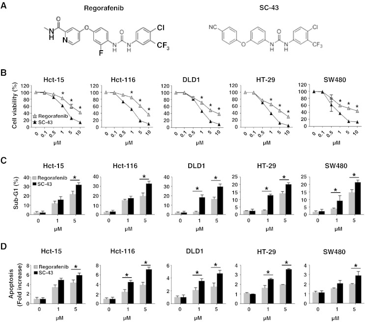
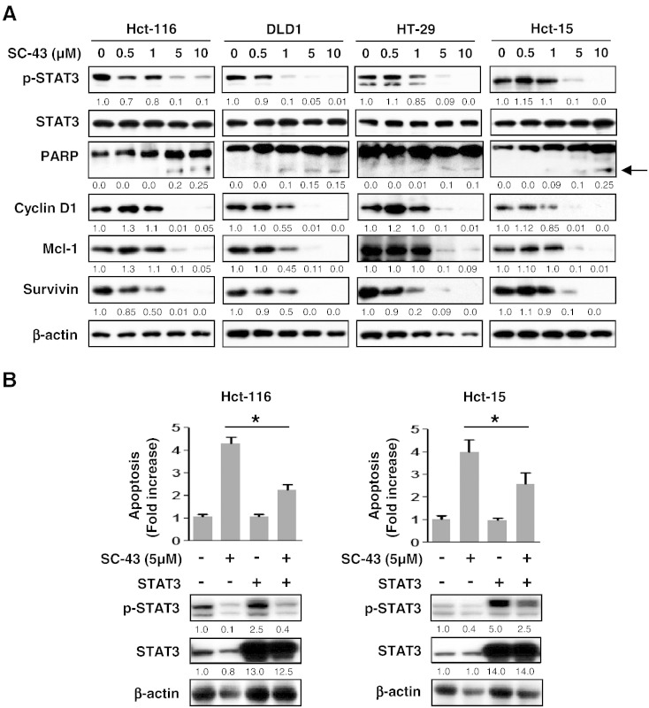
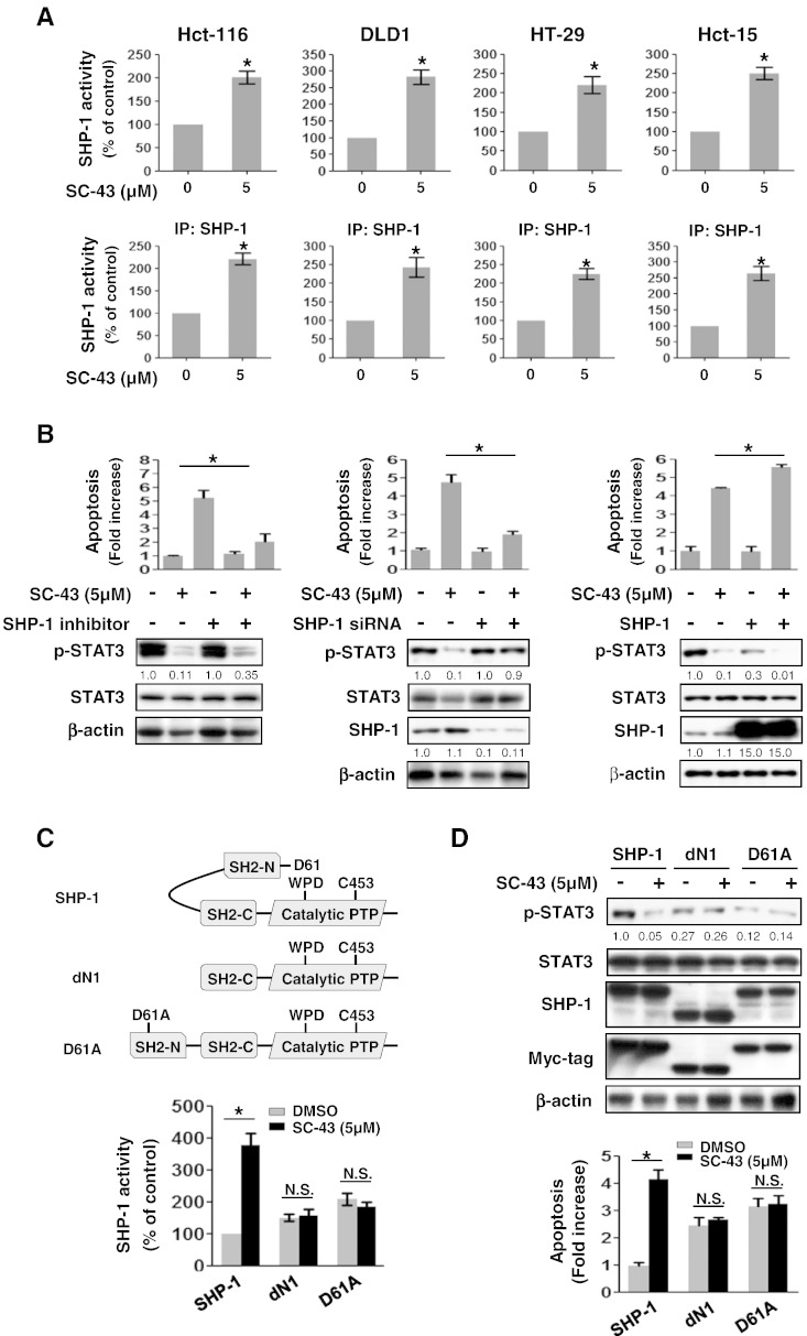
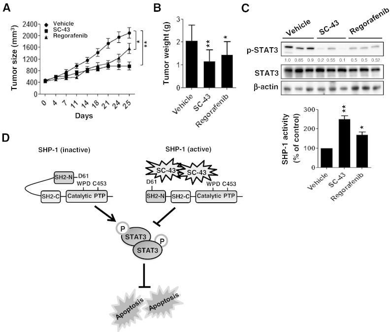
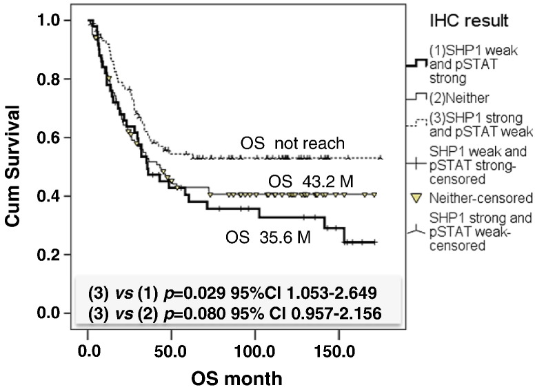
STAT3 activation is associated with poor prognosis in human colorectal cancer (CRC). Our previous data demonstrated that regorafenib (Stivarga) is a pharmacological agonist of SH2 domain-containing phosphatase 1 (SHP-1) that enhances SHP-1 activity and induces apoptosis by targeting STAT3 signals in CRC. This study aimed to find a therapeutic drug that is more effective than regorafenib for CRC treatment. Here, we showed that SC-43 was more effective than regorafenib at inducing apoptosis in vitro and suppressing tumorigenesis in vivo. SC-43 significantly increased SHP-1 activity, downregulated p-STAT3(Tyr705) level, and induced apoptosis in CRC cells. An SHP-1 inhibitor or knockdown of SHP-1 by siRNA both significantly rescued the SC-43-induced apoptosis and decreased p-STAT3(Tyr705) level. Conversely, SHP-1 overexpression increased the effects of SC-43 on apoptosis and p-STAT3(Tyr705) level. These data suggest that SC-43-induced apoptosis mediated through the loss of p-STAT3(Tyr705) was dependent on SHP-1 function. Importantly, SC-43-enhanced SHP-1 activity was because of the docking potential of SC-43, which relieved the autoinhibited N-SH2 domain of SHP-1 and inhibited p-STAT3(Tyr705) signals. Importantly, we observed that a significant negative correlation existed between SHP-1 and p-STAT3(Tyr705)expression in CRC patients (P = .038). Patients with strong SHP-1 and weak p-STAT3(Tyr705) expression had significantly higher overall survival compared with patients with weak SHP-1 and strong p-STAT3(Tyr705) expression (P = .029). In conclusion, SHP-1 is suitable to be a useful prognostic marker and a pharmacological target for CRC treatment. Targeting SHP-1-STAT3 signaling by SC-43 may serve as a promising pharmacotherapy for CRC.
Copyright © 2015 The Authors. Published by Elsevier Inc. All rights reserved.
Figures
Similar articles
-
Alteration of SHP-1/p-STAT3 Signaling: A Potential Target for Anticancer Therapy.Int J Mol Sci. 2017 Jun 8;18(6):1234. doi: 10.3390/ijms18061234. Int J Mol Sci. 2017. PMID: 28594363 Free PMC article. Review.
-
SHP-1 is a target of regorafenib in colorectal cancer.Oncotarget. 2014 Aug 15;5(15):6243-51. doi: 10.18632/oncotarget.2191. Oncotarget. 2014. PMID: 25071018 Free PMC article.
-
Regorafenib (Stivarga) pharmacologically targets epithelial-mesenchymal transition in colorectal cancer.Oncotarget. 2016 Sep 27;7(39):64136-64147. doi: 10.18632/oncotarget.11636. Oncotarget. 2016. PMID: 27580057 Free PMC article.
-
Obatoclax analog SC-2001 inhibits STAT3 phosphorylation through enhancing SHP-1 expression and induces apoptosis in human breast cancer cells.Breast Cancer Res Treat. 2014 Jul;146(1):71-84. doi: 10.1007/s10549-014-3000-0. Epub 2014 Jun 6. Breast Cancer Res Treat. 2014. PMID: 24903225
-
Targeting key transcriptional factor STAT3 in colorectal cancer.Mol Cell Biochem. 2021 Sep;476(9):3219-3228. doi: 10.1007/s11010-021-04156-8. Epub 2021 Apr 18. Mol Cell Biochem. 2021. PMID: 33866491 Review.
Cited by
-
Consideration of SHP-1 as a Molecular Target for Tumor Therapy.Int J Mol Sci. 2023 Dec 26;25(1):331. doi: 10.3390/ijms25010331. Int J Mol Sci. 2023. PMID: 38203502 Free PMC article. Review.
-
Alteration of SHP-1/p-STAT3 Signaling: A Potential Target for Anticancer Therapy.Int J Mol Sci. 2017 Jun 8;18(6):1234. doi: 10.3390/ijms18061234. Int J Mol Sci. 2017. PMID: 28594363 Free PMC article. Review.
-
Interfering B cell receptor signaling via SHP-1/p-Lyn axis shows therapeutic potential in diffuse large B-cell lymphoma.Mol Med. 2022 Aug 8;28(1):93. doi: 10.1186/s10020-022-00518-0. Mol Med. 2022. PMID: 35941532 Free PMC article.
-
Induction of DNMT1-dependent demethylation of SHP-1 by the natural flavonoid compound Baicalein overcame Imatinib-resistance in CML CD34+ cells.Cell Commun Signal. 2023 Mar 3;21(1):47. doi: 10.1186/s12964-023-01049-9. Cell Commun Signal. 2023. PMID: 36869331 Free PMC article.
-
Sertindole, an Antipsychotic Drug, Curbs the STAT3/BCL-xL Axis to Elicit Human Bladder Cancer Cell Apoptosis In Vitro.Int J Mol Sci. 2023 Jul 24;24(14):11852. doi: 10.3390/ijms241411852. Int J Mol Sci. 2023. PMID: 37511611 Free PMC article.
References
-
- Center MM, Jemal A, Smith RA, Ward E. Worldwide variations in colorectal cancer. CA Cancer J Clin. 2009;59:366–378. - PubMed
-
- Grothey A, Van Cutsem E, Sobrero A, Siena S, Falcone A, Ychou M, Humblet Y, Bouche O, Mineur L, Barone C. Regorafenib monotherapy for previously treated metastatic colorectal cancer (CORRECT): an international, multicentre, randomised, placebo-controlled, phase 3 trial. Lancet. 2013;381:303–312. - PubMed
-
- Strumberg D, Schultheis B. Regorafenib for cancer. Expert Opin Investig Drugs. 2012;21:879–889. - PubMed
-
- Banville D, Stocco R, Shen SH. Human protein tyrosine phosphatase 1C (PTPN6) gene structure: alternate promoter usage and exon skipping generate multiple transcripts. Genomics. 1995;27:165–173. - PubMed
-
- Mok SC, Kwok TT, Berkowitz RS, Barrett AJ, Tsui FW. Overexpression of the protein tyrosine phosphatase, nonreceptor type 6 (PTPN6), in human epithelial ovarian cancer. Gynecol Oncol. 1995;57:299–303. - PubMed
Publication types
MeSH terms
Substances
LinkOut - more resources
Full Text Sources
Other Literature Sources
Medical
Miscellaneous
