

Incremental value of quantitative CMR including parametric mapping for the diagnosis of acute myocarditis
- PMID: 26476398
- PMCID: PMC4882886
- DOI: 10.1093/ehjci/jev246
Incremental value of quantitative CMR including parametric mapping for the diagnosis of acute myocarditis
Abstract
Aim: Cardiac magnetic resonance (CMR) can visualize inflammatory tissue changes in acute myocarditis. Several quantitative image-derived parameters have been described to enhance the diagnostic value of CMR, but no direct comparison of these techniques is available.
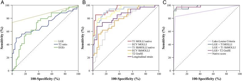
Methods and results: A total of 34 patients with suspected acute myocarditis and 50 control subjects underwent CMR. CMR protocol included quantitative assessment of T1 relaxation times using modified Look-Locker inversion recovery (MOLLI) and shortened MOLLI (ShMOLLI) acquisition schemes, extracellular volume fraction (ECV), T2 relaxation times, and longitudinal strain. Established Lake-Louise criteria (LLC) consisting of T2-weighted signal intensity ratio (T2-ratio), early gadolinium enhancement ratio (EGEr), and late gadolinium enhancement (LGE) were assessed. Receiver operating characteristics analysis was performed to compare diagnostic performance. Areas under the curve of native T1 (MOLLI: 0.95; ShMOLLI: 0.92) and T2 relaxation times (0.92) were higher compared with those of the other CMR parameters (T2-ratio: 0.71, EGEr: 0.71, LGE: 0.87, LLC: 0.90, ECV MOLLI: 0.77, ECV ShMOLLI: 0.80, longitudinal strain: 0.83). Combined with LGE, each native mapping technique outperformed the diagnostic performance of LLC (P < 0.01, respectively). A combination of native parameters (T1, T2, and longitudinal strain) significantly increased the diagnostic performance of CMR compared with LLC without need of contrast media application (0.99 vs. 0.90; P = 0.008).
Conclusion: In patients suspected of having acute myocarditis, diagnostic performance of CMR can be improved by implementation of quantitative CMR parameters. Especially, native mapping techniques have the potential to replace current LLC. CLINICALTRIALS.
Gov number: NCT02299856.
Keywords: Diagnosis; Inflammation; Magnetic resonance imaging; Mapping; Myocarditis.
Published on behalf of the European Society of Cardiology. All rights reserved. © The Author 2015. For permissions please email: journals.permissions@oup.com.
Figures

Comment in
-
Imaging myocardial inflammation by CMR mapping: good getting better?Eur Heart J Cardiovasc Imaging. 2016 Feb;17(2):134-5. doi: 10.1093/ehjci/jev308. Epub 2015 Nov 20. Eur Heart J Cardiovasc Imaging. 2016. PMID: 26590400 Free PMC article. No abstract available.
References
-
- Baccouche H, Mahrholdt H, Meinhardt G, Merher R, Voehringer M, Hill S, et al. Diagnostic synergy of non-invasive cardiovascular magnetic resonance and invasive endomyocardial biopsy in troponin-positive patients without coronary artery disease. Eur Heart J 2009;30:2869–79. - PubMed
-
- Drory Y, Turetz Y, Hiss Y, Lev B, Fisman EZ, Pines A, et al. Sudden unexpected death in persons less than 40 years of age. Am J Cardiol 1991;68:1388–92. - PubMed
-
- Liu PP, Mason JW. Advances in the understanding of myocarditis. Circulation 2001;104:1076–82. - PubMed
-
- Luetkens JA, Doerner J, Thomas DK, Dabir D, Gieseke J, Sprinkart AM, et al. Acute myocarditis: multiparametric cardiac MR imaging. Radiology 2014;273:383–92. - PubMed
MeSH terms
Substances
Associated data
LinkOut - more resources
Full Text Sources
Other Literature Sources
Medical
