PDBe: improved accessibility of macromolecular structure data from PDB and EMDB
- PMID: 26476444
- PMCID: PMC4702783
- DOI: 10.1093/nar/gkv1047
PDBe: improved accessibility of macromolecular structure data from PDB and EMDB
Abstract
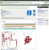
The Protein Data Bank in Europe (http://pdbe.org) accepts and annotates depositions of macromolecular structure data in the PDB and EMDB archives and enriches, integrates and disseminates structural information in a variety of ways. The PDBe website has been redesigned based on an analysis of user requirements, and now offers intuitive access to improved and value-added macromolecular structure information. Unique value-added information includes lists of reviews and research articles that cite or mention PDB entries as well as access to figures and legends from full-text open-access publications that describe PDB entries. A powerful new query system not only shows all the PDB entries that match a given query, but also shows the 'best structures' for a given macromolecule, ligand complex or sequence family using data-quality information from the wwPDB validation reports. A PDBe RESTful API has been developed to provide unified access to macromolecular structure data available in the PDB and EMDB archives as well as value-added annotations, e.g. regarding structure quality and up-to-date cross-reference information from the SIFTS resource. Taken together, these new developments facilitate unified access to macromolecular structure data in an intuitive way for non-expert users and support expert users in analysing macromolecular structure data.
© The Author(s) 2015. Published by Oxford University Press on behalf of Nucleic Acids Research.
Figures


References
-
- Berman H., Henrick K., Nakamura H. Announcing the worldwide Protein Data Bank. Nat. Struct. Biol. 2003;10:980. - PubMed
-
- Tagari M., Newman R., Chagoyen M., Carazo J., Henrick K. New electron microscopy database and deposition system. Trends Biochem. Sci. 2002;27:589. - PubMed
-
- Lawson C.L., Baker M.L., Best C., Bi C., Dougherty M., Feng P., van Ginkel G., Devkota B., Lagerstedt I., Ludtke S.J., et al. EMDataBank.org: unified data resource for CryoEM. Nucleic Acids Res. 2011;39:D456–D464. - PMC - PubMed
-
- Franklin R., Gosling R.G. Molecular configuration in sodium thymonucleate. Nature. 1953;171:740–741. - PubMed
Publication types
MeSH terms
Grants and funding
- MR/L007835/1/MRC_/Medical Research Council/United Kingdom
- BB/K016970/1/BB_/Biotechnology and Biological Sciences Research Council/United Kingdom
- 104948/WT_/Wellcome Trust/United Kingdom
- R01 GM079429/GM/NIGMS NIH HHS/United States
- BB/M013146/1/BB_/Biotechnology and Biological Sciences Research Council/United Kingdom
- GM079429/GM/NIGMS NIH HHS/United States
- BB/M011674/1/BB_/Biotechnology and Biological Sciences Research Council/United Kingdom
- BB/G022577/1/BB_/Biotechnology and Biological Sciences Research Council/United Kingdom
- 88944/WT_/Wellcome Trust/United Kingdom
- BB/J007471/1/BB_/Biotechnology and Biological Sciences Research Council/United Kingdom
LinkOut - more resources
Full Text Sources
Other Literature Sources

