. 2015 Nov 16;43(20):9587-99.
doi: 10.1093/nar/gkv1057. Epub 2015 Oct 17.

Selection of 2'-deoxy-2'-fluoroarabinonucleotide (FANA) aptamers that bind HIV-1 reverse transcriptase with picomolar affinity

Affiliations

Selection of 2'-deoxy-2'-fluoroarabinonucleotide (FANA) aptamers that bind HIV-1 reverse transcriptase with picomolar affinity

Irani Alves Ferreira-Bravo et al. Nucleic Acids Res. .

Abstract

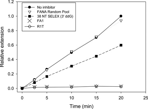
Using a Systematic Evolution of Ligands by Exponential Enrichment (SELEX) protocol capable of selecting xeno-nucleic acid (XNA) aptamers, a 2'-deoxy-2'-fluoroarabinonucleotide (FANA) aptamer (referred to as FA1) to HIV-1 reverse transcriptase (HIV-1 RT) was selected. FA1 bound HIV-1 RT with KD,app values in the low pM range under different ionic conditions. Comparisons to published HIV-1 RT RNA and DNA aptamers indicated that FA1 bound at least as well as these aptamers. FA1 contained a 20 nucleotide 5' DNA sequence followed by a 57 nucleotide region of FANA nucleotides. Removal of the fourteen 5' DNA nucleotides did not affect binding. FA1's predicted structure was composed of four stems and four loops. All stem nucleotides could be modified to G-C base pairs (14 total changes) with a small effect on binding. Eliminating or altering most loop sequences reduced or abolished tight binding. Overall, results suggested that the structure and the sequence of FA1 were important for binding. FA1 showed strong inhibition of HIV-1 RT in extension assays while no specific binding to avian myeloblastosis or Moloney murine leukemia RTs was detected. A complete DNA version of FA1 showed low binding to HIV-1 RT, emphasizing the unique properties of FANA in HIV-1 RT binding.


Figures

Figure 1.
Figure 1.
SELEX approach used to select FANA aptamers with tight binding to HIV-1 RT. The protocol used a DNA template containing a 40 nucleotide random region flanked by two fixed sequences at the 5′ end (19 nucleotide) and 3′ end (38 nucleotides). The FANA random pool produced using primer 1 was 79 nucleotides long, composed of 20 DNA nucleotides on the 5′ end and 59 FANA nucleotides. DNA template and FANA random pool were separated based on size on a denaturing polyacrylamide gel. Refer to Material and Methods for further details.
Figure 2.
Figure 2.
Titration of FANA with HIV-1 RT. A gel-shift experiment performed using 10 nM of selected material from round 5 of the SELEX protocol (Figure 1) or random pool, and different amounts of HIV-1 RT is shown. The concentrations of RT used were from left to right 0, 50, 100, 200, 300, 400 and 500 nM. Positions of unshifted and shifted material are indicated.
Figure 3.
Figure 3.
(A) Full length sequences of FA1, FA2 and FA3 aptamers recovered from Round 5 of SELEX. FA1 and FA3 lost two nucleotides and FA2 lost one nucleotide from the 40 nucleotide random starting pool region (non-underlined nucleotides) during the SELEX process. The 5′ and 3′ underlined nucleotides were derived from fixed sequence primers and were composed of DNA and FANA, respectively (see Figure 1). (B) Pairwise (using BioEdit) alignment of nucleotides derived from the 40 nucleotide random starting pool region of FA1, FA2 and FA3 aptamers. FA1/FA2 showed 75% identity while FA1/FA3 and FA2/FA3 each showed 63% identity in this region.
Figure 4.
Figure 4.
Structure of FANA aptamers (denoted FA) recovered from round 5 of FANA SELEX (see Figure 1) and 38 NT SELEX aptamer. For FANA aptamers, red indicates DNA nucleotides that were derived from the SELEX 5′ primer. Blue nucleotides are FANA nucleotides corresponding to the SELEX 3′ primer while sequence derived from the 40 nucleotide random region are in black. Various derivatives of FA1 and FA1GCstems are also shown (see text for a description). Folding was performed with mfold using standard conditions for RNA. Biochemical analysis of the various aptamers is reported in Table 1 and the Result section. *38 NT SELEX is a previously reported DNA aptamer (11).
Figure 5.
Figure 5.
Gel-shift analysis of FA1 with HIV-1 RT. (A) Radiolabeled FANA aptamer FA1 (2 pM final concentration, see Figure 4 for a depiction of FA1) was incubated at room temperature for 10 minutes with increasing concentrations of HIV-1 RT (as indicated) in 50 μl of buffer containing 50 mM Tris–HCl pH 8, 80 mM KCl, 2 mM MgCl2, 1 mM DTT, and 0.1 mg/ml BSA. Ten μl of 6× native gel loading buffer was added and the samples were loaded on a 6% native PAGE gel and electrophoresed and processed as described in Material and Methods. The positions of unshifted and shifted FA1 aptamer are indicated. (B) A graph of shifted material versus [HIV-1 RT] is shown. RT concentrations beyond 64 pM are not shown on the graph to emphasize the area where an increase in shifted material was observed. Beyond 32 pM the values for shifted material were essentially constant. The results are an average of 3 exp. ± standard deviations (error bars). The curve was generated by fitting the data to an equation for ligand binding with one-site saturation using SigmaPlot. The r2 value for this fit was 0.96. The KD,app value for gel-shift experiments in Table 2 of 8 ± 1 pM was determined from the mean of the individual experiments ± the standard deviation.
Figure 6.
Figure 6.
Inhibition of HIV-1 RT primer extension by selected aptamers. Assays included 50 nM primer-template (33 nucleotide primer: 50 nucleotide template) and 0.25 nM HIV-1 RT. One nM of the indicated aptamer was included. Reactions were initiated by adding dNTPs (50 μM final) and aliquots were removed at the indicated time points. Primer extension was monitored on denaturing polyacrylamide gels and quantified using a phosphorimager. All values for ‘Relative Extension’ are relative to the level of extension at 20 minutes in the ‘No inhibitor’ sample. The 3′ nucleotide of 38 NT SELEX was replaced with ddG using Klenow for this assay. FANA random pool was used for selection in round 1 of SELEX and has random nucleotides in the 40 base random region (Figure 1). Refer to Table 1 for information on the DNA aptamers, 38 NT SELEX and R1T. The structures of FA1 and 38 NT SELEX are shown in Figure 4.

References

    1. Telesnitsky A., Goff S.P. In: Retroviruses. Coffin JM, Hughes SH, Varmus HE, editors. NY: Cold Spring Harbor Laboratory Press; 1997. pp. 121–160. - PubMed
    1. Brody E.N., Gold L. Aptamers as therapeutic and diagnostic agents. J. Biotechnol. 2000;74:5–13. - PubMed
    1. Deisingh A.K. Aptamer-based biosensors: biomedical applications. Handb. Exp. Pharmacol. 2006:341–357. - PubMed
    1. Gold L. The SELEX process: a surprising source of therapeutic and diagnostic compounds. Harvey Lect. 1995;91:47–57. - PubMed
    1. James W. Aptamers in the virologists’ toolkit. J. Gen. Virol. 2007;88:351–364. - PubMed

Publication types

MeSH terms

Substances