. 2016 Oct;53(8):5607-27.
doi: 10.1007/s12035-015-9474-x. Epub 2015 Oct 17.

BDNF in Lower Brain Parts Modifies Auditory Fiber Activity to Gain Fidelity but Increases the Risk for Generation of Central Noise After Injury

Affiliations

BDNF in Lower Brain Parts Modifies Auditory Fiber Activity to Gain Fidelity but Increases the Risk for Generation of Central Noise After Injury

Tetyana Chumak et al. Mol Neurobiol. 2016 Oct.

Abstract

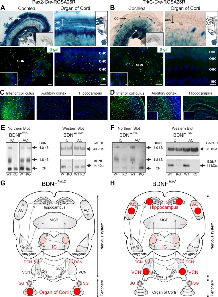
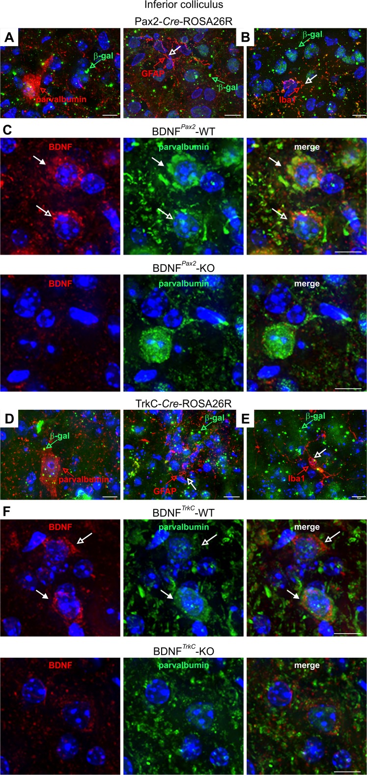
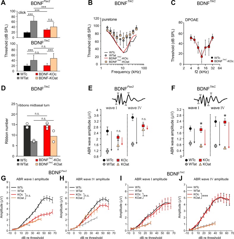
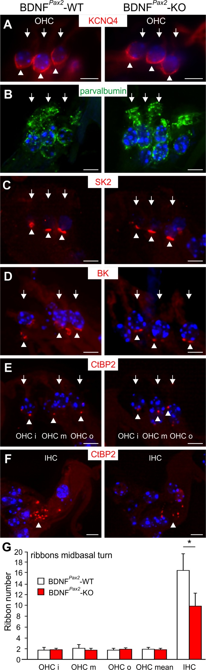
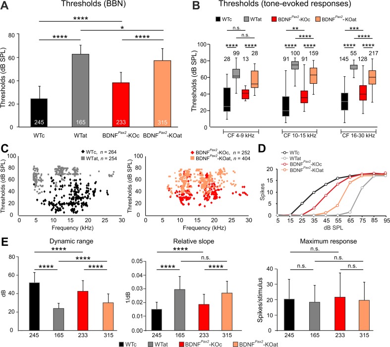
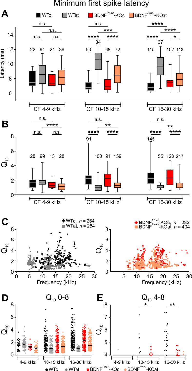
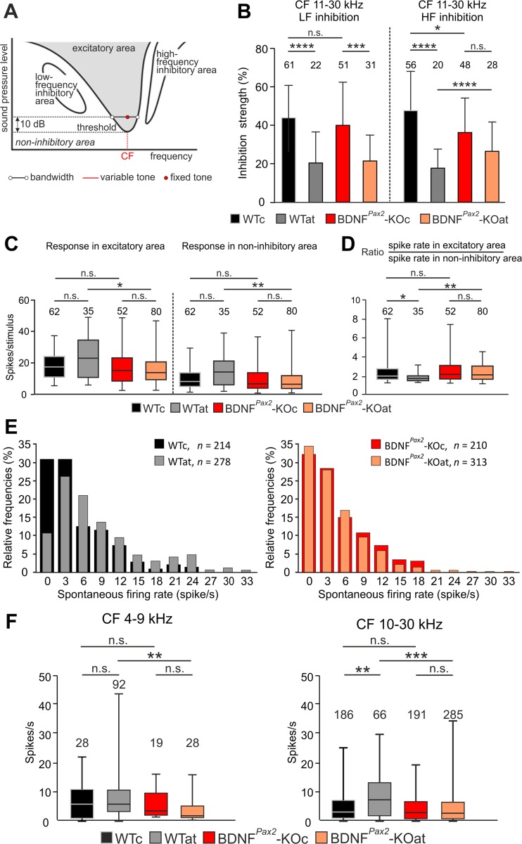
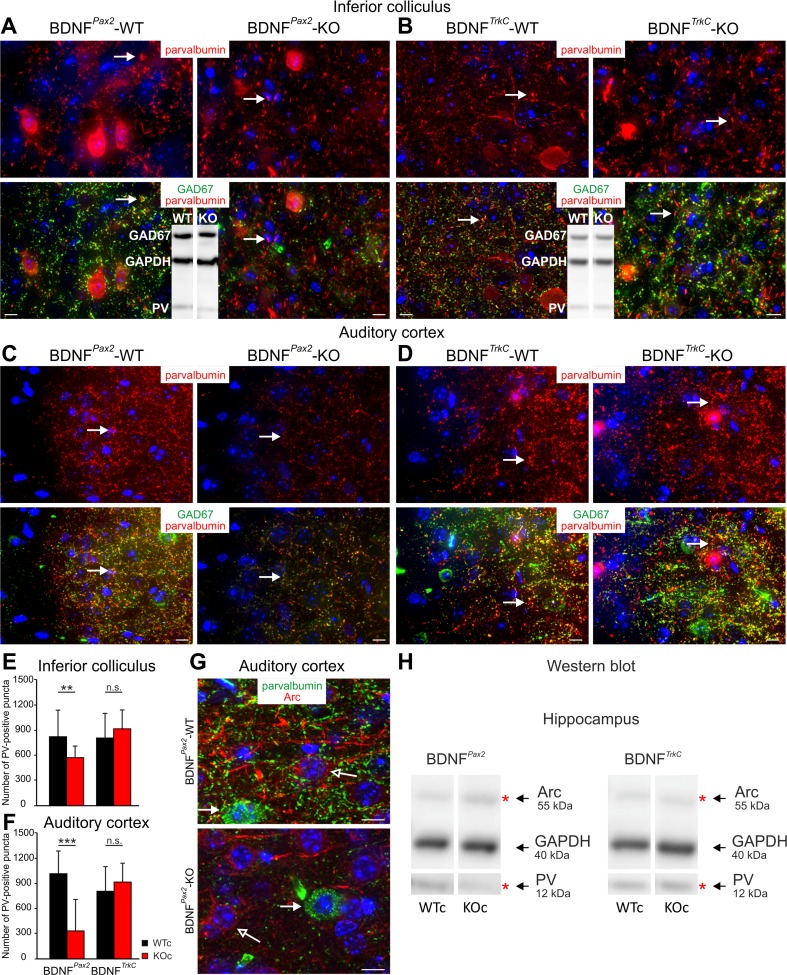
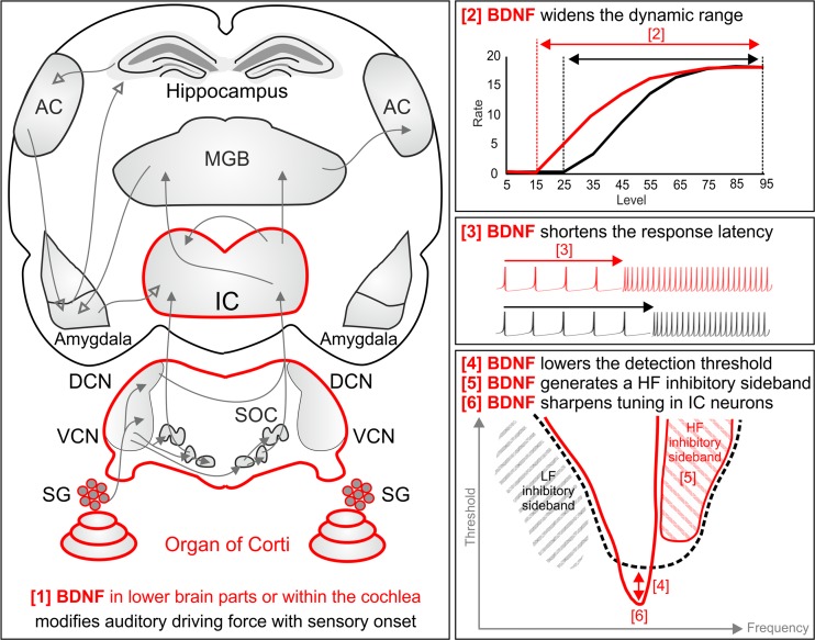
For all sensory organs, the establishment of spatial and temporal cortical resolution is assumed to be initiated by the first sensory experience and a BDNF-dependent increase in intracortical inhibition. To address the potential of cortical BDNF for sound processing, we used mice with a conditional deletion of BDNF in which Cre expression was under the control of the Pax2 or TrkC promoter. BDNF deletion profiles between these mice differ in the organ of Corti (BDNF (Pax2) -KO) versus the auditory cortex and hippocampus (BDNF (TrkC) -KO). We demonstrate that BDNF (Pax2) -KO but not BDNF (TrkC) -KO mice exhibit reduced sound-evoked suprathreshold ABR waves at the level of the auditory nerve (wave I) and inferior colliculus (IC) (wave IV), indicating that BDNF in lower brain regions but not in the auditory cortex improves sound sensitivity during hearing onset. Extracellular recording of IC neurons of BDNF (Pax2) mutant mice revealed that the reduced sensitivity of auditory fibers in these mice went hand in hand with elevated thresholds, reduced dynamic range, prolonged latency, and increased inhibitory strength in IC neurons. Reduced parvalbumin-positive contacts were found in the ascending auditory circuit, including the auditory cortex and hippocampus of BDNF (Pax2) -KO, but not of BDNF (TrkC) -KO mice. Also, BDNF (Pax2) -WT but not BDNF (Pax2) -KO mice did lose basal inhibitory strength in IC neurons after acoustic trauma. These findings suggest that BDNF in the lower parts of the auditory system drives auditory fidelity along the entire ascending pathway up to the cortex by increasing inhibitory strength in behaviorally relevant frequency regions. Fidelity and inhibitory strength can be lost following auditory nerve injury leading to diminished sensory outcome and increased central noise.

Keywords: BDNF; Central hyperactivity; High-spontaneous rate, low-threshold fibers; Homeostatic plasticity; Inferior colliculus; Sound detection threshold.


Conflict of interest statement

The authors declare that they have no competing interests.

Figures

Fig. 1
Fig. 1
Differential BDNF deletion patterns under the Pax2 or the TrkC promoter. a, b X-gal staining and β-gal immunolabeling of mice carrying the Pax2-Cre (a) or the TrkC-Cre transgene (b) on a ROSA26R background. a In the mature cochlea, β-galactosidase activity in Pax2-Cre-ROSA26R mice was detected in inner (IHC) and outer hair cells (OHC) of the organ of Corti (OC) and in spiral ganglion neurons (SGN). b β-Galactosidase activity in TrkC-Cre-ROSA26R mice was detected mainly in SGNs, but not in hair cells, as demonstrated by immunohistochemistry. c Immunohistochemistry for β-gal in the inferior colliculus (IC), auditory cortex (AC), and hippocampus of Pax2-Cre-ROSA26R mice. In the IC, β-gal staining can be detected, whereas no expression is observed in the AC and hippocampus. d Immunohistochemistry for β-gal in the IC, AC, and hippocampus of TrkC-Cre-ROSA26R mice. Clear β-gal staining is observed in the IC, AC, and hippocampus. In Cre-negative mice, no β-gal staining is seen (insets). Scale bars = a, b 10 μm; c, d 100 μm. e Northern and Western blots from IC and AC tissues of wild-type (WT) and BDNFPax2 knockout (KO) mice demonstrating a deletion of BDNF mRNA isoforms (1.8 and 4 kb) and BDNF protein (14 kDa) in the IC but not in the AC of BDNFPax2-KO mice. f Northern and Western blots from IC and AC tissues of WT and BDNFTrkC-KO mice demonstrating a deletion of BDNF mRNA isoforms (1.8 and 4 kb) and protein (14 kDa) in the IC and AC of BDNFTrkC-KO mice. For Northern blots, cyclophilin (CP) was used as a reference (0.8 kb); for Western blots, GAPDH (40 kDa) was used as a loading control. g, h Diagrams of the auditory pathway: the area of BDNF deletion exclusively in either the BDNFPax2-KO or the BDNFTrkC-KO is marked in dark red; the area of BDNF deletion in both BDNF-KO mouse lines is marked in bright red. g BDNF deletion in BDNFPax2-KO mice, h BDNF deletion in BDNFTrkC-KO mice
Fig. 2
Fig. 2
Immunohistochemistry of the IC of Pax2-Cre-ROSA26R and TrkC-Cre-ROSA26R mice and BDNFPax2 and BDNFTrkC WT and KO mice. a, d Immunostaining with anti-β-galactosidase (β-gal, green) and anti-parvalbumin (red) showing coexpression of β-galactosidase and parvalbumin in IC sections from both Pax2- and TrkC-Cre-ROSA26R mice. b, e Co-immunostaining with anti-β-galactosidase (β-gal, green) and either the oligodendrocyte marker anti-GFAP (red, open arrow) or the microglia marker anti-Iba1 (red, open arrow) in IC sections from both Pax2- and TrkC-Cre-ROSA26R mice shows no coexpression of β-galactosidase and GFAP or Iba1. Therefore, β-galactosidase is detected in neuronal cells. c, f Immunohistochemistry of IC sections stained with anti-BDNF (red) and anti-parvalbumin (green) antibodies, showing BDNF immunoreactivity in PV-positive and PV-negative neurons in BDNFPax2-WT (c, upper row) and BDNFTrkC-WT (f, upper row) mice. Closed arrows indicate cells positive for BDNF (red) and parvalbumin (green). Open arrows indicate cells expressing only BDNF (red). The specificity of the BDNF antibody is shown by the lack of BDNF immunostaining in BDNFPax2-KO and BDNFTrkC-KO mice (c, f, lower rows). Scale bars = 10 μm
Fig. 3
Fig. 3
Hearing function of BDNFPax2-KO and BDNFTrkC-KO mice before and after acoustic trauma. Auditory thresholds of BDNFPax2-KO (a, upper panel) and BDNFTrkC-KO (a, lower panel) mice analyzed by click (a) and tone-burst-evoked ABR (b) before (KOc) and after acoustic trauma (KOat). Compared to WT mice (WTc, WTat), BDNFPax2-KO are less vulnerable [25]. BDNFTrkC-KO mice exhibited normal hearing thresholds (a, lower panel, WTc, ears/mice: n = 22/11; KOc, n = 20/10; p > 0.999, two-way ANOVA) and show no significant difference to WT mice after acoustic trauma (WTat, ears/mice: n = 8/8; KOat, n = 7/7; p = 0.543, two-way ANOVA). Error bars, SD. c DPOAE thresholds in BDNFTrkC-WT and BDNFTrkC-KO mice were similar (WT, ears/mice: n = 19/10; KOn = 20/10; p = 0.482, two-way ANOVA). Error bars, SD. d IHC ribbon counts of midbasal cochlear turns in BDNFTrkC-WT and BDNFTrkC -KO mice before (WTc, KOc) and after noise exposure (WTat, KOat). The ribbon number of BDNFTrkC-KO mice was not significantly different from that of BDNFTrkC-WT animals. Error bars, SEM, n.s. p > 0.05; WTc, sections/mice: n = 5/2; WTat, n = 6/3; KOc, n = 11/4; KOat, n = 7/2. e, f Comparison of click-evoked ABR wave amplitudes in BDNFPax2-WT and BDNFPax2-KO mice (e) and BDNFTrkC-WT (WTc, WTat) and BDNFTrkC-KO (KOc, KOat) mice (f) before (WTc, KOc) and after noise exposure (WTat, KOat). In BDNFPax2-KO, suprathreshold amplitudes of wave I (auditory nerve) and wave IV (IC) are less reduced after noise exposure than in BDNFPax2-WT mice (compare black and red arrows in e for different reductions in WT and KO, respectively). In BDNFTrkC-KO, the reduction was not different from the reduction in BDNFTrkC-WT mice (compare black and red arrows in f for similar reduction in WT and KO, respectively). Two-way ANOVA with Bonferroni’s post hoc test (e) wave I, n.s. p = 0.254; wave IV, n.s. p = 0.893; WTat, ears/mice: n = 8/4; KOat, n = 7/4; f wave I, *p = 0.04; wave IV, *p = 0.02; WTat, mice/ears: n = 8/16; KOat, n = 8/16; error bars, SEM. g–j Suprathreshold ABR amplitude at the level of the auditory nerve (wave I) and IC (wave IV). Analysis was performed before and after acoustic trauma in BDNFPax2-WT (WTc, n = 16/8 ears/mice; WTat, n = 16/8 ears/mice) and BDNFPax2-KO mice (KOc, n = 16/8 ears/mice; KOat, n = 16/8 ears/mice) (g, h) compared to BDNFTrkC-WT (WTc, n = 17/8 ears/mice; WTat, n = 8/4 ears/mice) and BDNFTrkC-KO mice (KOc, n = 15/8 ears/mice; KOat, n = 7/4 ears/mice) (i, j). Note the near-complete convergence of growth functions of ABR wave I (g, n.s. p = 0.275; i, **p = 0.008) and IV (h, n.s. p = 0.420; j, ***p < 0.001) before and after AT in BDNFPax2-KO but not in BDNFTrkC-KO mice. Two-way ANOVA with Bonferroni’s post hoc test, error bars, SEM
Fig. 4
Fig. 4
Immunohistochemistry of outer hairs cells (OHC, arrows) in BDNFPax2-WT (n = 4, left panels) and BDNFPax2-KO (n = 4, right panels) mice. OHCs are stained for the voltage-gated potassium channel KCNQ4 (a), parvalbumin (b), the small conductance calcium-activated potassium channel SK2 (c), the large-conductance calcium-activated potassium channel BK (d), and CtBP2, a marker of ribbon synapses (e). f In contrast to OHCs, IHCs show reduced number of CtBP2-stained ribbons in BDNFPax2-KO mice. Arrowheads indicate antibody staining. g Quantification of ribbons stained with anti-CtBP2. No differences are observed in any row of the OHCs or in the mean ribbon number of OHCs, although a significantly reduced ribbon number can be seen in IHCs. Two-tailed unpaired Student’s t test with α = 5 (*p < 0.05). Scale bars = 5 μm
Fig. 5
Fig. 5
Response thresholds of IC neurons to broadband noise (BBN) and tone stimulation for control or sound-exposed BDNFPax2 wild-type (WTc, WTat) and BDNFPax2 knockout (KOc, KOat) animals. a Averaged thresholds of responses to BBN stimulation. b Box and whisker plot for thresholds of tone-evoked responses for neurons of individual CF frequency ranges (4–9, 10–15, 16–30 kHz). c Scatter plots of pure tone thresholds as a function of neuronal CF, shown separately for BDNFPax2-WT (WTc, n = 4; WTat, n = 5) and BDNFPax2-KO (KOc, n = 4; KOat, n = 5) animals. d, e Parameters of the rate-intensity function (RIF) of responses to BBN stimulation for control or sound-exposed BDNFPax2-WT (WTc, WTat) and BDNFPax2-KO (KOc, KOat) animals. d Typical examples of RIFs for all groups of animals. e Average dynamic range, relative slope, and maximum response for all groups of mice. One-way ANOVA with Bonferroni’s post hoc test and Kruskal-Wallis test with Dunn’s multiple comparison test, n.s. p > 0.05, *p < 0.05, **p < 0.01, ***p < 0.001, ****p < 0.0001. Data are presented as mean ± SD or median with interquartile range and extremes. The numbers in the graphs indicate the numbers of neurons
Fig. 6
Fig. 6
Minimum first spike latency (mFSL) and quality factor (Q 10) of IC neurons. a mFSL of responses to 80 dB SPL BBN bursts for control and sound-exposed BDNFPax2-WT (WTc, n = 4; WTat, n = 5) and BDNFPax2-KO (KOc, n = 4; KOat, n = 5) animals. Box and whisker plot for mFSL for neurons of individual CF frequency ranges (4–9, 10–15, 16–30 kHz). Kruskal-Wallis test with Dunn’s post hoc test: n.s. p > 0.05, *p < 0.05, ***p < 0.001, ****p < 0.0001. Data are presented as medians with interquartile ranges and extremes. be Q 10 of IC neuron responses to pure tone stimulation for control and sound-exposed BDNFPax2-WT (WTc, n = 4; WTat, n = 5) and BDNFPax2-KO (KOc, n = 4; KOat, n = 5) animals. b Box and whisker plot for Q 10 for neurons of individual CF frequency ranges (4–9, 10–15, 16–30 kHz). c Scatter plots illustrating the Q 10 as a function of neuronal CF, shown separately for BDNFPax2-WT and BDNFPax2-KO animals. d, e Scatter plots for Q 10 with y scale 0–8 (d) and scale 4–8 (e) for the neurons of individual frequency ranges (4–9, 10–15, 16–30 kHz). Note that IC neurons >10 kHz with Q 10 >5 are absent in BDNFPax2-KO mice (e). Kruskal-Wallis test with Dunn’s multiple comparison test: n.s. p > 0.05, **p < 0.01, ****p < 0.0001. Data are presented as medians with interquartile ranges and extremes. The numbers in the graphs indicate the numbers of neurons
Fig. 7
Fig. 7
Inhibition characteristics in control or sound-exposed BDNFPax2-WT (WTc, n = 4; WTat, n = 5) and BDNFPax2-KO (KOc, n = 4; KOat, n = 5) animals. a Schematic of response map to two-tone stimulation showing excitatory, inhibitory, and noninhibitory areas. b Comparison of low- and high-frequency sideband inhibition strength in middle and high CF (11–30 kHz) IC neurons with inhibitory strength of 1 % and higher. One-way ANOVA with Bonferroni’s post hoc test. c Spike rates of IC neurons with middle and high CF in response to two-tone stimulation, determined 20 dB above threshold in the excitatory area and noninhibitory area. Numbers in the graph indicate number of neurons. d Ratio of spike rates in the excitatory field 20 dB above threshold to spike rates in the noninhibitory area. The numbers of neurons are given above the corresponding boxes. Kruskal-Wallis test with Dunn’s multiple comparison test: n.s. p > 0.05, *p < 0.05, ***p < 0.001, ****p < 0.0001. Data are presented as mean ± SD or median with interquartile range and extremes. e Distribution of IC neurons according to their spontaneous firing rate (spikes/s) for control and sound-exposed BDNFPax2-WT (WTc, n = 4; WTat, n = 5) and BDNFPax2-KO (KOc, n = 4; KOat, n = 5) animals. f Spontaneous firing rates for neurons with CF in the high-frequency band (10–30 kHz) were significantly higher in BDNFPax2-WTat than in BDNFPax2-WTc but were similar in BDNFPax2-KOat and BDNFPax2-KOc. Kruskal-Wallis test with Dunn’s post hoc test demonstrated significant differences between WTc and WTat (**p < 0.01) and WTat and KOat (***p < 0.001) groups of animals. Data are presented as mean ± SD or median with interquartile range and extremes. The numbers in the graphs indicate the numbers of neurons
Fig. 8
Fig. 8
Immunohistochemistry of the inferior colliculus and auditory cortex of BDNFPax2-KO and BDNFTrkC-KO mice. a–d Immunohistochemistry of the IC and AC of BDNFPax2-KO (a, c) and BDNFTrkC-KO mice (b, d) immunolabeled with anti-parvalbumin (red) and anti-GAD67 (green). A reduction of PV- and GAD67-immunoreactive puncta in both IC (a) and AC (c) in BDNFPax2-KO mice compared to BDNFPax2-WT mice is observed. Arrows point to PV-immunoreactive puncta. No changes in PV and GAD67 expression are observed in the IC (b) and AC (d) of BDNFTrkC-KO mice compared to BDNFTrkC-WT mice. Arrows point to PV-immunoreactive puncta. Reduced PV levels in BDNFPax2-KO but not BDNFTrkC-KO mice in the IC are confirmed by Western blot (a, b insets). e, f Quantification of PV-immunoreactive puncta in the IC (e) of BDNFPax2-WT, BDNFPax2-KO, BDNFTrkC-WT, and BDNFTrkC-KO mice (10–20 slices of 3–6 independent experiments of n = 3 animals) and in the AC (f) of BDNFPax2-WT, BDNFPax2-KO, BDNFTrkC-WT, and BDNFTrkC-KO mice (10–20 slices of 3–6 independent experiments of n = 3 animals). Two-way ANOVA with Bonferroni’s post hoc test (**p < 0.01, ***p < 0.001). Data are presented as mean ± SD. g Immunohistochemistry of AC sections of BDNFPax2-WT (upper image) and BDNFPax2-KO (lower image) mice immunolabeled with anti-Arc (red) and anti-parvalbumin (green) antibodies. In BDNFPax2-KO mice, Arc (red) expression is reduced. Closed arrows point to PV-positive cells. Open arrows indicate PV-immunoreactive puncta surrounding Arc-positive neurons. Scale bars = 10 μm. h Parvalbumin (PV) and Arc levels in the hippocampus of BDNFPax2-WT and BDNFPax2-KO mice and BDNFTrkC-WT and BDNFTrkC-KO mice, respectively, detected by Western blots (exemplarily for n = 3–5 mice). Note that the reduction of PV is associated to an increase of Arc in BDNFPax2-KO but not BDNFTrkC-KO mice
Fig. 9
Fig. 9
Schematic summary of the results that illustrates our hypothesis. An auditory driving force which is modified with hearing onset and depends on BDNF in the lower parts of the auditory system or within the cochlea [1] widens the dynamic range above which spike rates can be detected [2]. This is achieved upon shortening of the response latency [3], leading to lowering of the detection threshold [4] through generation of a high-frequency inhibitory sideband [5] in, e.g., very narrowly tuned IC neurons [6]. Through these events, the probability to detect a spike above the noise floor may be improved. This BDNF activity on auditory fibers in high-frequency cochlear turns can be lost after acoustic trauma. In this case, spontaneous spike rates in central auditory pathways are elevated (hyperactivity). As a consequence, due to loss of basal inhibitory strength within the ascending circuit of affected frequency regions, the capacity to adapt to sensory deprivation might be reduced. HF high frequency, IC inferior colliculus, IHC inner hair cell, LF low frequency

Similar articles

Cited by

References

    1. Poo MM. Neurotrophins as synaptic modulators. Nat Rev Neurosci. 2001;2:24–32. doi: 10.1038/35049004. - DOI - PubMed
    1. Barde YA. The nerve growth factor family. Prog Growth Factor Res. 1990;2:237–248. doi: 10.1016/0955-2235(90)90021-B. - DOI - PubMed
    1. Rauskolb S, Zagrebelsky M, Dreznjak A, Deogracias R, Matsumoto T, Wiese S, Erne B, Sendtner M, Schaeren-Wiemers N, Korte M, et al. Global deprivation of brain-derived neurotrophic factor in the CNS reveals an area-specific requirement for dendritic growth. J Neurosci. 2010;30:1739–1749. doi: 10.1523/JNEUROSCI.5100-09.2010. - DOI - PMC - PubMed
    1. Bramham CR, Panja D. BDNF regulation of synaptic structure, function, and plasticity. Neuropharmacology. 2014;76 Pt C:601–602. doi: 10.1016/j.neuropharm.2013.08.012. - DOI - PubMed
    1. Maffei A, Lambo ME, Turrigiano GG. Critical period for inhibitory plasticity in rodent binocular V1. J Neurosci. 2010;30:3304–3309. doi: 10.1523/JNEUROSCI.5340-09.2010. - DOI - PMC - PubMed

Publication types