

N-cadherin functions as a growth suppressor in a model of K-ras-induced PanIN
- PMID: 26477318
- PMCID: PMC4837100
- DOI: 10.1038/onc.2015.382
N-cadherin functions as a growth suppressor in a model of K-ras-induced PanIN
Abstract
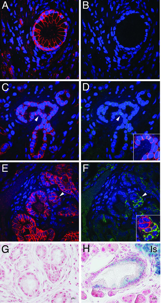
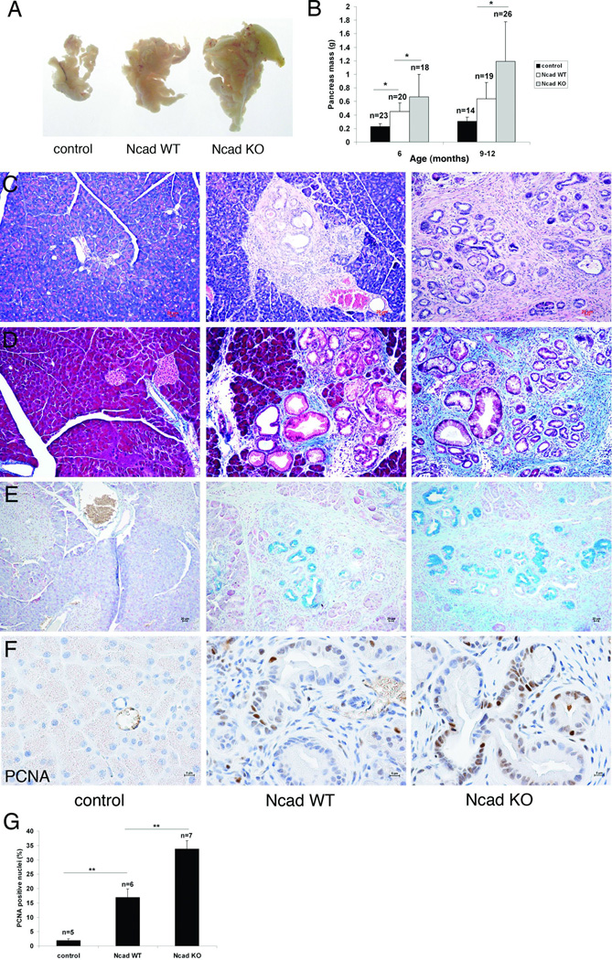
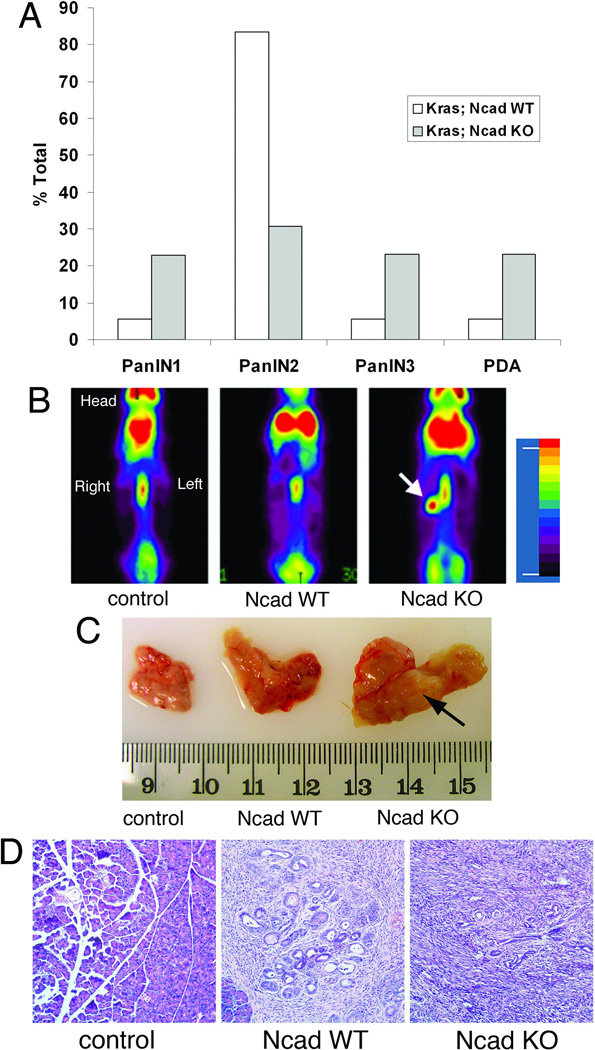
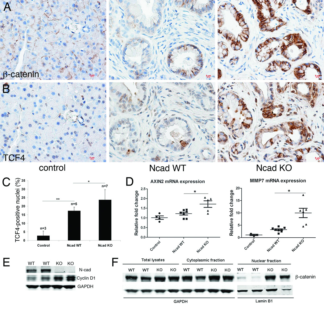
Cadherin subtype switching from E-cadherin to N-cadherin is associated with the epithelial-to-mesenchymal transition (EMT), a process required for invasion and dissemination of carcinoma cells. We found that N-cadherin is expressed in human and mouse pancreatic intraepithelial neoplasia (PanIN), suggesting that N-cadherin may also have a role in early-stage pancreatic cancer. To investigate the role of N-cadherin in mouse PanIN (mPanIN), we simultaneously activated oncogenic K-ras(G12D) and deleted the N-cadherin (Cdh2) gene in the murine pancreas. Genetic ablation of N-cadherin (N-cad KO) caused hyperproliferation, accelerated mPanIN progression, and early tumor development in K-ras(G12D) mice. Decreased E-cadherin and redistribution of β-catenin accompanied the loss of N-cadherin in pancreatic ductal epithelial cells (PDEC). Nuclear accumulation of β-catenin and its transcription co-activator Tcf4 led to activation of Wnt/β-catenin target genes. Unexpectedly, loss of N-cadherin in the K-ras(G12D) model resulted in increased mPanIN progression and tumor incidence. These in vivo results demonstrate for the first time that N-cadherin functions as a growth suppressor in the context of oncogenic K-ras.
Conflict of interest statement
The authors declare no conflicts of interest.
Figures
Similar articles
-
E-cadherin expression in obesity-associated, Kras-initiated pancreatic ductal adenocarcinoma in mice.Surgery. 2015 Dec;158(6):1564-72. doi: 10.1016/j.surg.2015.07.023. Epub 2015 Aug 18. Surgery. 2015. PMID: 26297056 Free PMC article.
-
Spontaneous induction of murine pancreatic intraepithelial neoplasia (mPanIN) by acinar cell targeting of oncogenic Kras in adult mice.Proc Natl Acad Sci U S A. 2008 Dec 2;105(48):18913-8. doi: 10.1073/pnas.0810097105. Epub 2008 Nov 21. Proc Natl Acad Sci U S A. 2008. PMID: 19028870 Free PMC article.
-
Requirement of NEMO/IKKγ for effective expansion of KRAS-induced precancerous lesions in the pancreas.Oncogene. 2013 May 23;32(21):2690-5. doi: 10.1038/onc.2012.272. Epub 2012 Jul 2. Oncogene. 2013. PMID: 22751123
-
E-cadherin/β-catenin complex and the epithelial barrier.J Biomed Biotechnol. 2011;2011:567305. doi: 10.1155/2011/567305. Epub 2011 Oct 11. J Biomed Biotechnol. 2011. PMID: 22007144 Free PMC article. Review.
-
Cdc6 as a novel target in cancer: Oncogenic potential, senescence and subcellular localisation.Int J Cancer. 2020 Sep 15;147(6):1528-1534. doi: 10.1002/ijc.32900. Epub 2020 Feb 17. Int J Cancer. 2020. PMID: 32010971 Free PMC article. Review.
Cited by
-
Analysis of lncRNA-mRNA networks after MEK1/2 inhibition based on WGCNA in pancreatic ductal adenocarcinoma.J Cell Physiol. 2020 Apr;235(4):3657-3668. doi: 10.1002/jcp.29255. Epub 2019 Oct 3. J Cell Physiol. 2020. PMID: 31583713 Free PMC article.
-
Heterocellular N-cadherin junctions enable nontransformed cells to inhibit the growth of adjacent transformed cells.Cell Commun Signal. 2022 Feb 17;20(1):19. doi: 10.1186/s12964-021-00817-9. Cell Commun Signal. 2022. PMID: 35177067 Free PMC article.
-
Yin Yang 1 promotes the neuroendocrine differentiation of prostate cancer cells via the non-canonical WNT pathway (FYN/STAT3).Clin Transl Med. 2023 Oct;13(10):e1422. doi: 10.1002/ctm2.1422. Clin Transl Med. 2023. PMID: 37771187 Free PMC article.
-
Distribution of E- and N-cadherin in subgroups of non-functioning pituitary neuroendocrine tumours.Endocrine. 2022 Jun;77(1):151-159. doi: 10.1007/s12020-022-03051-6. Epub 2022 Jun 8. Endocrine. 2022. PMID: 35674926 Free PMC article.
-
N-cadherin in cancer metastasis, its emerging role in haematological malignancies and potential as a therapeutic target in cancer.BMC Cancer. 2018 Oct 1;18(1):939. doi: 10.1186/s12885-018-4845-0. BMC Cancer. 2018. PMID: 30285678 Free PMC article. Review.
References
-
- Ryan DP, Hong TS, Bardeesy N. Pancreatic adenocarcinoma. The New England journal of medicine. 2014 Sep 11;371(11):1039–1049. PubMed PMID: 25207767. - PubMed
-
- van Roy F. Beyond E-cadherin: roles of other cadherin superfamily members in cancer. Nature reviews Cancer. 2014 Feb;14(2):121–134. PubMed PMID: 24442140. - PubMed
-
- Hotz B, Arndt M, Dullat S, Bhargava S, Buhr HJ, Hotz HG. Epithelial to mesenchymal transition: expression of the regulators snail, slug, and twist in pancreatic cancer. Clin Cancer Res. 2007 Aug 15;13(16):4769–4776. PubMed PMID: 17699854. - PubMed
Publication types
MeSH terms
Substances
Grants and funding
LinkOut - more resources
Full Text Sources
Other Literature Sources
Medical
Molecular Biology Databases
Research Materials
Miscellaneous
