. 2016 Jun 30;35(26):3365-75.
doi: 10.1038/onc.2015.395. Epub 2015 Oct 19.

Radiation promotes colorectal cancer initiation and progression by inducing senescence-associated inflammatory responses

Affiliations

Radiation promotes colorectal cancer initiation and progression by inducing senescence-associated inflammatory responses

S B Kim et al. Oncogene. .

Abstract

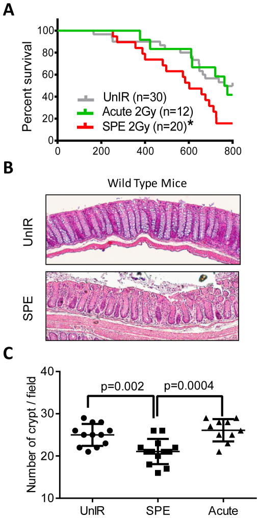
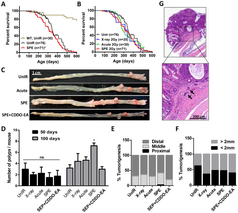
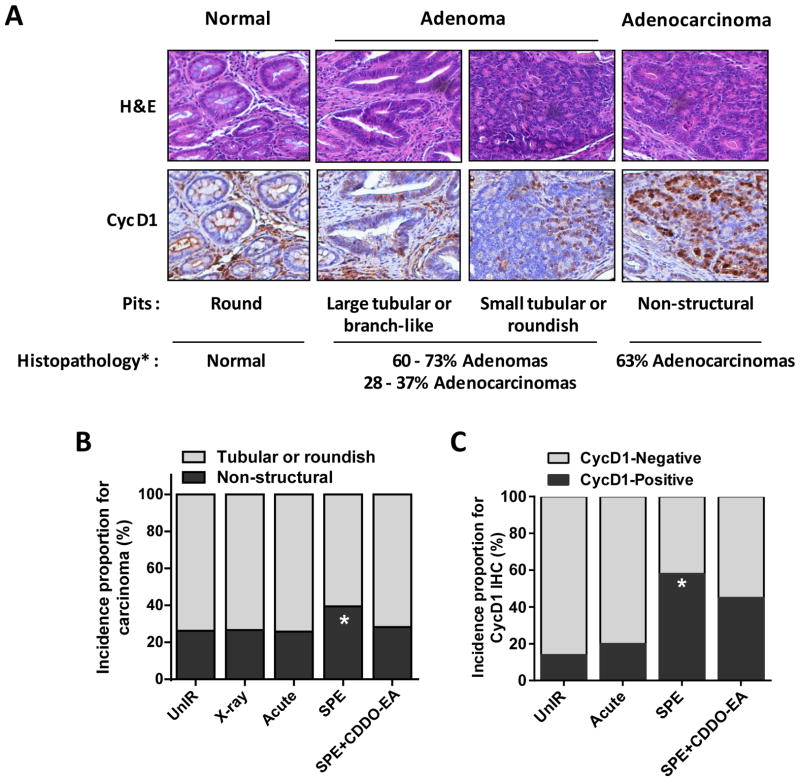
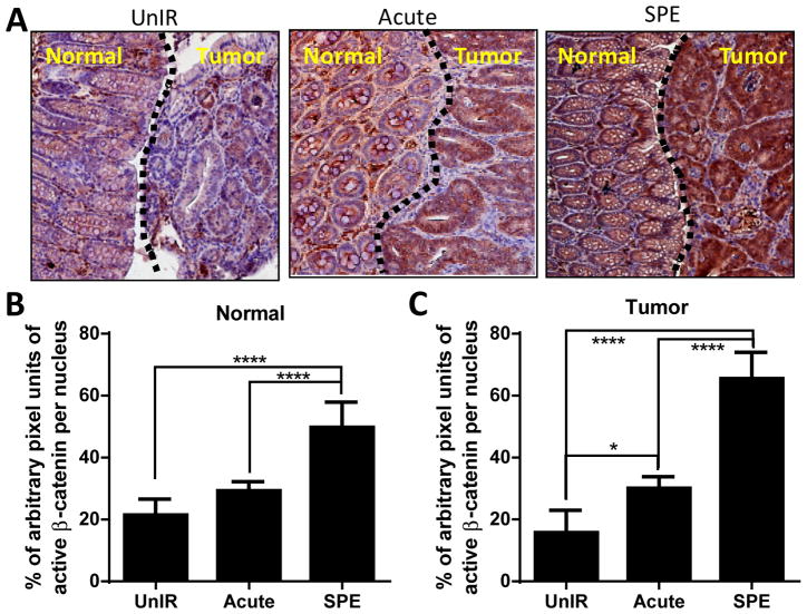
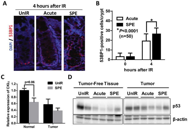
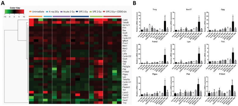
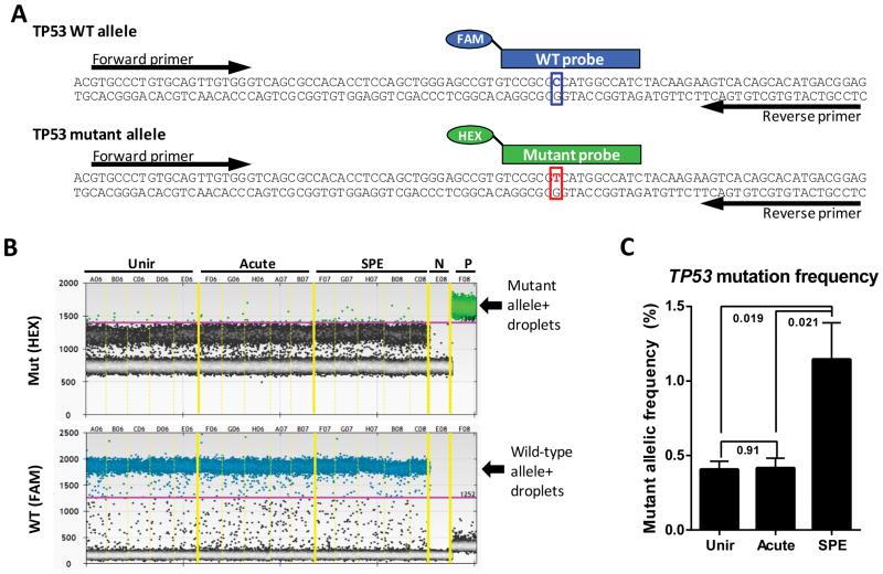
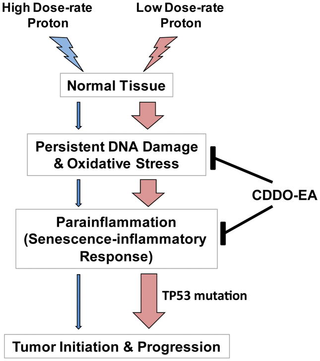
Proton radiotherapy is becoming more common as protons induce more precise DNA damage at the tumor site with reduced side effects to adjacent normal tissues. However, the long-term biological effects of proton irradiation in cancer initiation compared with conventional photon irradiation are poorly characterized. In this study, using a human familial adenomatous polyposis syndrome susceptible mouse model, we show that whole-body irradiation with protons are more effective in inducing senescence-associated inflammatory responses (SIRs), which are involved in colon cancer initiation and progression. After proton irradiation, a subset of SIR genes (Troy, Sox17, Opg, Faim2, Lpo, Tlr2 and Ptges) and a gene known to be involved in invasiveness (Plat), along with the senescence-associated gene (P19Arf), are markedly increased. Following these changes, loss of Casein kinase Iα and induction of chronic DNA damage and TP53 mutations are increased compared with X-ray irradiation. Proton irradiation also increases the number of colonic polyps, carcinomas and invasive adenocarcinomas. Pretreatment with the non-steroidal anti-inflammatory drug, 2-cyano-3,12-dioxooleana-1,9(11)-dien-28-oic acid-ethyl amide (CDDO-EA), reduces proton irradiation-associated SIR and tumorigenesis. Thus exposure to proton irradiation elicits significant changes in colorectal cancer initiation and progression that can be mitigated using CDDO-EA.


Conflict of interest statement

Competing interests

JWS is on the SAB of Reata Pharmaceuticals (Irving, Texas).

Figures

Figure 1
Figure 1. Effect of low dose-rate protons (SPE simulation) on lifespan and crypt number in wild type mice
(A) Kaplan-Meier survival plot of unirradiated or irradiated wild type mice demonstrating that irradiation with only 2 Gy sSPE significantly decreases survival compared with acute 2 Gy proton or unirradiated controls. *, P<0.05 in the log-rank test. (B) Representative images of H&E-stained colon crypts in unirradiated and sSPE-irradiated wild type mice demonstrating less crypt numbers with sSPE irradiation. (C) Quantification of crypt numbers on the 3 fields of vision with a magnification of 10X from 4 mice per group. Significant difference was evaluated by two-way ANOVA with Multiple comparisons.
Figure 2
Figure 2. Tumorigenic effect of simulated SPE in CPC;Apc mice
(A–B) Kaplna-Meier survival plot of unirradiated or irradiated CPC;Apc mice. Unirradiated wild type mice (brown); unirradiated CPC;Apc mice (black); sSPE-irradiated CPC;Apc mice (red); x-ray irradiated CPC;Apc mice (blue); acute proton-irradiated CPC;Apc mice (green). *, P=0.0322 in Gehan-Breslow-Wilcoxon test compared to unirradiated CPC;Apc mice survival. (C) Representative images of polyps 100 days after irradiated or age-matched CPC;Apc mice colon. Scale bar, 1 cm. Quantification of polyp number (D), segmental distribution (E), and size (F) difference of the CPC;Apc mice 100 days after irradiation or age-matched unirradiated control. (n=6 mice per group). *, P<0.05 in the Student’s t-test compared with unirradiated control. (G) Representative image for invasion foci (black arrows) in sSPE-irradiated CPC;Apc mice. Scale bar, 200 μm.
Figure 3
Figure 3. Tumor grade in irradiated CPC;Apc colons comparable with unirradiated controls 100 days after irradiation
(A) Modified classification of colorectal cancer based on pit pattern and Cyclin D1 expression (–27, 50). (B) Quantification of overall incidences proportion for non-structural adenocarcinoma in colons of unirradiated or irradiated CPC;Apc mice demonstrates increased incidence of adenocarcinoma with sSPE exposure. (C) Quantification of overall incidence proportion for cyclin D1-positive tumors in colons demonstrating that irradiation with only 2 Gy sSPE significantly increase incidence of cyclin D1-positive carcinomas compared with unirradiated controls. *, P<0.05 compared with unirradiated control.
Figure 4
Figure 4. Prolonged activation of β-catenin in normal colons 100 days after sSPE radiation
(A) Representative images of β-catenin (brown) stained tumor free (normal) or tumor areas of colon sections from unirradiated control, acute proton, and sSPE irradiated CPC;Apc mice. Hematoxylin (blue) was used for counterstaining. (B) Quantification of total β-catenin staining in tumor free (normal) areas of control, acute proton, and sSPE irradiated sections. (C) Quantification of total β-catenin staining in tumor areas of control, acute proton, and sSPE irradiated sections. Intensity of β-catenin staining was normalized with intensity of hematoxylin staining. At least six fields of vision from the normal and tumor areas were captured in each section and were analyzed. Average data from 4 mice are presented graphically. *, P<0.05 and ****, P<0.001 in two-way ANOVA with Multiple comparisons.
Figure 5
Figure 5. sSPE radiation triggers DNA damage, oxidation, and loss of CKIα and p53 expression
(A) Representative images of 53BP1 immunostaining in normal colonic crypts 4 hours after irradiation. anti-53BP1 (red); DAPI (blue). (B) Quantification of 53BP1-positive cells per crypt demonstrating exposure to sSPE showed higher 53BP1-positive cells 4 hours after irradiation relative to acute proton irradiated groups. n=50 crypts were counted from 3 mice per group. (C) Quantitative RT-PCR shows lower expression of CKIα in normal tissue as well as tumor 100 days after sSPE irradiation relative to unirradiated control. (n=3 mice) (D) Tumor-free normal tissues irradiated with proton radiation show lower expression of p53 relative to unirradiated control.
Figure 6
Figure 6. Quantitative RT-PCR analysis of tumor-free distal colons reveals activation of a set of senescence-associated inflammatory response (SIR) genes after sSPE irradiation
(A) Hierarchical clustering and associated heatmap demonstrating capacity of 33 SIR genes to segregate experimental cohorts. Color bar indicates relative fold change. (B) 9 SIR genes showing significant increase in expression 100 days after sSPE irradiation. (*, P<0.05, ANOVA).
Figure 7
Figure 7. ddPCR-base detection of a single-base mutation of TP53 in colon tumors
(A) Strategy for detection of mouse TP53 mutation (p.A156V/c.467C>T) showing a common primer pair and allele-specific probes conjugated with different fluorophores (FAM and HEX) are indicated. (B) ddPCR for the TP53 mutant and wild-type allelic discrimination was performed with colon tumors from unirradiated (Unir), acute proton irradiated (Acute), and sSPE irradiated (SPE) groups. Green and blue dots represent droplets containing the mutant and the wild-type alleles, respectively. Note that while the probes weakly cross-detected the wild-type and mutant alleles, this cross-hybridization can be excluded from quantitative analysis in ddPCR by setting the threshold (pink line) of detection high enough to discriminate true-positive droplets from false-positive droplets, as shown here. N, negative control (no template); P, positive control (mutant template only). (C) Quantificatioin of ddPCR shows the average frequency of the TP53 mutant allele. P values calculated by Student’s t- test (n=5) are shown on the plot.
Figure 8
Figure 8. A schematic model of low dose-rate proton-induced tumorigenesis through para-inflammation
Exposure to low dose-rate proton induces persistent DNA damage responses and oxidative stress, which triggers senescence-inflammatory response (SIR). In p53-mutated tissues, SIR results in a breach of homeostasis, hyperproliferation, invasion, and carcinogenesis. Nonsteroidal anti-inflammatory drug, CDDO-EA, treatment moderates DNA damage, oxidative stress, and para-inflammation, thus helping to regain homeostasis.

References

    1. Verhey LJ, Munzenrider JE. Proton beam therapy. Annu Rev Biophys Bioeng. 1982;11:331–57. - PubMed
    1. Suit HD, Goitein M, Tepper J, Koehler AM, Schmidt RA, Schneider R. Explorotory study of proton radiation therapy using large field techniques and fractionated dose schedules. Cancer. 1975 Jun;35(6):1646–57. - PubMed
    1. D’angio GJ, Lawrence JH. Medical research with high-energy heavy particles. Nucleonics. 1963;21:56–61.
    1. Lawrence JH, Tobias CA, Born JL, Linfoot JA, Kling RP, Gottschalk A. Alpha and Proton Heavy Particles and the Bragg Peak in Therapy. Trans Am Clin Climatol Assoc. 1964;75:111–6. - PMC - PubMed
    1. Moravek Z, Bogner L. Analysis of the physical interactions of therapeutic proton beams in water with the use of Geant4 Monte Carlo calculations. Zeitschrift fur medizinische Physik. 2009;19(3):174–81. - PubMed

Publication types

MeSH terms

Substances