. 2015 Nov 17;31(45):12339-47.
doi: 10.1021/acs.langmuir.5b02902. Epub 2015 Oct 30.

Biofunctionalization of Large Gold Nanorods Realizes Ultrahigh-Sensitivity Optical Imaging Agents

Affiliations

Biofunctionalization of Large Gold Nanorods Realizes Ultrahigh-Sensitivity Optical Imaging Agents

Elliott D SoRelle et al. Langmuir. .

Erratum in

  • Langmuir. 2015 Nov 17;31(45):12347

Abstract

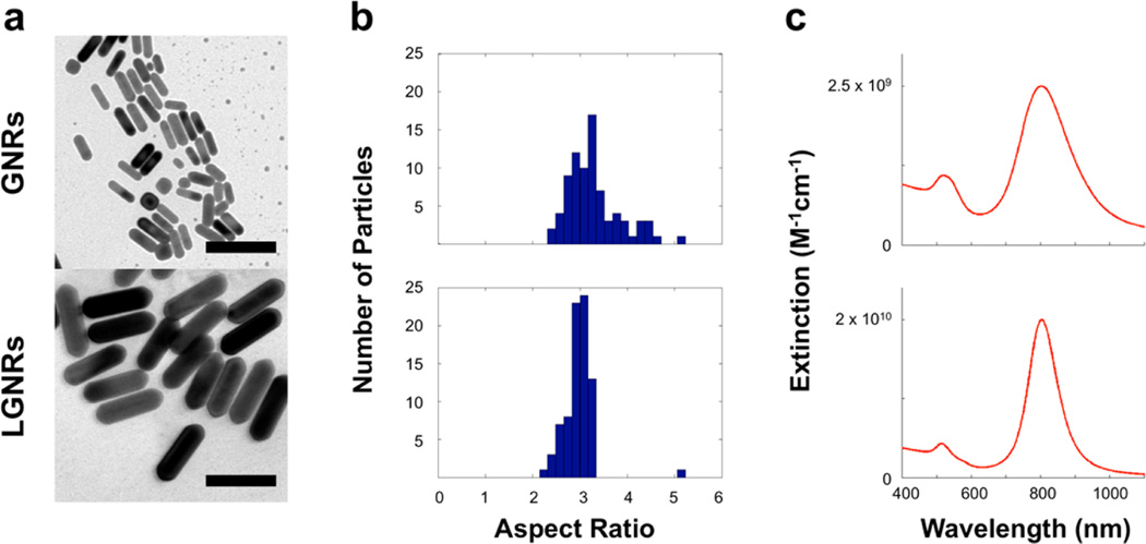
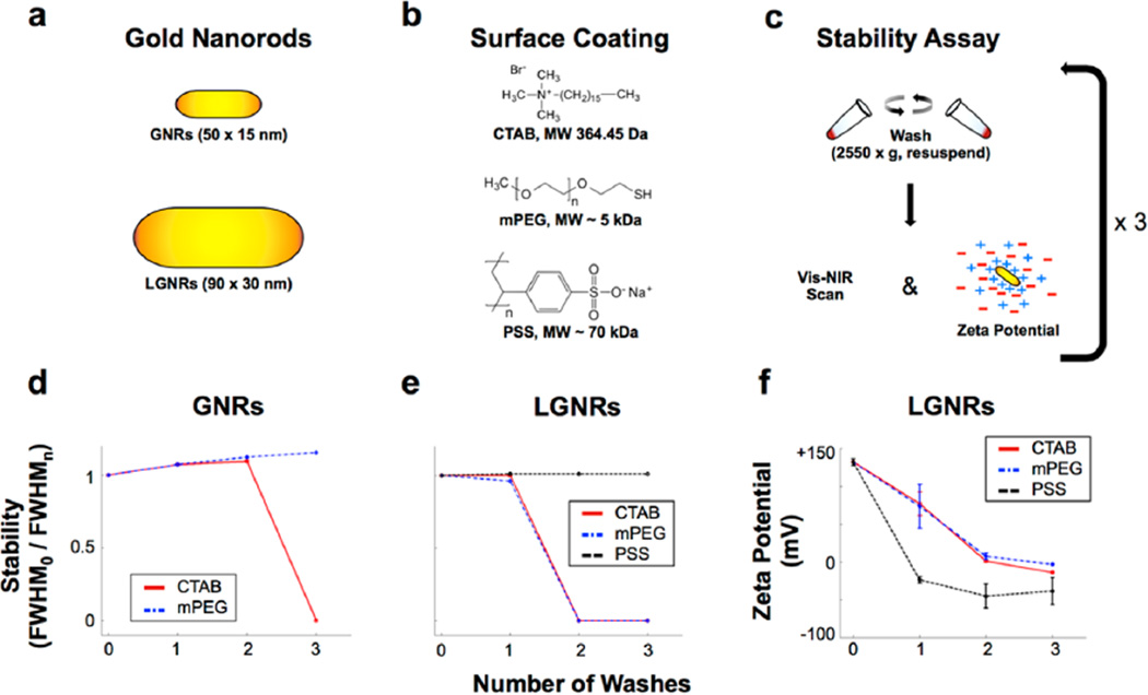
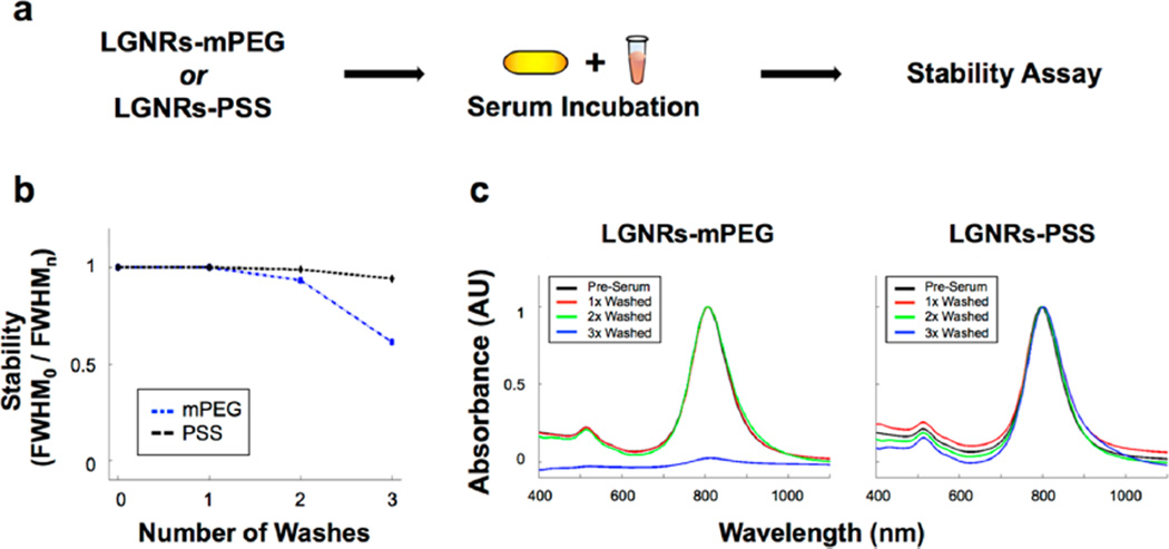
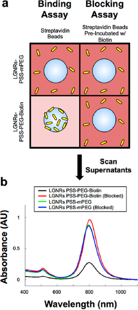
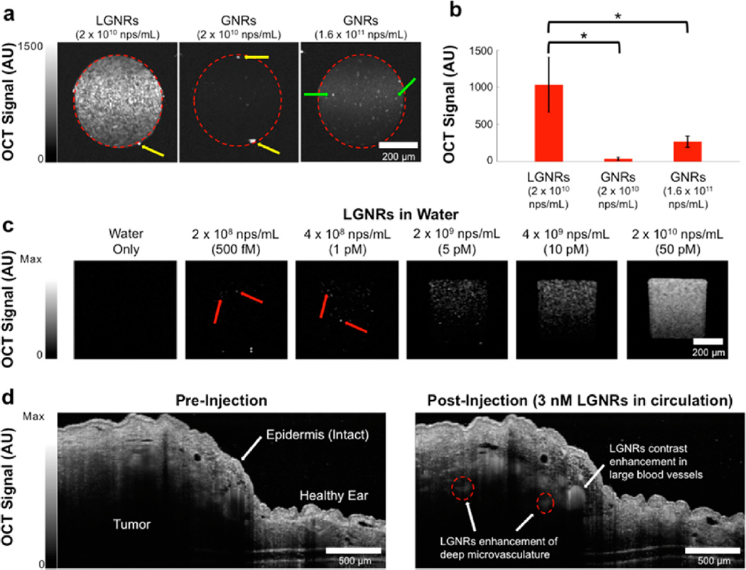
Gold nanorods (GNRs, ∼ 50 × 15 nm) have been used ubiquitously in biomedicine for their optical properties, and many methods of GNR biofunctionalization have been described. Recently, the synthesis of larger-than-usual GNRs (LGNRs, ∼ 100 × 30 nm) has been demonstrated. However, LGNRs have not been biofunctionalized and therefore remain absent from biomedical literature to date. Here we report the successful biofunctionalization of LGNRs, which produces highly stable particles that exhibit a narrow spectral peak (FWHM ∼100 nm). We further demonstrated that functionalized LGNRs can be used as highly sensitive scattering contrast agents by detecting individual LGNRs in clear liquids. Owing to their increased optical cross sections, we found that LGNRs exhibited up to 32-fold greater backscattering than conventional GNRs. We leveraged these enhanced optical properties to detect LGNRs in the vasculature of live tumor-bearing mice. With LGNR contrast enhancement, we were able to visualize tumor blood vessels at depths that were otherwise undetectable. We expect that the particles reported herein will enable immediate sensitivity improvements in a wide array of biomedical imaging and sensing techniques that rely on conventional GNRs.


Figures

Figure 1
Figure 1
Initial particle characterization. (a) TEM images of GNRs (length: 45 ± 7 nm, width: 14 ± 2 nm) and LGNRs (length: 93 ± 7 nm, width: 33 ± 1 nm) produced by separate methods (scale bars = 100 nm). Additional TEM images are provided in Figure S1. (b) Particle aspect ratio (AR) distributions for GNR and LGNR batches (n = 80 for each) were determined. GNRs AR: 3.3 ± 0.6, and LGNRs AR: 3.0 ± 0.3. (c) Absorbance spectra of each GNR batch were also measured. GNRs peak wavelength: 802 nm, and LGNRs peak wavelength: 804 nm. LGNRs exhibited a narrower spectrum (FWHM = 100 nm) than GNRs (FWHM = 150 nm).
Figure 2
Figure 2
Characterization of GNR stability trends as a function of size and surface coating. (a,b) GNRs and LGNRs were initially characterized by Vis-NIR spectrometry and Electrophoretic Light Scattering (ELS) and subsequently prepared with one of three different surface coating molecules. (c) Coated GNRs were then subjected to consecutive rounds of washing with distilled deionized water and centrifugation. GNRs were analyzed by Vis-NIR spectrometry and ELS after each wash to evaluate particle stability. (d,e) Particle stability was measured as spectral peak broadening by dividing the longitudinal absorbance peak full width at half-maximum before washing (FWHM0) by peak full width at half-maximum after each of the washes (FWHMn, where n = 0–3). Plotting FWHM0/FWHMn for each GNR size and coating reveals trends in stability for comparison. Original absorbance spectra for all GNRs are presented in Figures S2 and S3. (f) Zeta potential measurements for LGNRs are consistent with spectral stability trends, and they also provide validation of successful surface coating. Error bars represent standard error of the mean (s.e.m.) from triplicate measurements. Zeta potential measurements for all GNRs are presented in Figure S4 and Table S1.
Figure 3
Figure 3
LGNRs are robustly stable in biological serum. (a) LGNRs-mPEG and LGNRs-PSS were prepared, incubated with fetal bovine serum (FBS) for 3 h, and subjected to three rounds of washing by centrifugation. (b) The normalized absorbance spectrum of each GNR type was taken after each wash to assess serum stability. Measurements of absorbance peak FWHM0/FWHMn demonstrate that LGNRs-PSS are more stable in biological serum than LGNRs-mPEG. (c) Raw absorbance spectra for each particle type demonstrate this difference in stability more clearly, as LGNRs-mPEG exhibit virtually no plasmonic peak after the third wash. Unlike LGNRs-mPEG, LGNRs-PSS exhibit little change in absorbance properties.
Figure 4
Figure 4
LGNRs-PSS can be subsequently functionalized to achieve high-specificity molecular binding properties. (a) LGNRs-PSS-PEG-Biotin and LGNRs-PSS-mPEG were prepared and incubated with FBS to mimic biological environments. FBS-incubated GNRs were then used in biotin–streptavidin binding and blocking assays. For the binding assay, LGNRs-PSS-PEG-Biotin and LGNRs-PSS-mPEG were incubated with streptavidin-coated polystyrene beads (3 µm diameter) and centrifuged for 10s at 1000g to separate beads from free GNRs. The same process was repeated in the blocking assay, except that the streptavidin-coated beads were preincubated with excess free biotin to preclude specific binding of Large-GNRs-PSS-PEG-Biotin. (b) Absorbance measurements of the supernatant from each of the four bead-GNR combinations were taken after incubation. The same concentration of GNRs (OD 1) was used in each incubation, but only the supernatants from the incubation of LGNRs-PSS-PEG-Biotin and streptavidin beads exhibited a significant decrease in GNR concentration. These results demonstrate the proof of principle that LGNRs-PSS can be functionalized with ligands that retain molecular binding specificity in the presence of nonspecific proteins, which will be advantageous for future applications to targeted molecular imaging. Photographs from these molecular specificity assays are presented in Figure S5.
Figure 5
Figure 5
LGNRs are highly effective OCT contrast agents that enable single particle sensitivity in vitro and nanomolar sensitivity for noninvasive in vivo imaging. (a) Linear-scale OCT B-scans of LGNRs and two concentrations of GNRs show that LGNRs scatter significantly more near-infrared light (~800 nm) than GNRs of equivalent plasmonic resonance per nanoparticle. LGNRs also display stronger scattering than GNRs even when LGNRs and GNRs are prepared to equal mass concentration (2 × 10¹⁰ nps/mL for LGNRs and 1.6 × 10¹¹ nps/mL for GNRs; the 8-fold increased GNR concentration accounts for the particle volume difference between LGNRs and GNRs). (b) Region of interest analysis shows that LGNRs exhibit ~4-fold greater scattering than GNRs prepared to equivalent mass concentration. Considering the 4-fold greater LGNRs signal as well as the 8-fold difference in particle concentrations, LGNRs can produce up to ~32-fold greater OCT contrast than GNRs per particle (*p < 0.0001). Yellow arrows indicate specular reflections from capillary tubes. Green arrows show examples of spherical impurities (d ~ 50 nm) present in GNR solutions. (c) LGNRs produce enough scattering to enable detection of individual LGNRs in water. When prepared to 500 fM, the number of LGNRs expected to be present within the imaged capillary volume is ~70. This number is roughly consistent with the number of discrete puncta observed in the sample tube. The total signal increases for higher LGNR concentrations. Red arrows point to selected single LGNRs. (d) LGNRs-PSS-mPEG (250 µL of 23.5 nM) were tail vein-injected into a nude mouse bearing a U87MG tumor xenograft in the right ear pinna to achieve a particle concentration of 3 nM in circulation. OCT B-scans were acquired (completely noninvasively) before and after injection. LGNR-PSS-mPEG contrast results in increased OCT signal in blood vessels. LGNR-PSS-mPEG contrast-enhancement reveals small blood vessels (red dashed circles) deep within the tumor that cannot be visualized prior to injection.

References

    1. Huang X, El-Sayed IH, Qian W, El-Sayed MA. Cancer Cell Imaging and Photothermal Therapy in the Near-Infrared Region by Using Gold Nanorods. J. Am. Chem. Soc. 2006;128:2115–2120. - PubMed
    1. Hauck TS, Jennings TL, Yatsenko T, Kumaradas JC, Chan WCW. Enhancing the Toxicity of Cancer Chemotherapeutics with Gold Nanorod Hyperthermia. Adv. Mater. 2008;20:3832–3838.
    1. von Maltzahn G, Park JH, Agrawal A, Bandaru NK, Das SK, Sailor MJ, Bhatia SN. Computationally Guided Photothermal Tumor Therapy Using Long-Circulating Gold Nanorod Antennas. Cancer. Res. 2009;69:3892–3900. - PMC - PubMed
    1. Huang HC, Barua S, Kay DB, Rege K. Simultaneous Enhamcenent of Photothermal Stability and Gene Delivery Efficacy of Gold Nanorods Using Polyelectrolytes. ACS Nano. 2009;3:2941–2952. - PMC - PubMed
    1. Choi WI, Kim JY, Kang C, Byeon CC, Kim YH, Tae G. Tumor Regression in vivo by Photothermal Therapy Based on Gold-Nanorod-Loaded, Functional Nanocarriers. ACS Nano. 2011;5:1995–2003. - PubMed

Publication types

MeSH terms

LinkOut - more resources