. 2016 Jan 15:125:446-455.
doi: 10.1016/j.neuroimage.2015.10.018. Epub 2015 Oct 20.

Tracer kinetic modelling for DCE-MRI quantification of subtle blood-brain barrier permeability

Affiliations

Tracer kinetic modelling for DCE-MRI quantification of subtle blood-brain barrier permeability

Anna K Heye et al. Neuroimage. .

Abstract

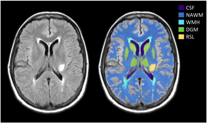
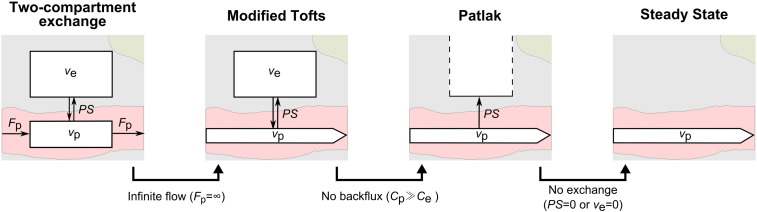
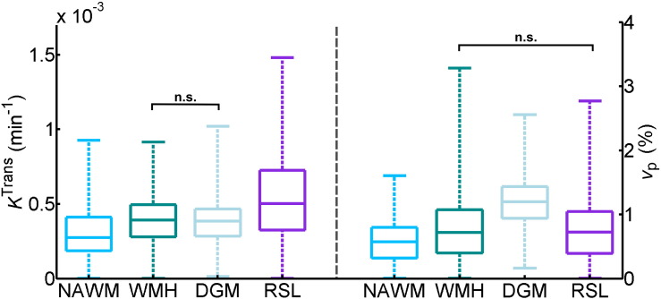
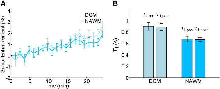
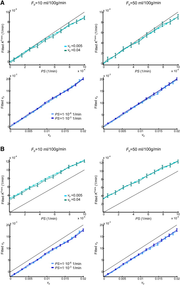
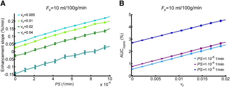
There is evidence that subtle breakdown of the blood-brain barrier (BBB) is a pathophysiological component of several diseases, including cerebral small vessel disease and some dementias. Dynamic contrast-enhanced MRI (DCE-MRI) combined with tracer kinetic modelling is widely used for assessing permeability and perfusion in brain tumours and body tissues where contrast agents readily accumulate in the extracellular space. However, in diseases where leakage is subtle, the optimal approach for measuring BBB integrity is likely to differ since the magnitude and rate of enhancement caused by leakage are extremely low; several methods have been reported in the literature, yielding a wide range of parameters even in healthy subjects. We hypothesised that the Patlak model is a suitable approach for measuring low-level BBB permeability with low temporal resolution and high spatial resolution and brain coverage, and that normal levels of scanner instability would influence permeability measurements. DCE-MRI was performed in a cohort of mild stroke patients (n=201) with a range of cerebral small vessel disease severity. We fitted these data to a set of nested tracer kinetic models, ranking their performance according to the Akaike information criterion. To assess the influence of scanner drift, we scanned 15 healthy volunteers that underwent a "sham" DCE-MRI procedure without administration of contrast agent. Numerical simulations were performed to investigate model validity and the effect of scanner drift. The Patlak model was found to be most appropriate for fitting low-permeability data, and the simulations showed vp and K(Trans) estimates to be reasonably robust to the model assumptions. However, signal drift (measured at approximately 0.1% per minute and comparable to literature reports in other settings) led to systematic errors in calculated tracer kinetic parameters, particularly at low permeabilities. Our findings justify the growing use of the Patlak model in low-permeability states, which has the potential to provide valuable information regarding BBB integrity in a range of diseases. However, absolute values of the resulting tracer kinetic parameters should be interpreted with extreme caution, and the size and influence of signal drift should be measured where possible.

Keywords: Blood–brain barrier; Cerebral small vessel disease; Dynamic contrast-enhanced MRI; Tracer kinetic modelling.


Figures

Fig. 1
Fig. 1
Representative MRI data and tissue segmentation. FLAIR image (left) and tissue masks superimposed on FLAIR image (CSF: cerebrospinal fluid, NAWM: normal-appearing white matter, WMH: white matter hyperintensities, DGM: deep grey matter, RSL: recent stroke lesion).
Fig. 2
Fig. 2
Set of nested tracer kinetic models. Target parameters of DCE-MRI modelling are the fractional plasma volume vp, the fractional interstitial volume ve, the plasma flow Fp and the permeability-surface area product PS. The four models are related by a series of simplifying assumptions.
Fig. 3
Fig. 3
Cohort average signal enhancement curves. Post-contrast signal enhancement versus time obtained from the median signal intensity in each tissue type (NAWM: normal-appearing white matter, WMH: white matter hyperintensities, DGM: deep grey matter, RSL: recent stroke lesion, SS: sagittal sinus) and averaged over all patients (n = 201). Y-axis scales for the tissue and sagittal sinus enhancement curves are shown on the left and right, respectively.
Fig. 4
Fig. 4
Comparison of model fits to the patient data. (A) Example concentration–time curve for normal-appearing white matter in a single patient. In general, the steady-state model does not fit the data well; while both the Patlak and modified Tofts (mTofts) models typically fit the data similarly well, the Patlak model has a higher Akaike weight (AW) than the modified Tofts model in most cases due to the lower number of free parameters. (B) Comparison of AW for the three models in normal-appearing white matter (NAWM), white matter hyperintensities (WMH), deep grey matter (DGM) and recent stroke lesions (RSL). In most patients, the Patlak model had the highest AW for all tissue types (legend as in A).
Fig. 5
Fig. 5
Comparison of fitted Patlak parameters between tissue types. Box plots showing the distribution of KTrans (left) and vp (right) in normal-appearing white matter (NAWM), white matter hyperintensities (WMH), deep grey matter (DGM) and recent stroke lesions (RSL). Brackets with n.s. indicate non-significant differences with p > 0.5; all other differences between tissue types are significant with p < 0.001 (brackets omitted for clarity).
Fig. 6
Fig. 6
Contrast-free measurements in healthy volunteers. (A) Average signal enhancement curves (n = 15) in normal-appearing white matter (NAWM) and deep grey matter (DGM), showing a drift in signal intensity; error bars indicate the mean ± standard error. (B) T1 measurements obtained before and after the DCE-MRI sequence using the inversion recovery method (n = 7); error bars indicate the mean ± 1.96 standard deviations.
Fig. 7
Fig. 7
Simulated accuracy of Patlak parameters. (A) Relationship of permeability-surface area product PS (top row) and blood plasma volume vp (bottom row) values, with corresponding fitted Patlak parameters. Results are shown for two different blood plasma flow (Fp), PS and vp values. For all simulations, the interstitial volume was set to 0.2. Error bars indicate the mean ± 1.96 standard deviations; the grey line represents the identity line. (B) As above but including a 0.08%/min signal drift.
Fig. 8
Fig. 8
Relationship between simulated semi-quantitative parameters and tissue properties. (A) Relationship between the signal enhancement slope and the permeability-surface area product PS for different blood plasma volumes vp. (B) Relationship between normalised area under the signal enhancement curve and vp for different PS values. A signal drift of 0.08%/min was added to the synthetic data; results are shown for blood plasma flow Fp = 10 ml/100 g/min and interstitial volume ve = 0.2.

Similar articles

Cited by

References

    1. Ahearn T.S., Staff R.T., Redpath T.W., Semple S.I.K. The use of the Levenberg–Marquardt curve-fitting algorithm in pharmacokinetic modelling of DCE-MRI data. Phys. Med. Biol. 2005;50:N85–N92. - PubMed
    1. Akaike H. A new look at the statistical model identification. IEEE Trans. Autom. Control. 1974;19:716–723.
    1. Armitage P., Behrenbruch C., Brady M., Moore N. Extracting and visualizing physiological parameters using dynamic contrast-enhanced magnetic resonance imaging of the breast. Med. Image Anal. 2005;9:315–329. - PubMed
    1. Armitage P.A., Farrall A.J., Carpenter T.K., Doubal F.N., Wardlaw J.M. Use of dynamic contrast-enhanced MRI to measure subtle blood–brain barrier abnormalities. Magn. Reson. Imaging. 2011;29:305–314. - PMC - PubMed
    1. Barnes S.R., Ng T.S.C., Montagne A., Law M., Zlokovic B.V., Jacobs R.E. Optimal acquisition and modeling parameters for accurate assessment of low K trans blood–brain barrier permeability using dynamic contrast-enhanced MRI. Magn. Reson. Med. 2015 - PMC - PubMed

Publication types