. 2015 Oct 22;163(3):594-606.
doi: 10.1016/j.cell.2015.09.055. Epub 2015 Oct 17.

Evolving new protein-protein interaction specificity through promiscuous intermediates

Affiliations

Evolving new protein-protein interaction specificity through promiscuous intermediates

Christopher D Aakre et al. Cell. .

Abstract

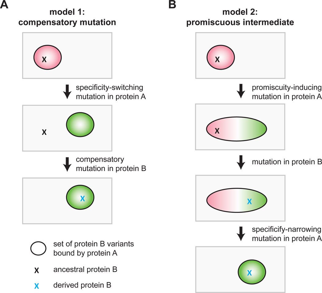
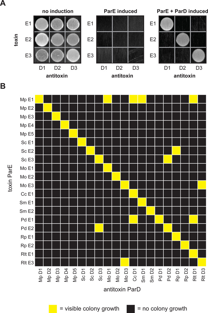
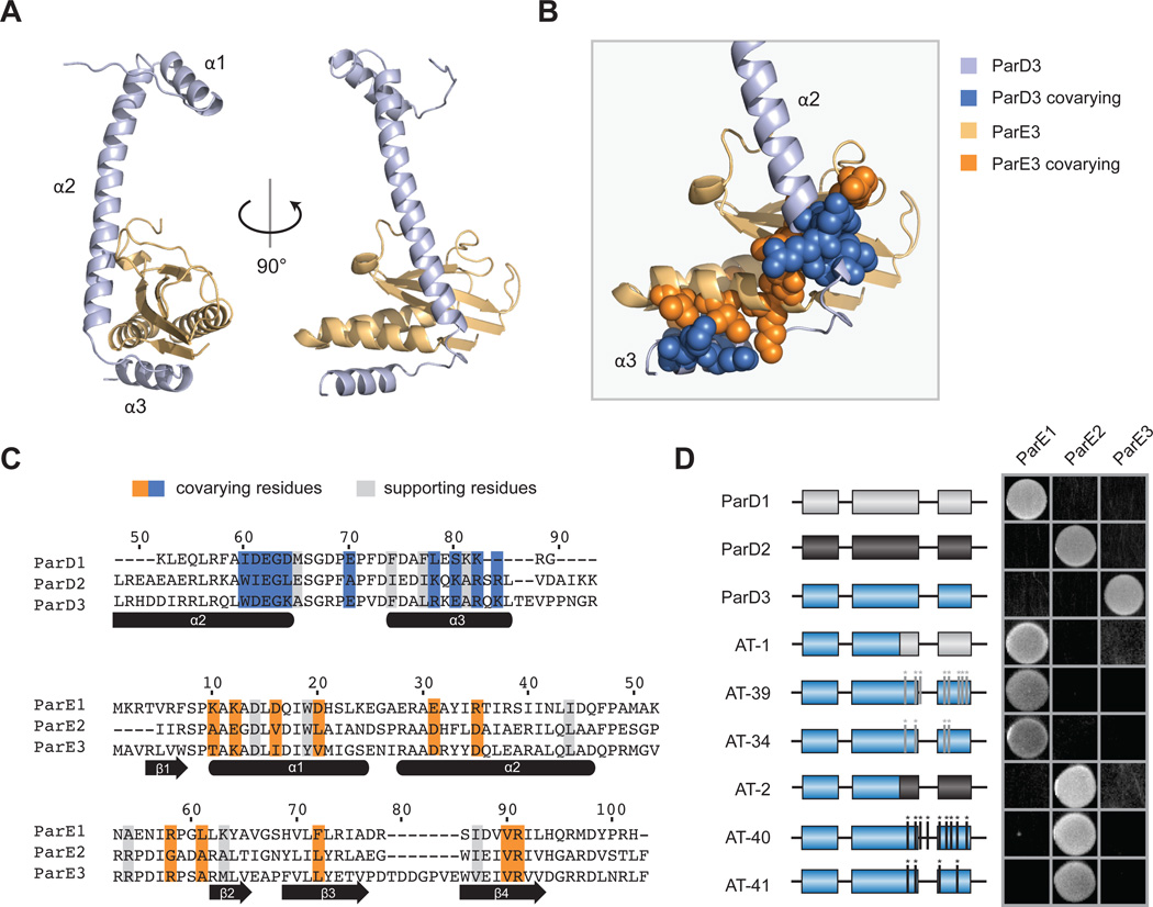
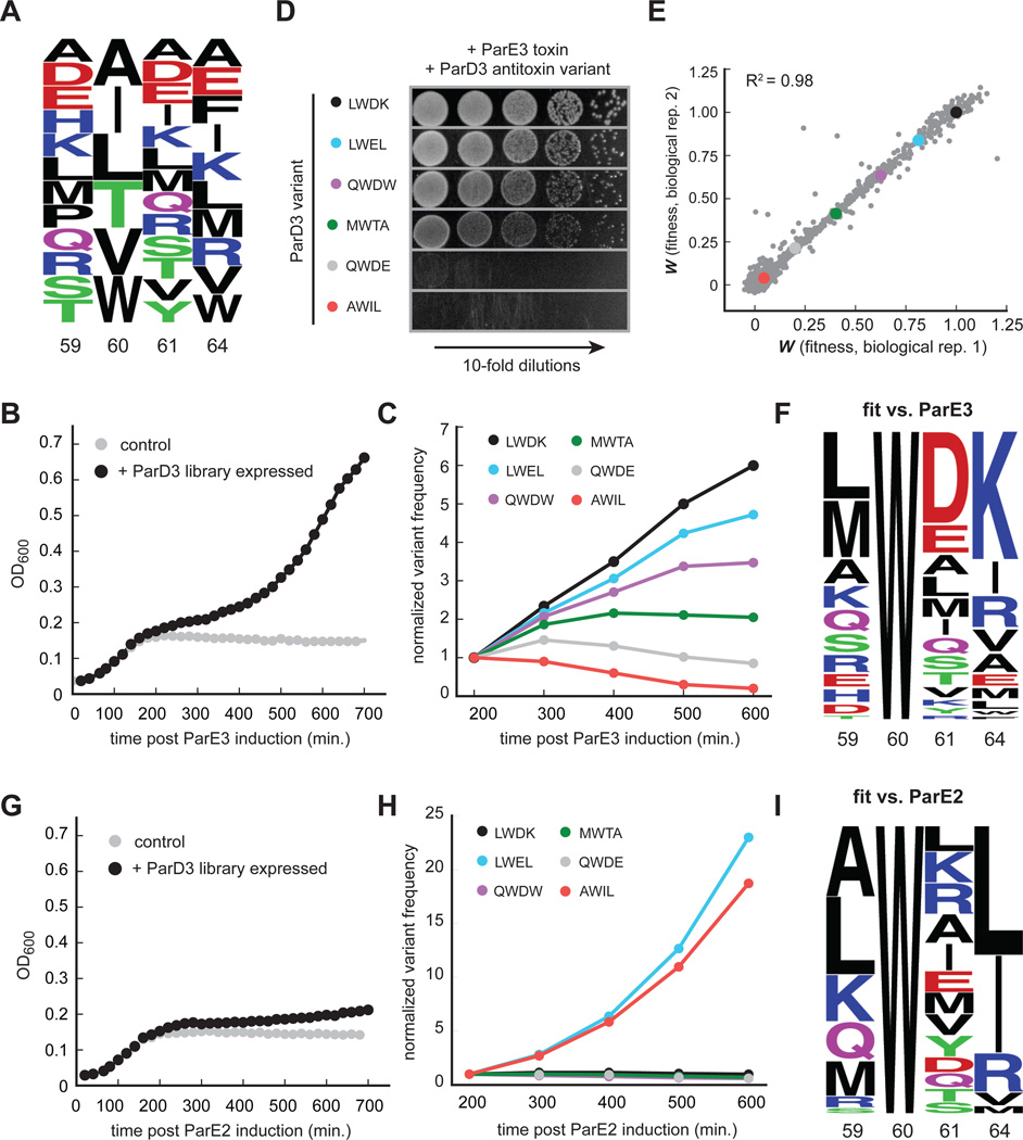
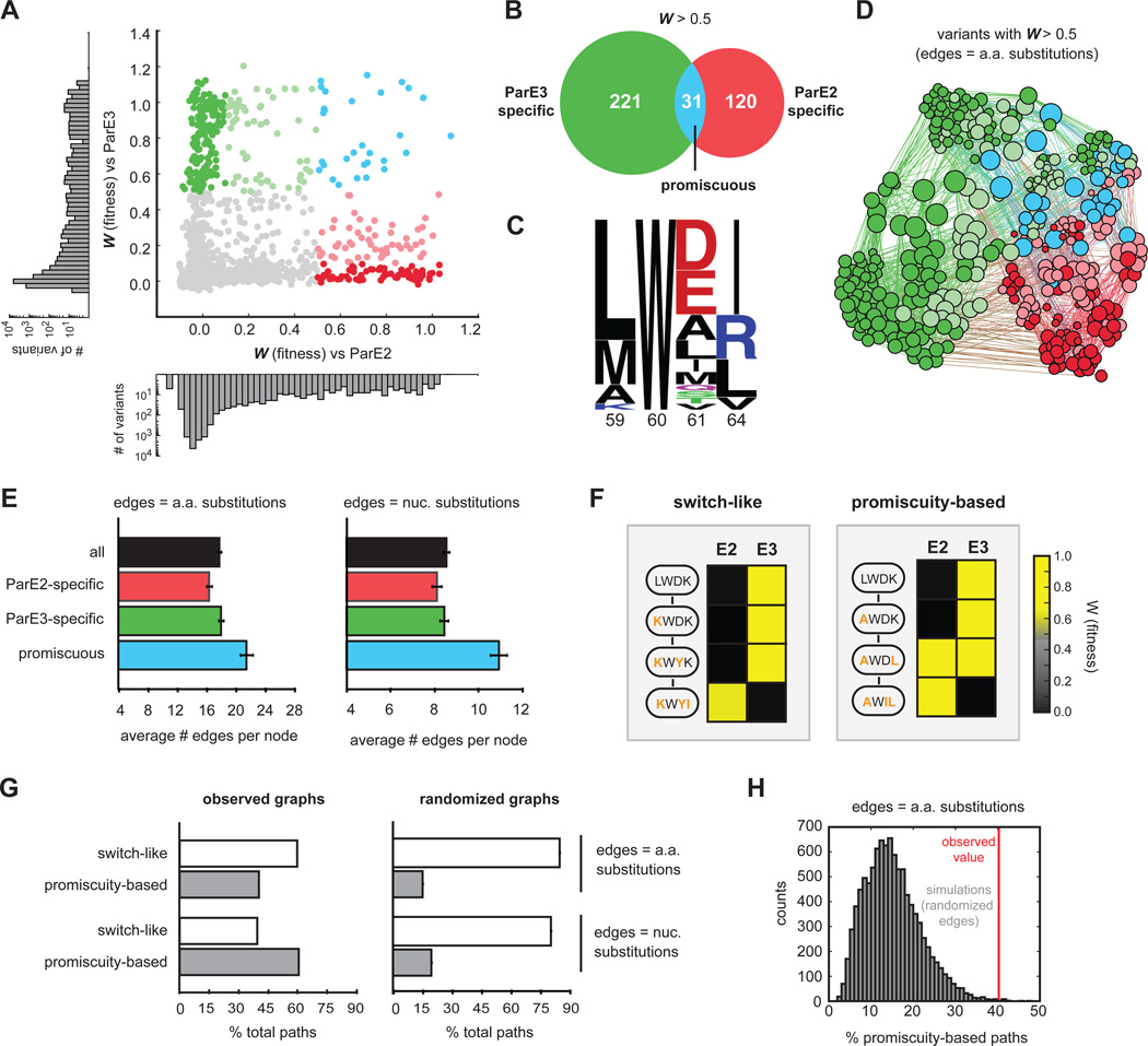
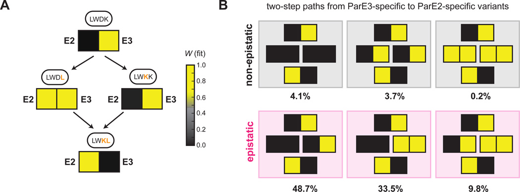
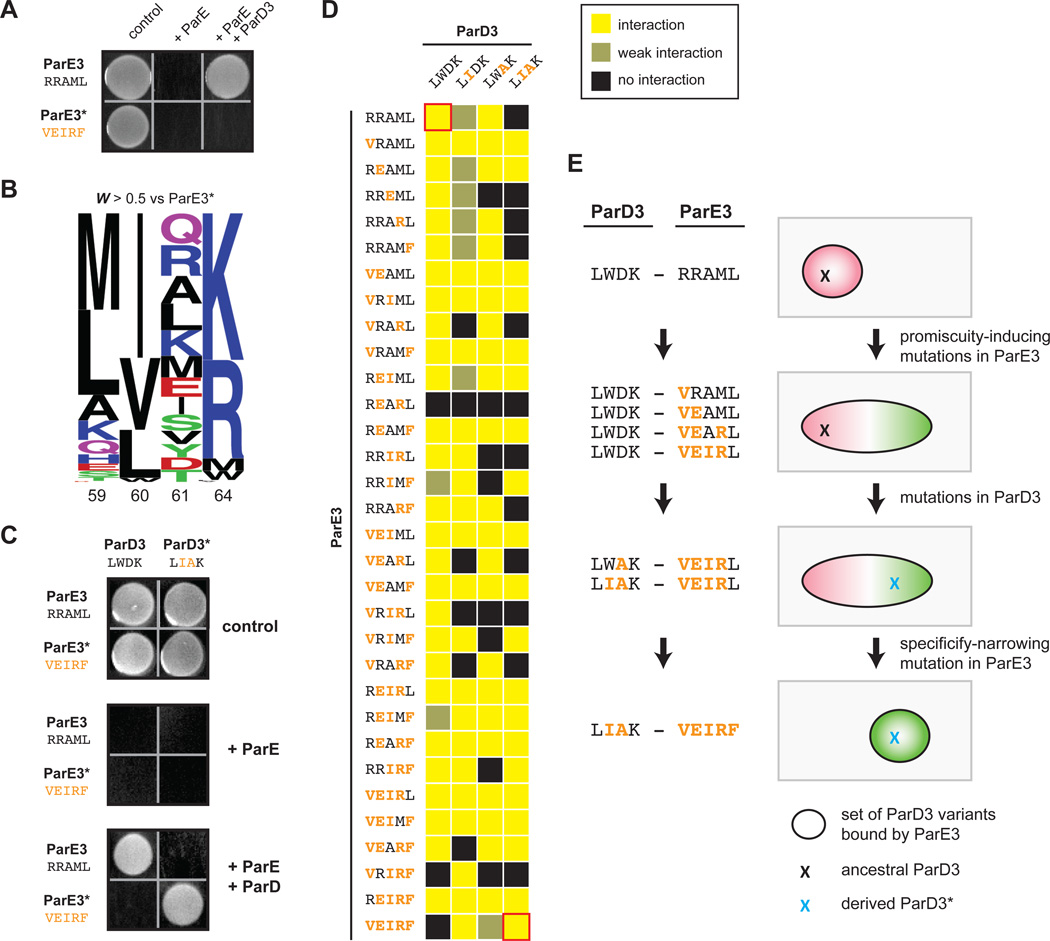
Interacting proteins typically coevolve, and the identification of coevolving amino acids can pinpoint residues required for interaction specificity. This approach often assumes that an interface-disrupting mutation in one protein drives selection of a compensatory mutation in its partner during evolution. However, this model requires a non-functional intermediate state prior to the compensatory change. Alternatively, a mutation in one protein could first broaden its specificity, allowing changes in its partner, followed by a specificity-restricting mutation. Using bacterial toxin-antitoxin systems, we demonstrate the plausibility of this second, promiscuity-based model. By screening large libraries of interface mutants, we show that toxins and antitoxins with high specificity are frequently connected in sequence space to more promiscuous variants that can serve as intermediates during a reprogramming of interaction specificity. We propose that the abundance of promiscuous variants promotes the expansion and diversification of toxin-antitoxin systems and other paralogous protein families during evolution.


Figures

Figure 1
Figure 1. Models for the evolution of new protein-protein interaction specificity
(A) In a model of coevolution through compensatory mutations, an initial mutation in protein A that disrupts the A-B interaction is rescued by a compensatory mutation in protein B. Ovals represent the set of protein B variants that are bound by protein A, and Xs indicate particular protein B variants. Note that the intermediate state is a non-functional interaction. (B) In an alternative model for protein coevolution, protein A first accumulates a mutation that broadens its specificity, followed by a second mutation in protein B that retains its interaction with the new form of A but that would have disrupted its interaction with the ancestral form of protein A. In a final step, protein A mutates to narrow its specificity to include the derived, and not ancestral, form of protein B.
Figure 2
Figure 2. Toxins and antitoxins from the ParD-ParE family exhibit high interaction specificity
(A) Testing of interaction specificity for ParD antitoxins and ParE toxins from Mesorhizobium opportunistum. Plasmids harboring the toxins and antitoxins indicated were cotransformed into E. coli with ParD and ParE induced as indicated. (B) Comprehensive testing of interaction specificity for 20 ParD and ParE pairs from eight different species. Cells containing each possible ParD-ParE pair were grown on plates that induce the toxin and antitoxin, respectively, and grown overnight at 37°C. Yellow, visible colonies following serial dilution; black, no visible colonies. Also see Fig. S1.
Figure 3
Figure 3. Covarying residues dictate interaction specificity in the ParD-ParE family
(A) Structure of the M. opportunistum ParD3-ParE3 complex. Light orange, ParE3 monomer; light blue, ParD3 monomer. (B) A section of the ParD3-ParE3 structure from (A) magnified; covarying residues shown in space-filling representation. (C) Alignment of M. opportunistum ParD and ParE paralogs with coevolving residues highlighted in blue or orange for ParD or ParE, respectively. Supporting residues, which coevolve with the interfacial coevolving residues, are highlighted in grey. (D) Mutations in the C-terminus of ParD3 can reprogram interaction specificity. The indicated ParD3 mutants were tested against each ParE homolog from M. opportunistum using the E. coli toxicity-rescue assay. Also see Figs. S2–S3.
Figure 4
Figure 4. High-throughput mapping of mutant fitness at co-evolving interface
(A) Composition of the ParD3 antitoxin library at the four variable positions. (B) Library growth following ParE3 toxin induction. (C) Frequency changes over time for the indicated ParD3 variants following ParE3 induction. (D) Testing of individual variants from (C) using the toxicity rescue assay. 10-fold serial dilutions were plated from cultures expressing the ParD3 variant indicated and the ParE3 toxin. (E) Two biological replicates of fitness measurements derived from screening the ParD3 library against the ParE3 toxin. (F) Frequency logo for ParD3 library variants with high fitness against ParE3 (WE3 > 0.5). (G) Library growth following induction of the non-cognate ParE2 toxin. (H) Frequency changes over time for the indicated ParD3 library variants. (I) Frequency logo for ParD3 library variants with high fitness against ParE2 (WE2 > 0.5). Also see Fig. S4.
Figure 5
Figure 5. Specificity-reprogramming paths are highly enriched for promiscuous variants
(A) Fitness of ParD3 variants against ParE2 and ParE3. Green, specific for ParE3; blue, capable of antagonizing both ParE2 and ParE3; red, specific for ParE2. Histograms of fitness values against ParE2 and ParE3 are shown. (B) Venn diagram of ParD3 variants reactive against ParE3, ParE2, or both. (C) Frequency logo of promiscuous ParD3 variants (WE2 > 0.5, WE3 > 0.5). (D) Force-directed graph of all ParD3 variants reactive against ParE3 or ParE2 (W > 0.5). Nodes represent individual variants and edges represent single amino acid substitutions. Node size scales with increasing degree and color corresponds to the specificity classes in (A). (E) Average number of edges per node for the indicated categories of ParD3 variants. Error bars indicate SEM. (F) Examples of ‘switch-like’ and ‘promiscuity-based’ mutational paths from an E3-specific variant to an E2-specific variant with the fitness against each variant color-coded based on the scale shown. (G) Left, percentage of ‘switch-like’ and ‘promiscuity-based’ paths from the wild-type ParD3 sequence (LWDK) to each of the 66 ParE2-specific variants (WE2 > 0.5, WE3 < 0.1). Right, same as left panel but for 10,000 simulations in which the graph edges were randomly shuffled while keeping the total edge count and degree distribution constant. Error bars represent SEM. (H) Histogram representing percentage of ‘promiscuity-based’ paths in 10,000 edge shuffling simulations; red line indicates percentage for the observed amino acid graph. Also, see Fig. S5.
Figure 6
Figure 6. Mutational order dictates specificity class of intermediate variants
(A) Mutational paths from LWDK to LWKL for ParD3 with fitness of each variant against ParE2 and ParE3 shown as a heatmap: yellow, high fitness; black low fitness. (B) The six path types that reprogram ParD3 specificity in two mutational steps. Percentage of mutational paths in each category is indicated for a threshold of 0.5 used to define a positive interaction. Also see Fig. S6.
Figure 7
Figure 7. Mutational trajectories to an orthogonal ParD3*-ParE3* pair
(A) ParE3* is insulated from antitoxin ParD3. A plasmid containing either ParE3 or ParE3* was co-transformed into E. coli with a plasmid expressing ParD3, and cells were plated on medium that induces or represses expression of the toxin and antitoxin. (B) Frequency logo for ParD3 library variants with high fitness against ParE3* (WE3* > 0.5). (C) ParE3*-ParD3* is insulated from the wild-type ParD3-ParE3 pair. (D) Toxicity-rescue interaction assays for all ParD3 and ParE3 mutant combinations. Top left, wild-type ParD3-ParE3 pair; bottom right, orthogonal ParD3*-ParE3* pair. Promiscuous ParE3 intermediates are those capable of interacting with both ParD3 and ParD3*. (E) Example of a series of single substitutions that lead to the insulated ParE3*-ParD3* system while retaining the toxin-antitoxin interaction at each step by first expanding the specificity of ParE3, followed by changes in ParD3, and finally by restricting the specificity of ParE3. Also see Fig. S7.

Comment in

Similar articles

Cited by

References

    1. Aharoni A, Gaidukov L, Khersonsky O, Mc QGS, Roodveldt C, Tawfik DS. The 'evolvability' of promiscuous protein functions. Nat Genet. 2005;37:73–76. - PubMed
    1. Anderson DW, McKeown AN, Thornton JW. Intermolecular epistasis shaped the function and evolution of an ancient transcription factor and its DNA binding sites. eLife. 2015;4 - PMC - PubMed
    1. Bloom JD, Arnold FH. In the light of directed evolution: pathways of adaptive protein evolution. PNAS. 2009;(106 Suppl 1):9995–10000. - PMC - PubMed
    1. Capra EJ, Perchuk BS, Skerker JM, Laub MT. Adaptive mutations that prevent crosstalk enable the expansion of paralogous signaling protein families. Cell. 2012;150:222–232. - PMC - PubMed
    1. Dalton KM, Crosson S. A conserved mode of protein recognition and binding in a ParD-ParE toxin-antitoxin complex. Biochemistry. 2010;49:2205–2215. - PMC - PubMed

Publication types