

Lymphangioleiomyomatosis
Atlas Genet Cytogenet Oncol Haematol.
2009.
No abstract available
Figures

Chest CT scan of a patient with LAM (A) showing numerous thin-walled cysts distributed throughout the lungs. (B) The lung parenchyma is almost completely replaced by very small cysts.
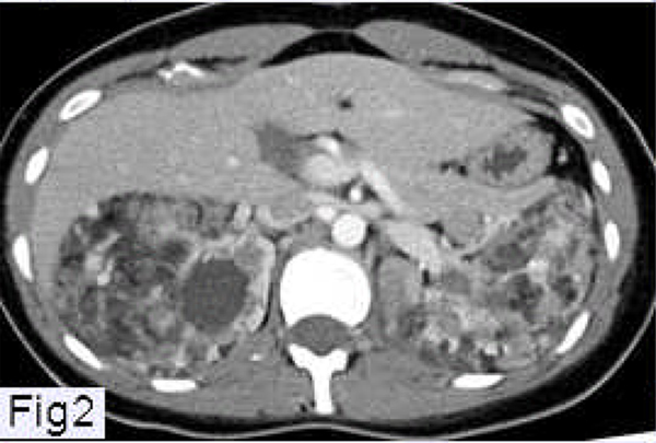
Abdominal CT scan of a patient with LAM showing angiomyolipomas involving both kidneys.
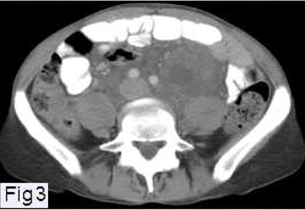
Abdominal CT scan of a patient with LAM showing a large lymphangioleiomyoma located in the retroperitoneal area and surrounding the aorta and inferior vena cava.

A and B. LAM nodule comprising spindle-shaped cells and larger epithelioid cells (A). Nodules of various sizes (B) are seen in involved lung (hematoxylin-eosin; original magnification ×50).
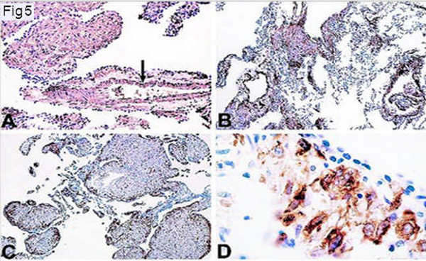
Immunohistochemistry of LAM cells. Immunoperoxidase method and counterstaining with hematoxylin. A and B: Immunoreactivity with a-smooth muscle actin antibodies. LAM cells show strong reactivity (A). Pulmonary vascular smooth muscle cells are also strongly positive (arrow). LAM cells in the walls of the lung cysts are also strongly reactive (arrow) (B) (original magnification ×250 for each). C: Immunoreactivity with monoclonal antibody HMB-45. Immunoreactive cells are distributed in the periphery of LAM lung nodules (arrow) (original magnification ×250). D: Immunoreactivity with monoclonal antibody HMB-45. Higher-magnification view of tissue shown in C. A strong granular reaction is present in large epithelioid LAM cells adjacent to epithelial cells covering LAM lung nodules (arrow) (D) (original magnification ×1000).

Left panel: close-up of LAM nodule (hematoxylin-eosin). Right panel: same nodule showing positive immunocytochemistry stain for HMB 45 (original magnification ×200).

Characteristics of LAM cells (A–C). Reaction of LAM cells cultured from lung and pulmonary artery smooth muscle cells (PASM) with monoclonal antibody against SMA (A). Reaction of cultured LAM cells and melanoma cells (MALME-3M) with monoclonal antibody HMB-45 (B). Fluorescence in situ hybridization (FISH) for TSC1 (green) and TSC2 (red) in LAM cells showing normal presence of two of each alleles as well as abnormal presence of TSC2 alleles (left) (C). FISH for TSC1 (green, arrow) and TSC2 (red, arrowhead) in LAM cell with one (right) or two TSC2 (left) alleles (C). Bar, 20 µm.
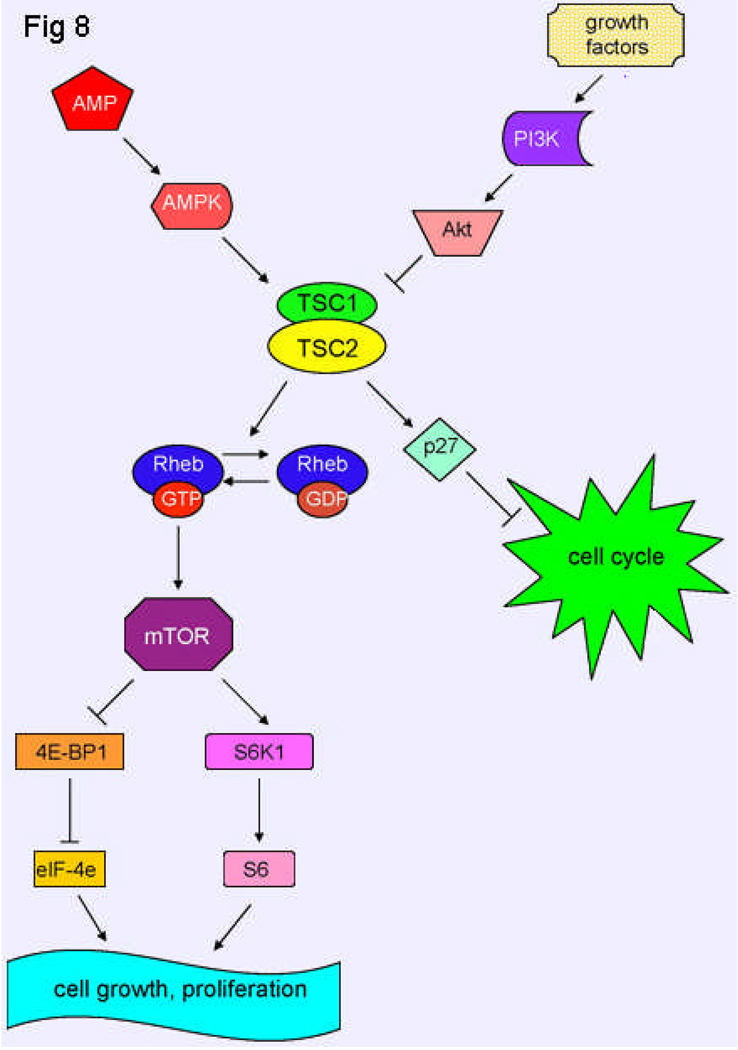
Schematic model of TSC1 and TSC2 pathways. The TSC1/TSC2 complex has roles in cell cycle progression and in cell growth and proliferation. Tuberin binds p27KIP1, a cyclin-dependent kinase inhibitor, stabilizing it and resulting in inhibition of cell cycle progression. Tuberin also has Rheb GAP activity, which converts Rheb-GTP to Rheb-GDP, resulting in inactive Rheb. Rheb controls mTOR, which is a kinase that controls translation through the phosphorylation of 4E-BP1 and S6K1. Akt, when activated by growth factors, phosphorylates tuberin, leading to an inhibition of tuberin and resulting in cell growth and proliferation. However, when a state of low cellular energy exists, AMPK phosphorylates tuberin, activating it, and thereby inhibiting cell growth.
References
-
- Berger U, Khaghani A, Pomerance A, Yacoub MH, Coombes RC. Pulmonary lymphangioleiomyomatosis and steroid receptors. Am J Clin Pathol. 1990 May;93(5):609–614. PMID 2183584. - PubMed
-
- European Chromosome 16 Tuberous Sclerosis Consortium. Identification and characterization of the tuberous sclerosis gene on chromosome 16. Cell. 1993 Dec 31;75(7):1305–1315. PMID 8269512. - PubMed
-
- Soucek T, Pusch O, Wienecke R, DeClue JE, Hengstschlager M. Role of the tuberous sclerosis gene-2 product in cell cycle control. Loss of the tuberous sclerosis gene-2 induces quiescent cells to enter S phase. J Biol Chem. 1997 Nov 14;272(46):29301–29308. PMID 9361010. - PubMed
-
- Xiao G-H, Shoarinejad F, Jin F, Golemis EA, Yeung RS. The tuberous sclerosis 2 gene product, tuberin, functions as a Rab5 GTPase activating protein (GAP) in modulating endocytosis. J Biol Chem. 1997 Mar 7;272(10):6097–6100. PMID 9045618. - PubMed
-
- Henry KW, Yuan X, Koszewski NJ, Onda H, Kwiatkowski DJ, Noonan DJ. Tuberous sclerosis gene 2 product modulates transcription mediated by steroid hormone receptor family members. J Biol Chem. 1998 Aug 7;273(32):20535–20539. PMID 9685410. - PubMed
Grants and funding
LinkOut - more resources
Full Text Sources