. 2015 Summer;7(3):1871.

Tumor-Induced Osteomalacia

Affiliations

Tumor-Induced Osteomalacia

Rajiv Kumar et al. Transl Endocrinol Metab. 2015 Summer.
No abstract available


Figures

Figure 1
Figure 1
Figure 1A. Skeletal radiograph of the patient with nutritional osteomalacia. Note the presence of pseudofractures of the metatarsals (arrows). Figure 1B. Technetium-99m methylene diphosphonate (Tc-99m MDP) bone scan of patient with osteomalacia. Note uptake over the ribs and sacro-iliac region.
Figure 1
Figure 1
Figure 1A. Skeletal radiograph of the patient with nutritional osteomalacia. Note the presence of pseudofractures of the metatarsals (arrows). Figure 1B. Technetium-99m methylene diphosphonate (Tc-99m MDP) bone scan of patient with osteomalacia. Note uptake over the ribs and sacro-iliac region.
Figure 2
Figure 2
Photomicrograph of bone (Goldner’s stain) showing normal bone (top left panel) and osteomalacic bone (top right panel) and normal mineralization (bottom left panel) and a mineralization defect (bottom right panel) after the administration of tetracycline labels.
Figure 3
Figure 3
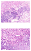
Low power photomicrograph (10x) of a phosphaturic mesenchymal tumor, showing the characteristic admixture of bland spindled cells, small blood vessels, adipocytes and calcified matrix. High power photomicrograph (40x:) of a phosphaturic mesenchymal tumor, with an osteoclastic reaction to the distinctive calcified matrix produced by the lesional cells.
Figure 4
Figure 4
Figure 4A. Magnetic resonance scan of a patient with TIO. The arrow points to the tumor. Figure 4B. Technetium 99m sestamibi scans of a patient with TIO (same patient as in figure 4B). Arrow and arrow head point to tumor.
Figure 4
Figure 4
Figure 4A. Magnetic resonance scan of a patient with TIO. The arrow points to the tumor. Figure 4B. Technetium 99m sestamibi scans of a patient with TIO (same patient as in figure 4B). Arrow and arrow head point to tumor.
Figure 5
Figure 5
Phosphorus homeostasis in humans.
Figure 6
Figure 6
Adaptations to changes in dietary phosphate intake.
Figure 7
Figure 7
Mechanisms of hypophosphatemia in TIO and the inherited rickets

References

    1. McCance RA. Osteomalacia with Looser’s nodes (Milkman’s Syndrome) due to a raised resistance to Vitamin D acquired about the age of 15 years. Quarterly Journal of Medicine. 1947;16:33–46. - PubMed
    1. Prader A, Illig R, Uehlinger RE, Stalder G. Rachitis infolge knochentumors (Rickets caused by bone tumors) Helv Pediatr Acta. 1959;14:554–565. - PubMed
    1. Salassa RM, Jowsey J, Arnaud CD. Hypophosphatemic osteomalacia associated with “nonendocrine” tumors. N Engl J Med. 1970;283:65–70. - PubMed
    1. Evans DJ, Azzopardi JG. Distinctive tumours of bone and soft tissue causing acquired vitamin-D- resistant osteomalacia. Lancet. 1972;1:353–354. - PubMed
    1. Olefsky J, Kempson R, Jones H, Reaven G. “Tertiary” hyperparathyroidism and apparent “cure” of vitamin-D- resistant rickets after removal of an ossifying mesenchymal tumor of the pharynx. N Engl J Med. 1972;286:740–745. - PubMed

LinkOut - more resources