. 2015 Oct;2(10):960-71.
doi: 10.1002/acn3.248. Epub 2015 Sep 11.

Characterization of IgG4 anti-neurofascin 155 antibody-positive polyneuropathy

Affiliations

Characterization of IgG4 anti-neurofascin 155 antibody-positive polyneuropathy

Hidenori Ogata et al. Ann Clin Transl Neurol. 2015 Oct.

Abstract

Objective: To investigate anti-neurofascin 155 (NF155) antibody-positive chronic inflammatory demyelinating polyneuropathy (CIDP).

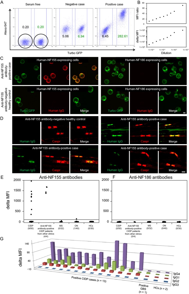
Methods: Sera from 50 consecutive CIDP patients diagnosed in our clinic, 32 patients with multiple sclerosis, 40 patients with other neuropathies including 26 with Guillain-Barré syndrome (GBS)/Fisher syndrome, and 30 healthy controls were measured for anti-NF antibodies by flow cytometry using HEK293 cell lines stably expressing human NF155 or NF186. Four additional CIDP patients with anti-NF155 antibodies referred from other clinics were enrolled for clinical characterization.

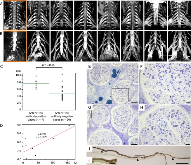
Results: The positivity rate for anti-NF155 antibodies in CIDP patients was 18% (9/50), who all showed a predominance of IgG4 subclass. No other subjects were positive, except one GBS patient harboring IgG1 anti-NF155 antibodies. No anti-NF155 antibody carriers had anti-NF186 antibodies. Anti-NF155 antibody-positive CIDP patients had a significantly younger onset age, higher frequency of drop foot, gait disturbance, tremor and distal acquired demyelinating symmetric phenotype, greater cervical root diameter on magnetic resonance imaging neurography, higher cerebrospinal fluid protein levels, and longer distal and F-wave latencies than anti-NF155 antibody-negative patients. Marked symmetric hypertrophy of cervical and lumbosacral roots/plexuses was present in all anti-NF155 antibody-positive CIDP patients examined by neurography. Biopsied sural nerves from two patients with anti-NF155 antibodies demonstrated subperineurial edema and occasional paranodal demyelination, but no vasculitis, inflammatory cell infiltrates, or onion bulbs. Among anti-NF155 antibody-positive patients, treatment responders more frequently had daily oral corticosteroids and/or immunosuppressants in addition to intravenous immunoglobulins than nonresponders did.

Interpretation: Anti-NF155 antibodies occur in a subset of CIDP patients with distal-dominant involvement and symmetric nerve hypertrophy.


Figures

Figure 1
Figure 1
Anti-NF1155 and anti-NF186 antibody assays by flow cytometry and cell- and tissue-based immunohistochemistry. (A) Representative flow cytometry assay results showing serum-free (left), negative (middle), and positive (right) conditions. The MFI of cell-associated turbo GFP and Alexa 647 was measured for each sample. Cells expressing NF155-turbo GFP and naive cells are easily separable according to the MFI of turbo GFP. The MFI of cell-associated Alexa 647 was measured to detect human IgG bound to NF155, using cells without NF155 expression as a negative control. Note that positive and negative patients are clearly separable. For each serum sample, the MFI ratio was calculated by (Alexa 647 MFI of NF155-transfected cells/Alexa 647 MFI of NF155-untransfected cells), and delta MFI was calculated by (Alexa 647 MFI of NF155-transfected cells − Alexa-647 MFI of NF155-untransfected cells). Cutoff points for MFI ratio and delta MFI were set at 10 and 100, respectively, based on preliminary experiments. Antibodies against human recombinant NF186 protein were also measured using the same method. (B) Calibration curves using serum from a representative CIDP patient with anti-NF155 antibodies show proportionate decreases in MFI ratio and delta MFI values, by serum serial dilution (1:20, 1:100, 1:200, 1:400, 1:800, 1:1600, 1:3200, 1:6400, and 1:12,800). (C) Immunohistochemistry of cells expressing human NF155-turbo GFP or NF186-turbo GFP shows that serum from a representative CIDP patient with anti-NF155 antibodies reacts with human NF155-expressing cells, but not with human NF186-expressing cells. Scale bar = 10 μm. (D) Double immunostaining of mouse teased sciatic nerve fibers with anti-Caspr antibody and sera from anti-NF155 antibody-positive CIDP patients or anti-NF155 antibody-negative healthy control. Similar paranodal staining patterns were observed for anti-Caspr antibody and patients’ sera. Scale bar = 2 μm. (E) Delta MFI for anti-NF155 antibodies. (F) Delta MFI for anti-NF186 antibodies. Numbers of positive samples/number of examined samples are expressed in parentheses. (G) Delta MFI for each IgG subclass in anti-NF155 antibodies. IgG4 antibodies are predominant in all 13 anti-NF155 antibody-positive CIDP patients, while IgG1 and IgG2 anti-NF155 antibodies are also present to a lesser extent. One GBS patient with anti-NF155 antibodies harbored IgG1 alone. CIDP, chronic inflammatory demyelinating polyneuropathy; GBS, Guillain–Barré syndrome; GFP, green fluorescence protein; HCs, healthy controls; MFI, mean fluorescence intensity; MS, multiple sclerosis; NF, neurofascin; ON, other neuropathies.
Figure 2
Figure 2
Cervical and lumbosacral neurography and pathology of sural nerve specimens from anti-NF155 antibody-positive CIDP patients. (A) Images of cervical roots and brachial plexuses of seven anti-NF155 antibody-positive CIDP patients and one representative anti-NF155 antibody-negative CIDP patient (the left end in a rectangle). (B) Images of lumbosacral roots and plexuses in the same patients as (A). Marked nerve hypertrophy was observed in all anti-NF155 antibody-positive CIDP patients. (C) Comparison of the largest cervical root diameters between CIDP patients with and without anti-NF155 antibodies. A significantly higher frequency of largest nerve roots >6.0 mm was observed in anti-NF155 antibody-positive CIDP patients than in anti-NF155 antibody-negative CIDP patients (100% vs. 25%, = 0.0009). Open circles indicate patients with DADS. (D) Relationship between cervical root diameter and disease duration in anti-NF155 antibody-positive CIDP patients. Open circles indicate patients with DADS. (E) Toluidine blue staining of the sural nerve from a 39-year-old female with a disease duration of 9 months. (F) Higher magnification of the rectangular field in (E). (G) Toluidine blue staining of the sural nerve from a 40-year-old male with a disease duration of 18 years. (H) Higher magnification of the rectangular field in (G). Subperineurial edema is present in the absence of vasculitis, infiltration of inflammatory cells, or onion bulb formation. Demyelinated fibers (arrow) and naked axons are infrequently present in both patients, while myelinated fiber loss is more evident in the patient with the longer disease duration, but is still not severe. (I) Teased nerve fiber specimens from a 40-year-old male with longer disease duration. Paranodal demyelination is partial. Arrowheads indicate nodes of Ranvier. (J) Higher magnification of the rectangular field in (I). Scale bars: E and G = 100 μm; F and H = 20 μm; I = 200 μm; J = 20 μm. CIDP, chronic inflammatory demyelinating polyneuropathy; DADS, distal acquired demyelinating symmetric neuropathy; NF, neurofascin.

Similar articles

Cited by

References

    1. Dalakas MC. Advances in the diagnosis, pathogenesis and treatment of CIDP. Nat Rev Neurol. 2011;7:507–517. - PubMed
    1. Querol L, Nogales-Gadea G, Rojas-Garcia R, et al. Antibodies to contactin-1 in chronic inflammatory demyelinating polyneuropathy. Ann Neurol. 2013;73:370–380. - PubMed
    1. Tait S, Gunn-Moore F, Collinson JM, et al. An oligodendrocyte cell adhesion molecule at the site of assembly of the paranodal axo-glial junction. J Cell Biol. 2000;150:657–666. - PMC - PubMed
    1. Sherman DL, Tait S, Melrose S, et al. Neurofascins are required to establish axonal domains for saltatory conduction. Neuron. 2005;48:737–742. - PubMed
    1. Pedraza L, Huang JK, Colman DR. Organizing principles of the axoglial apparatus. Neuron. 2001;30:335–344. - PubMed