. 2015 Sep 28;2(5):ENEURO.0056-15.2015.
doi: 10.1523/ENEURO.0056-15.2015. eCollection 2015 Sep.

Functional Integration of Adult-Born Hippocampal Neurons after Traumatic Brain Injury(1,2,3)

Affiliations

Functional Integration of Adult-Born Hippocampal Neurons after Traumatic Brain Injury(1,2,3)

Laura E Villasana et al. eNeuro. .

Abstract

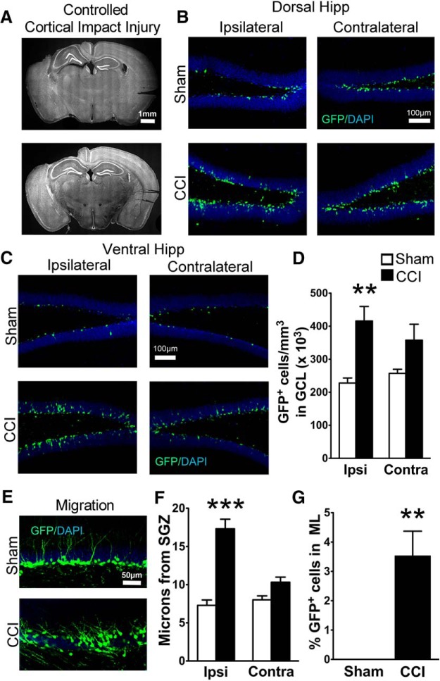
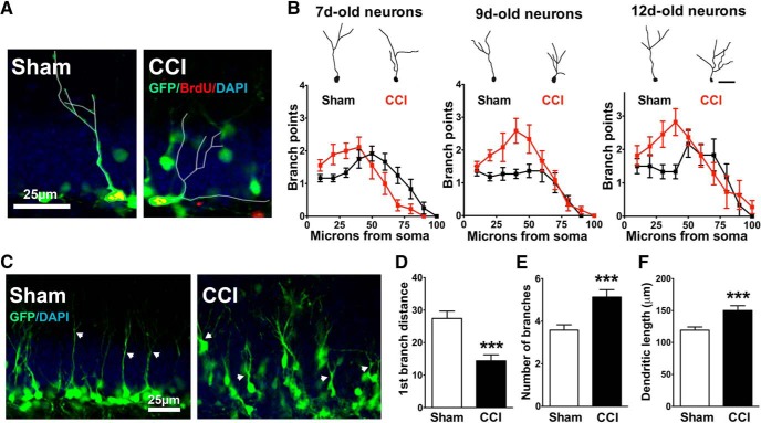
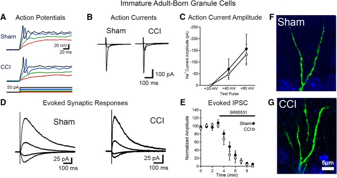
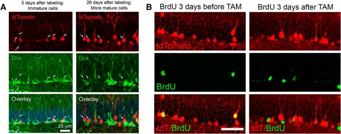
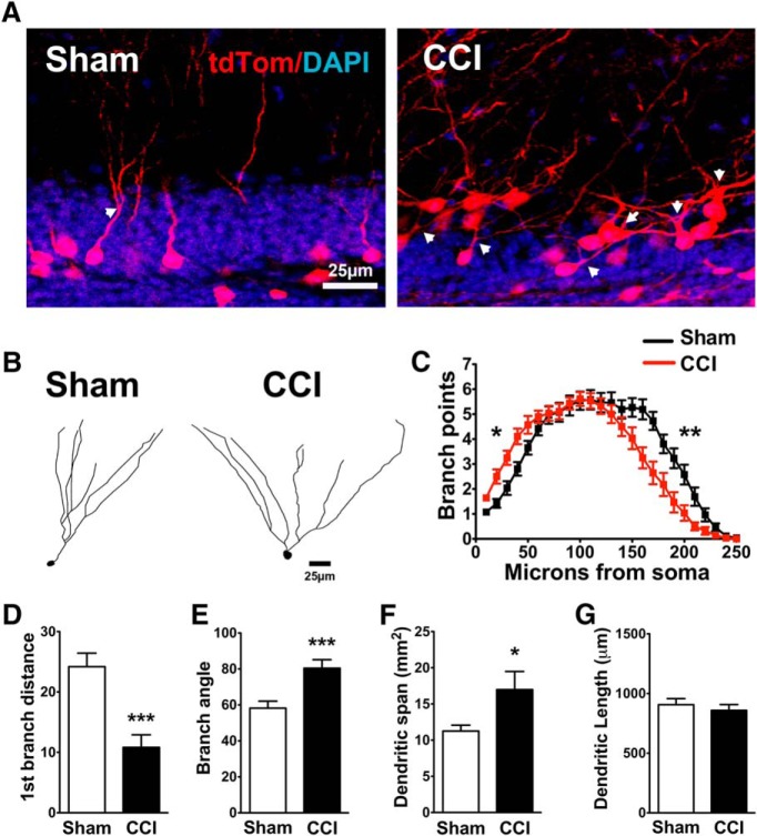
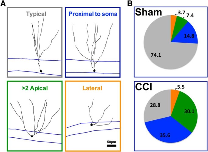
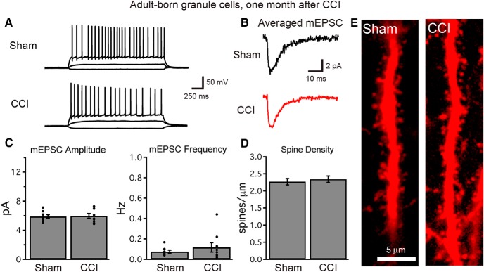
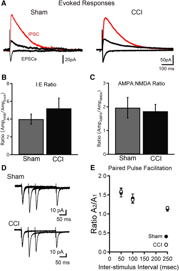
Traumatic brain injury (TBI) increases hippocampal neurogenesis, which may contribute to cognitive recovery after injury. However, it is unknown whether TBI-induced adult-born neurons mature normally and functionally integrate into the hippocampal network. We assessed the generation, morphology, and synaptic integration of new hippocampal neurons after a controlled cortical impact (CCI) injury model of TBI. To label TBI-induced newborn neurons, we used 2-month-old POMC-EGFP mice, which transiently and specifically express EGFP in immature hippocampal neurons, and doublecortin-CreER(T2) transgenic mice crossed with Rosa26-CAG-tdTomato reporter mice, to permanently pulse-label a cohort of adult-born hippocampal neurons. TBI increased the generation, outward migration, and dendritic complexity of neurons born during post-traumatic neurogenesis. Cells born after TBI had profound alterations in their dendritic structure, with increased dendritic branching proximal to the soma and widely splayed dendritic branches. These changes were apparent during early dendritic outgrowth and persisted as these cells matured. Whole-cell recordings from neurons generated during post-traumatic neurogenesis demonstrate that they are excitable and functionally integrate into the hippocampal circuit. However, despite their dramatic morphologic abnormalities, we found no differences in the rate of their electrophysiological maturation, or their overall degree of synaptic integration when compared to age-matched adult-born cells from sham mice. Our results suggest that cells born after TBI participate in information processing, and receive an apparently normal balance of excitatory and inhibitory inputs. However, TBI-induced changes in their anatomic localization and dendritic projection patterns could result in maladaptive network properties.

Keywords: adult neurogenesis; functional integration; hippocampus; maturation; synaptic integration; traumatic brain injury.


Conflict of interest statement

The authors declare no competing financial interests.

Figures

Figure 1.
Figure 1.
Transgenic POMC-EGFP mice demonstrate CCI-induced neurogenesis and increased dispersion of immature granule cells. A, Representative images of the extent of cortical damage 2 weeks following CCI. The noninjured (contralateral) sides are marked by notches in the cortical tissue placed during processing. B, C, Representative images of GFP+ cells in the dorsal (B) and ventral (C) hippocampus of sham and CCI-treated mice 2 weeks after surgery, both ipsilateral and contralateral to injury. D, CCI-treated mice had more GFP+ cells on the injured hemisphere compared to sham mice (**p = 0.00013, n = 7 and 8 mice/group). E, Representative images of GFP+ cell dispersion in the granule cell layer of the ipsilateral dentate gyrus. F, CCI-treated mice had increased cell migration away from the SGZ on the injured hemisphere compared with sham mice (***p = 0.00018, n = 6 mice/group; white and black bars represent sham and CCI-treated mice, respectively). G, A greater percentage of GFP+ cells from CCI-treated mice migrated into the molecular layer (ML) of the ipsilateral dentate gyrus (**p = 0.002811, n = 6 mice/group).
Figure 2.
Figure 2.
Granule cells born after CCI have accelerated dendritic growth. A, Confocal image of BrdU+ cells expressing GFP in POMC-EGFP mice, used to precisely date the birth of cells for morphologic analysis. In this example, BrdU was administered to POMC-EGFP mice 5 d after CCI or sham procedure and perfusion was fixed at 14 d, so that BrdU+ cells were 9 d postmitosis. Traced cells are outlined in white. B, Representative images (top) of dendritic tracings of 7-, 9-, and 12-d-old GFP+ cells in sham and CCI-treated mice, and the Sholl analyses (bottom) for group data at each time point. In each cohort, CCI increased the number of dendritic branch points of immature cells born during post-traumatic neurogenesis (p = 0.002713). Branches also occurred closer to the cell body in CCI-treated mice in each cohort, which was independent of cell age (p = 0.7615). C, Representative images of GFP+ cells and their dendritic outgrowth. White arrows point to the first dendritic branch points. D–F, GFP+ cells from CCI treated mice branched closer to their cell body (***p = 0.001216; D), had more cumulative branches (***p = 0.000517; E), and had greater total dendritic length (***p = 0.00118; F).
Figure 3.
Figure 3.
Immature granule cells born after TBI are excitable and integrated into the hippocampal circuitry. A, Current-clamp recordings from GFP-positive immature granule cells born after sham or CCI surgery demonstrate that cells born after TBI fire action potentials during depolarizing current injection. Respective voltage traces during current injections of +5 pA (red), +10 pA (green), +30 pA (blue), and +40 pA (black) for both sham and CCI-treated animals are shown. Summary data are given in the text. B, C, Single-cell, voltage-clamp recordings demonstrate that immature granule cells have similar voltage-dependent action currents. B, Averaged action currents from example cells during steps of +20, +40, and +60 mV from a holding potential of −70 mV. C, Summary data of the amplitude of the action current response in response to the various steps between groups (sham = 7 cells; CCI = 7 cells; p = 0.6554 with a +40 mV step; p = 0.7555 with a +60 mV step). D, Synaptic currents elicited in immature adult-born neurons in response to stimulation at the IML/MML border, while voltage clamping the cells to −70, −40, 0, and +40 mV. E, Summary data of response amplitudes of evoked currents (recorded while clamping cells at +40 mV), demonstrating almost complete block of the evoked synaptic response by the GABAAR antagonist SR95531 (10 μm; n = 5 cells per group). F, G, High-power imaging of dendrites from newborn neurons in sham and CCI-treated mice show no evidence of dendritic spines on immature GFP+ cells in either condition.
Figure 4.
Figure 4.
Tamoxifen administration pulse labels adult born neurons in DcxCre/tdTomato mice. A, Tamoxifen (TAM) was administered to adult DcxCre/tdTom mice, and they were fixed at two different time intervals thereafter. Early after TAM administration (left), tdTom+ cells have immature dendritic morphologies and stain for the immature marker Dcx, indicating recent mitosis. After 3 weeks (right), tdTom+ cells no longer costain for Dcx, suggesting that labeled cells mature, and that no new immature cells have been labeled. The minor overlap of red and green in the projected image at 26 d after TAM administration reflects the overlap of separate cells in the stack and not coexpression. B, DcxCre/tdTom mice received BrdU to mark cells born either before (left) or after (right) adult TAM administration, and were fixed 3 weeks later. Cells born before TAM administration are efficiently labeled by tdTom expression, but cells born after TAM did not express tdTom, indicating that neurons born just prior to tamoxifen administration are selectively pulse labeled. Scale bar, 50 μm.
Figure 5.
Figure 5.
Neurons born shortly after CCI survive and maintain their aberrant localization in maturity. A, Representative images of ∼4-week-old tdTom+ cells from the ipsilateral hippocampus of DcxCre/tdTom sham and CCI-treated mice, in which cells born after CCI were permanently pulse labeled with tdTomato. B, One month after injury, CCI-treated mice had more tdTom+ cells in the ipsilateral granule cell layer of the dentate gyrus compared with sham mice (**p = 0.008729, n = 6 mice/group). C, Representative images of tdTom+ cell dispersion in the granule cell layer of the ipsilateral dentate gyrus. D, CCI-treated mice had greater cell migration in the ipsilateral dentate gyrus compared with sham mice (*p = 0.04230, n = 6 mice/group).
Figure 6.
Figure 6.
CCI-induced changes in the dendritic morphology of newborn neurons persist as these cells mature. A, Representative images of 4-week-old tdTom+ cells from sham and CCI-treated mice. Arrows point to the first dendritic branch point of selected cells. B, Representative tracings of tdTom+ cells from sham and CCI-treated mice. C, Sholl analyses of dendritic arborization of tdTom+ cells reveal a CCI-induced persistent increase in the number of branch points observed near the soma (*p = 0.0001, 0.003, 0.004, 0.003, and 0.049 for each 10 μm increment between 10 and 50 μm from the soma34), but a reduction in more distal regions (**p = 0.038, 0.002, 0.001, 0.009, 0.003, 0.003, 0.004, and 0.038 for each 10 μm increment between 150 and 220 μm from the soma34; n = 26 cells from 7 sham mice; 28 cells from 8 CCI-treated mice). D–F, tdTom+ cells from CCI-treated mice branched closer to their cell body (***p = 0.000135; D), and had wider angles (***p = 0.000736; E) and area (*p = 0.04037; F) between the dendrites that were farthest apart. G, tdTom+ cells from sham and CCI-treated mice had similar cumulative dendritic lengths (p = 0.5056).
Figure 7.
Figure 7.
CCI increased the heterogeneity of dendritic branch morphology in cells born after injury. A, Representative morphology of tdTom+ cells categorized according to their dendritic phenotype. B, The majority of tdTom+ cells in sham mice were characterized by a single apical dendrite that branched farther away from the cell body (>10 μm from soma, typical cell), whereas the majority of tdTom+ cells in CCI-treated mice branched more proximal to their somata (<10 μm), had more than one apical dendrite, or had an apical dendrite that projected laterally from their soma (<30° from GCL). There were fewer typical tdTom+ cells in CCI-treated mice compared with sham mice (p = 0.02138, n = 5 mice/group).
Figure 8.
Figure 8.
Granule cells born after TBI become functionally integrated into the hippocampal circuit. A, At 1 month after injury, single-cell recordings demonstrate normal firing patterns and the excitability of cells born early after TBI. Voltage traces show the response of adult-born granule cells to current injections of −10, +10, and +50 pA. B, C, Whole cell, voltage-clamp recordings of mEPSCs demonstrate that cells born after TBI acquire excitatory synaptic connections (B), and that mEPSC amplitude and frequency (C) are similar to those recorded from adult-born cells of similar age in sham animals (n = 8 and 9 cells for sham and CCI, respectively; p > 0.4 each). Each graph depicts summary data (bars) and individual cell data (points). D, At 1 month after injury, cells born early following TBI possess dendritic spines within the dentate molecular layer, and dendritic spine density is similar in cells born after sham or CCI procedures (sham, n = 16 cells; CCI, n = 22 cells; p = 0.6053). E, Images of dendritic spines from cells born after CCI or sham procedure.
Figure 9.
Figure 9.
Granule cells born after TBI acquire perforant path inputs and maintain balanced functional connectivity between signaling pathways. A, Stimulation of the dentate molecular layer evokes both excitatory and inhibitory currents in cells born after sham and CCI procedures. Inhibitory currents were recorded while voltage clamping adult-born cells to 0 mV (red traces), and excitatory currents (black) were recorded at −70 and +40 mV in the presence of the GABAAR inhibitor SR95531, with the stimulation intensity and electrode position kept constant. B, The ratio of evoked inhibitory current amplitude (at 0 mV) to excitatory current amplitude (AMPAR mediated at −70 mV) was measured for single cells with the same stimulation parameters. This inhibition/excitation ratio (I:E ratio) was similar for cells born after sham or CCI (p = 0.4250; n = 9 and 13 cells). C, The ratio of AMPAR- to NMDAR-mediated currents at perforant path synapses was similar for cells born after CCI (p = 0.7751; n = 11 and 16 cells). D, E, Paired-pulse facilitation was not different at perforant path synapses for cells between groups. D, Example traces show overlaid pairs of responses with interpulse intervals of 50, 100, and 250 ms for cells born after both sham and CCI. E, Summary data show no difference in paired-pulse facilitation between groups (p = 0.8652, 0.8252, and 0.6852 for 50, 100, and 250 ms interspike intervals, respectively; sham, n = 8; CCI, n = 11 cells from 3 mice/condition).

References

    1. Aimone JB, Li Y, Lee SW, Clemenson GD, Deng W, Gage FH (2014) Regulation and function of adult neurogenesis: from genes to cognition. Physiol Rev 94:991-1026. 10.1152/physrev.00004.2014 - DOI - PMC - PubMed
    1. Blaiss CA, Yu TS, Zhang G, Chen J, Dimchev G, Parada LF, Powell CM, Kernie SG (2011) Temporally specified genetic ablation of neurogenesis impairs cognitive recovery after traumatic brain injury. J Neurosci 31:4906-4916. 10.1523/JNEUROSCI.5265-10.2011 - DOI - PMC - PubMed
    1. Cameron HA, Woolley CS, McEwen BS, Gould E (1993) Differentiation of newly born neurons and glia in the dentate gyrus of the adult rat. Neuroscience 56:337-344. - PubMed
    1. Carlson SW, Madathil SK, Sama DM, Gao X, Chen J, Saatman KE (2014) Conditional overexpression of insulin-like growth factor-1 enhances hippocampal neurogenesis and restores immature neuron dendritic processes after traumatic brain injury. J Neuropathol Exp Neurol 73:734-746. 10.1097/NEN.0000000000000092 - DOI - PMC - PubMed
    1. Chancey JH, Adlaf EW, Sapp MC, Pugh PC, Wadiche JI, Overstreet-Wadiche LS (2013) GABA depolarization is required for experience-dependent synapse unsilencing in adult-born neurons. J Neurosci 33:6614-6622. 10.1523/JNEUROSCI.0781-13.2013 - DOI - PMC - PubMed

LinkOut - more resources