

Malva sylvestris Inhibits Inflammatory Response in Oral Human Cells. An In Vitro Infection Model
- PMID: 26479870
- PMCID: PMC4610699
- DOI: 10.1371/journal.pone.0140331
Malva sylvestris Inhibits Inflammatory Response in Oral Human Cells. An In Vitro Infection Model
Abstract
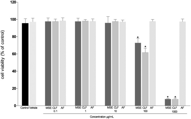
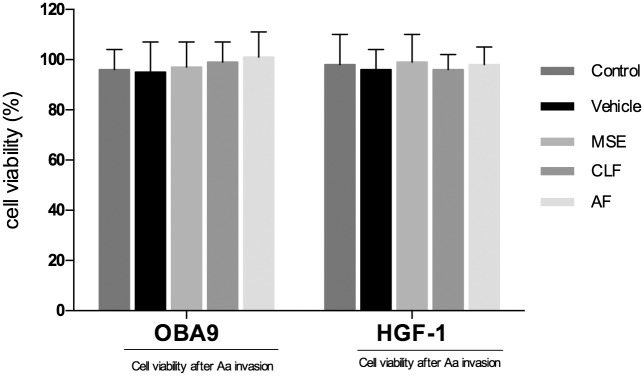
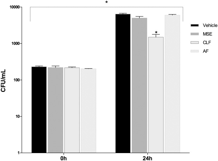
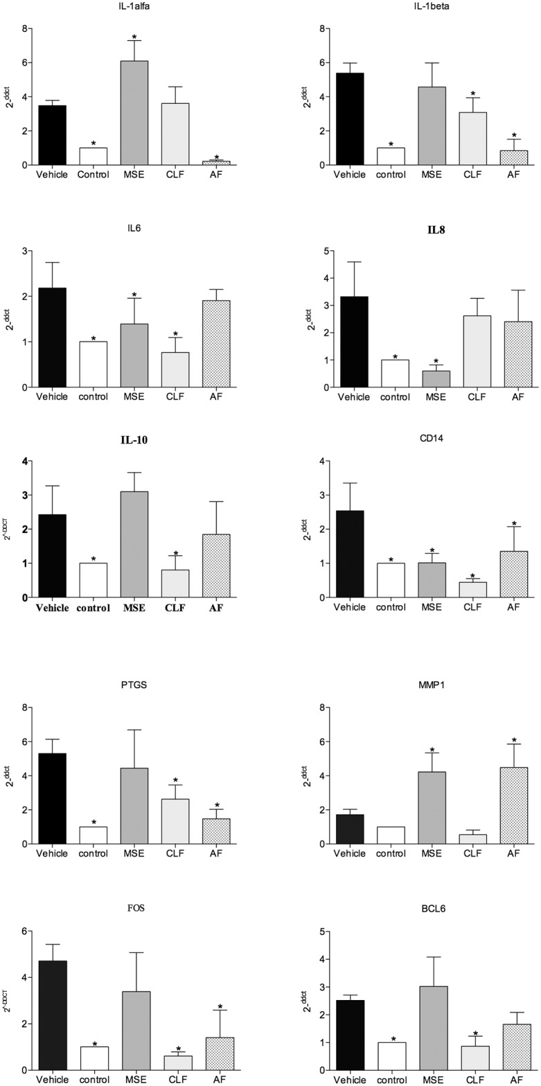
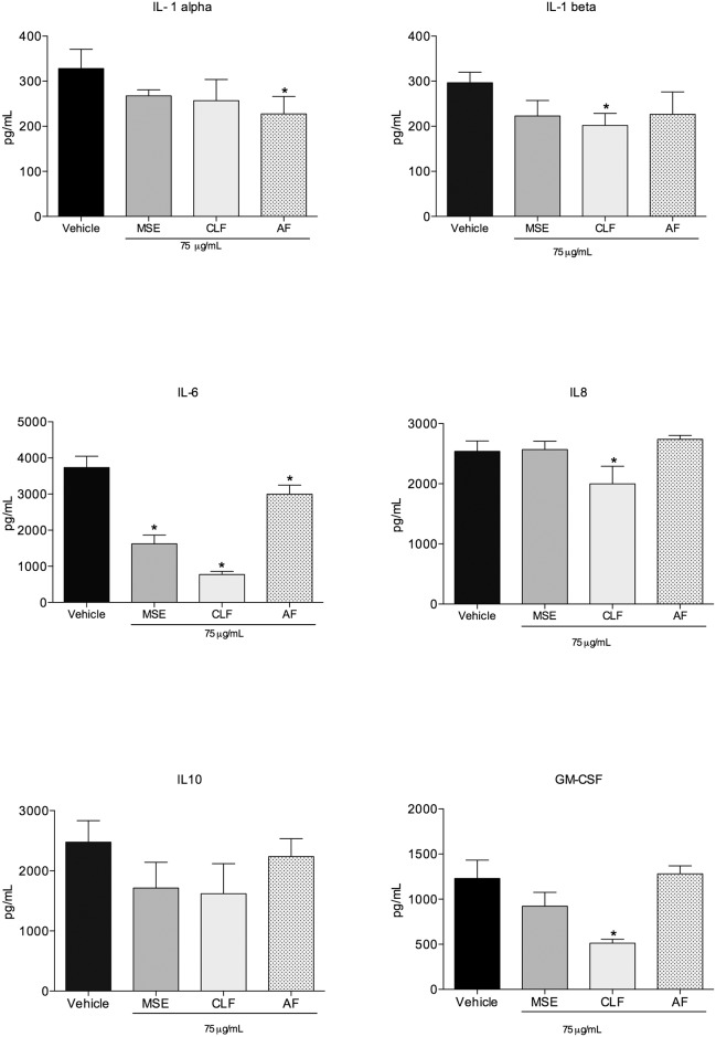
The aim of this study was to investigate the in vitro anti-inflammatory activity of Malva sylvestris extract (MSE) and fractions in a co-culture model of cells infected by Aggregatibacter actinomycetemcomitans. In addition, we evaluated the phytochemical content in the extract and fractions of M. sylvestris and demonstrated that polyphenols were the most frequent group in all samples studied. An in vitro dual-chamber model to mimic the periodontal structure was developed using a monolayer of epithelial keratinocytes (OBA-9) and a subepithelial layer of fibroblasts (HGF-1). The invasive periodontopathogen A. actinomycetemcomitans (D7S-1) was applied to migrate through the cell layers and induce the synthesis of immune factors and cytokines in the host cells. In an attempt to analyze the antimicrobial properties of MSE and fractions, a susceptibility test was carried out. The extract (MIC 175 μg/mL, MBC 500μg/mL) and chloroform fraction (MIC 150 μg/mL, MBC 250 μg/mL) were found to have inhibitory activity. The extract and all fractions were assessed using a cytotoxicity test and results showed that concentrations under 100 μg/mL did not significantly reduce cell viability compared to the control group (p > 0.05, viability > 90%). In order to analyze the inflammatory response, transcriptional factors and cytokines were quantified in the supernatant released from the cells. The chloroform fraction was the most effective in reducing the bacterial colonization (p< 0.05) and controlling inflammatory mediators, and promoted the down-regulation of genes including IL-1beta, IL-6, IL-10, CD14, PTGS, MMP-1 and FOS as well as the reduction of the IL-1beta, IL-6, IL-8 and GM-CSF protein levels (p< 0.05). Malva sylvestris and its chloroform fraction minimized the A. actinomycetemcomitans infection and inflammation processes in oral human cells by a putative pathway that involves important cytokines and receptors. Therefore, this natural product may be considered as a successful dual anti-inflammatory-antimicrobial candidate.
Conflict of interest statement
Figures
Similar articles
-
Oral microbe-host interactions: influence of β-glucans on gene expression of inflammatory cytokines and metabolome profile.BMC Microbiol. 2017 Mar 7;17(1):53. doi: 10.1186/s12866-017-0946-1. BMC Microbiol. 2017. PMID: 28270109 Free PMC article.
-
Anti-Inflammatory, Anti-Osteoclastogenic and Antioxidant Effects of Malva sylvestris Extract and Fractions: In Vitro and In Vivo Studies.PLoS One. 2016 Sep 19;11(9):e0162728. doi: 10.1371/journal.pone.0162728. eCollection 2016. PLoS One. 2016. PMID: 27643502 Free PMC article.
-
Malva sylvestris derivatives as inhibitors of HIV-1 BaL infection.Nat Prod Res. 2021 Mar;35(6):1064-1069. doi: 10.1080/14786419.2019.1619720. Epub 2019 Aug 20. Nat Prod Res. 2021. PMID: 31429300 Free PMC article.
-
The phytochemical profiling, pharmacological activities, and safety of malva sylvestris: a review.Naunyn Schmiedebergs Arch Pharmacol. 2023 Mar;396(3):421-440. doi: 10.1007/s00210-022-02329-w. Epub 2022 Nov 22. Naunyn Schmiedebergs Arch Pharmacol. 2023. PMID: 36418467 Free PMC article. Review.
-
Phytochemistry and Pharmacological Activity of Malva sylvestris L: A Detailed Insight.Comb Chem High Throughput Screen. 2024;27(16):2309-2322. doi: 10.2174/0113862073269336231009110313. Comb Chem High Throughput Screen. 2024. PMID: 37855358 Review.
Cited by
-
In vitro Antibacterial Effect of Hydroalcoholic Extract of Lawsonia inermis, Malva sylvestris, and Boswellia serrata on Aggregatibacter actinomycetemcomitans.Adv Biomed Res. 2019 Mar 20;8:22. doi: 10.4103/abr.abr_205_18. eCollection 2019. Adv Biomed Res. 2019. PMID: 31016180 Free PMC article.
-
Protective Effects of the Aqueous Extract of Malva sylvestris Plant against the Lethality and Histopathological Damage in Experimental Envenoming of Hemiscorpius lepturus Venom in Balb/c Mice.Curr Pharm Des. 2024;30(21):1699-1704. doi: 10.2174/0113816128296935240502070245. Curr Pharm Des. 2024. PMID: 38757320
-
Anti-Inflammatory Activity of Natural Products.Molecules. 2016 Oct 1;21(10):1321. doi: 10.3390/molecules21101321. Molecules. 2016. PMID: 27706084 Free PMC article. Review.
-
The Phytochemical Profile and Biological Activity of Malva neglecta Wallr. in Surgically Induced Endometriosis Model in Rats.Molecules. 2022 Nov 15;27(22):7869. doi: 10.3390/molecules27227869. Molecules. 2022. PMID: 36431970 Free PMC article.
-
Cell culture models of oral mucosal barriers: A review with a focus on applications, culture conditions and barrier properties.Tissue Barriers. 2018;6(3):1479568. doi: 10.1080/21688370.2018.1479568. Epub 2018 Sep 25. Tissue Barriers. 2018. PMID: 30252599 Free PMC article. Review.
References
Publication types
MeSH terms
Substances
Grants and funding
LinkOut - more resources
Full Text Sources
Other Literature Sources
Medical
Molecular Biology Databases
Research Materials
