. 2015 Nov;21(11):1272-9.
doi: 10.1038/nm.3962. Epub 2015 Oct 19.

Hair follicle-derived IL-7 and IL-15 mediate skin-resident memory T cell homeostasis and lymphoma

Affiliations

Hair follicle-derived IL-7 and IL-15 mediate skin-resident memory T cell homeostasis and lymphoma

Takeya Adachi et al. Nat Med. 2015 Nov.

Abstract

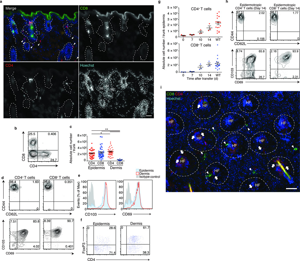
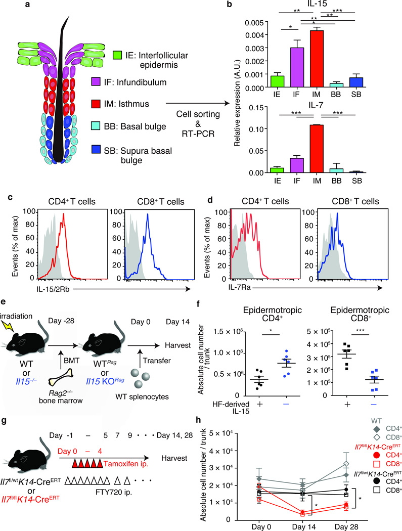
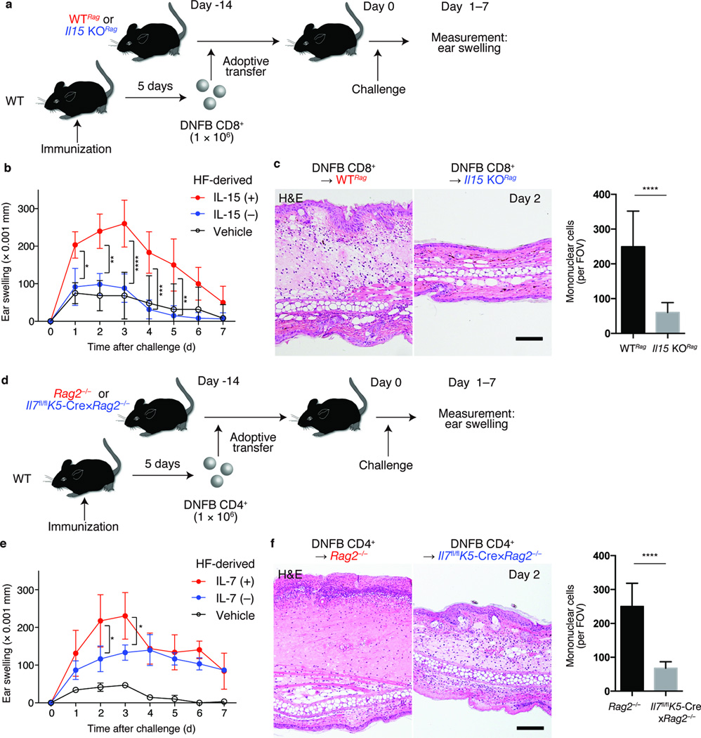
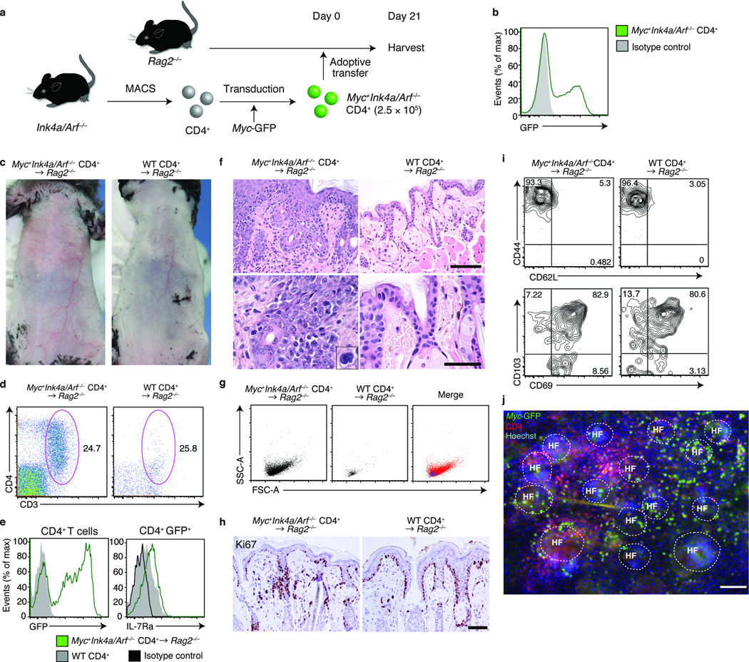
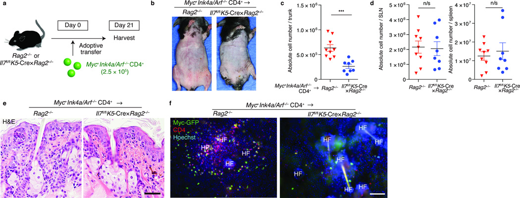
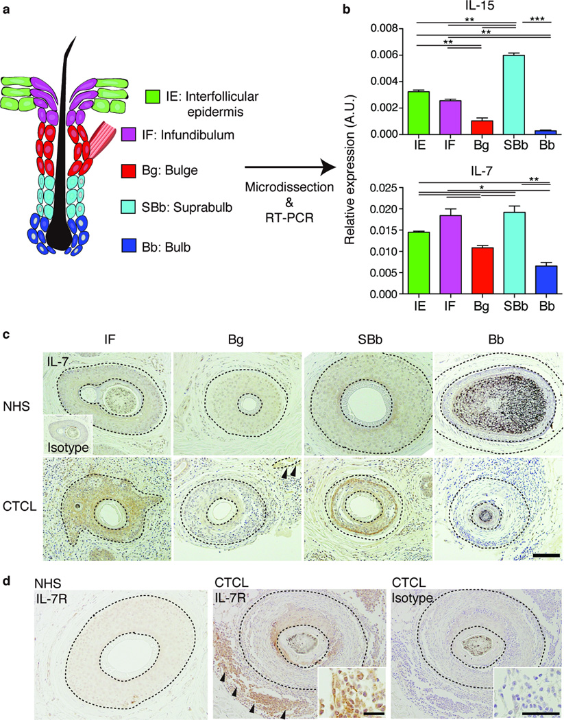
The skin harbors a variety of resident leukocyte subsets that must be tightly regulated to maintain immune homeostasis. Hair follicles are unique structures in the skin that contribute to skin dendritic cell homeostasis through chemokine production. We demonstrate that CD4(+) and CD8(+) skin-resident memory T cells (TRM cells), which are responsible for long-term skin immunity, reside predominantly within the hair follicle epithelium of the unperturbed epidermis. TRM cell tropism for the epidermis and follicles is herein termed epidermotropism. Hair follicle expression of IL-15 was required for CD8(+) TRM cells, and IL-7 for CD8(+) and CD4(+) TRM cells, to exert epidermotropism. A lack of either cytokine in the skin led to impaired hapten-induced contact hypersensitivity responses. In a model of cutaneous T cell lymphoma, epidermotropic CD4(+) TRM lymphoma cell localization depended on the presence of hair follicle-derived IL-7. These findings implicate hair follicle-derived cytokines as regulators of malignant and non-malignant TRM cell tissue residence, and they suggest that the cytokines may be targeted therapeutically in inflammatory skin diseases and lymphoma.


Figures

Figure 1
Figure 1. Epidermotropic CD4+ and CD8+ TRM associate with hair follicles
(a) Skin sections from WT mice, stained as indicated. Dotted lines delineate hair follicles. Asterisk and arrowheads depict CD4+ T cells in epidermis and dermis, respectively. Scale bar, 50 µm. (b) Flow cytometry analysis of CD45+CD3+MHCllTCRγδ epidermal cells from WT mice. Numbers in quadrants represent percentage among the gated subset (c) T cell numbers in the epidermis and dermis. Each symbol represents individual mice; horizontal lines depict the mean and error bars indicate s.e.m. *P < 0.05, **P < 0.01 (unpaired two-tailed Student’s t-test). (d) CD44 and CD62L, and CD103 and CD69 expression on T cells in the epidermis. (e) Expression of CD103 and CD69 expression on CD4+ T cells in the epidermis (red) and dermis (blue) with isotype controls (grey). (f) Frequency of CD4+FoxP3+ Treg cells in the epidermis and dermis. (g) Number of donor T cells in Rag2−/− recipient epidermis monitored via flow cytometry at indicated dates after adoptive transfer of WT splenocytes. (h) Epidermotropic T cells from (g) were analyzed via flow cytometry as in (d), 14 d after transfer. Shown are CD45+CD3+MHCllTCRγδ CD4+ (left panels) or CD8+ (right panels) populations. (i) Epidermal sheets from mice as in (g), day 14 after transfer, were stained as indicated. Dashed lines delineate hair follicles. Scale bar, 100 µm. Data are from one experiment representative of three independent experiments with three mice per group (a,b,d–k) or from ten independent experiments with a total of 42 mice (c).
Figure 2
Figure 2. Epidermotropic TRM require hair follicle-derived cytokines
(a) Schematic representation of mouse hair follicle keratinocytes in telogen. Keratinocytes were sorted into five subsets based on cell-surface marker expressions. (b) Real-time PCR analysis for il15 and il7 expressions by hair follicle keratinocyte subsets, presented in arbitrary units (A.U.), relative to Actb expression. (c) IL-15/2 receptor β (IL-15/2Rβ) and (d) IL-7 receptor α (IL-7Rα) expression by TRM, assessed via flow cytometry, gated on the CD45+CD3+MHCllTCRγδ population (n = 3). (e) Experimental scheme for the analysis of TRM dependence on hair follicle-derived IL-15. WT or il15−/− mice were reconstituted with Rag2−/− bone marrow (WTRag or Il15 KORag), and were transferred with WT splenocytes. (f) Epidermotropic TRM numbers in recipient WTRag or il15 KORag mice (n = 6). Symbols represent individual mice. (g) Experimental scheme for analyzing TRM dependence on hair follicle-derived IL-7. Indicated mice were treated with tamoxifen i.p. on days 0 to 4. FTY720 was injected i.p. from day -1 for 7 consecutive days and every other day thereafter, until tissue harvest. (h) Epidermotropic TRM numbers after tamoxifen-induced ablation of hair follicle IL-7 (n = 3). Data are representative of three (b–d) or two (f, h) independent experiments. *P < 0.05, **P < 0.01, ***P < 0.001 (unpaired two-tailed Student’s t-test). Horizontal lines depict the mean, and error bars indicate s.e.m. in all graphs.
Figure 3
Figure 3. Impaired contact hypersensitivity responses in the absence of hair follicle-derived cytokines
(a) Experimental scheme for the analysis of CD8+ TRM-mediated CHS in the absence of peripheral tissue (hair follicle)-derived IL-15. CD8+ T cells from DNFB (DNFB CD8+) or vehicle-immunized (Vehicle CD8+) WT mice were transferred into indicated lymphopenic mice. 14 days later, recipients were challenged with DNFB on their ears, and ear-swelling responses were measured. (b) Ear thickness (Δmm) of DNFB CD8+-transferred WTRag (red), Il15 KORag (blue), and vehicle CD8+-transferred WTRag (black) (n = 3). (c) Hematoxylin and eosin staining of ear sections from indicated mice, day 2 after challenge and quantification of skin-infiltrating mononuclear cells. (d) Experimental scheme for analyzing CD4+ TRM-mediated CHS in the absence of hair follicle-derived IL-7. DNFB CD4+ or vehicle CD4+ were adoptively transferred into indicated lymphopenic mice. (e) Ear thickness (Δ mm) of DNFB CD4+-transferred Rag2−/− (red), Il7fl/flK5-Cre×Rag2−/− (blue), or vehicle CD4+-transferred Rag2−/− (black) (n = 3). (f) Hematoxylin and eosin staining of ear sections from indicated mice, day 2 after challenge, and quantification of skin-infiltrating mononuclear cells. Data are from one experiment, representative of two independent experiments with three mice per group in each (b, c, e, f). Cell counts represent pools of three fields of view per section (100×) from three mice per group (c, f). Scale bar, 100 µm (c, f). *P < 0.05, **P < 0.01, ***P < 0.001, ****P < 0.0001 (two way ANOVA).
Figure 4
Figure 4. Generation of a model of T cell lymphoma with skin involvement
(a) Experimental scheme for a CD4+ T cell lymphoma model with skin involvement. CD4+ T cells isolated from Ink4a/Arf−/− mice were retrovirally transduced with Myc-GFP (Myc+Ink4a/Arf−/− CD4+) and were adoptively transferred into Rag2−/− mice. (b) Flow cytometry analysis of Myc-GFP expression on Myc+Ink4a/Arf−/− CD4+ T cells before transfer. (c) Skin phenotype of Myc+Ink4a/Arf−/− CD4+-transferred (left panel) or WT CD4+-transferred Rag2−/− mice mice (right panel) (n = 3). (d) Flow cytometry analysis of CD3+CD4+ epidermotropic cells in indicated mice, for (e) expression of GFP and IL-7Rα, (g) forward and side scatter profiles, (i) CD44 versus CD62L and CD103 versus CD69 expression. Gated on CD45+MHCllTCRγδ (d,g), CD45+MHCllTCRγδ CD3+CD4+ (left panel in e, i and j) or CD45+MHCllTCRγδ CD3+CD4+GFP+ populations (right panel in e) (n = 3). (f) Hematoxylin and eosin staining of lip sections from indicated mice and (h) immunohistochemical staining for Ki-67 (n = 3). (j) Immunofluorescence microscopy of epidermal sheets, visualized for Myc-GFP, CD4 and Hoechst (n = 3). Scale bar, 100 µm. Data are from one experiment representative of three independent experiments with three mice per group in each.
Figure 5
Figure 5. CD4+ TRM lymphoma cells depend on hair follicle-derived IL-7 to exhibit epidermotropism
(a) Experimental scheme. Myc+Ink4a/Arf−/− CD4+ T cells were adoptively transferred into Rag2−/− or Il7fl/flK5-Cre×Rag2−/− mice. Recipient mice were harvested approximately three weeks after transfer. (b) Skin phenotype of recipient mice of indicated genotype (n = 3). Quantification of (c) epidermotropic TRM or (d) T cells in skin-draining LNs (SLN) and spleen, assessed at day 21 after adoptive transfer (n = 8–9). ***P < 0.001 (unpaired two-tailed Student’s t-test). (e) Histology of skin sections from indicated mice, stained for hematoxylin and eosin, and (f) immunofluorescence microscopy of ear epidermal sheets of indicated mice, visualized for Myc-GFP, CD4 and Hoechst, assessed day 21 after adoptive transfer (n = 3). Scale bar, 100 µm. Data are from one experiment representative of three independent experiments with two to three mice per group in each (b,e,f) or an accumulation of three independent experiments (c,d).
Figure 6
Figure 6. IL-7 and IL-15 expression in human hair follicles from normal scalp and cutaneous T cell lymphoma
(a) Schematic representation of human hair follicle keratinocytes. (b) Real-time PCR analysis for IL15 and IL7 expression at distinct sites in the hair follicle. *P < 0.05, **P < 0.01, ***P < 0.001 (unpaired two-tailed Student’s t-test). (c) Immunohistochemistry of scalp sections from normal humans (NHS) (n = 3 samples from three subjects) and from subjects with CTCL (n = 6 samples from 3 subjects), stained with an anti-IL-7 antibody or with an isotype control. Scale bar, 100 µm. Arrowheads indicate arrector pili muscle (reflecting the bulge region). (d) Immunohistochemistry of scalp sections from patients with CTCL, stained with an anti-IL-7R antibody or with an isotype control (n = 6 samples from three subjects). Scale bar, 100µm in low power field, 50 µm in high power field. Dashed lines delineate hair follicles. Arrowheads indicate IL-7R-positive lymphoma cells. Data are from one experiment representative of three (b,c) or two (d) independent experiments.

Similar articles

Cited by

References

    1. Cotsarelis G, Sun TT, Lavker RM. Label-retaining cells reside in the bulge area of pilosebaceous unit: implications for follicular stem cells, hair cycle, and skin carcinogenesis. Cell. 1990;61:1329–1337. - PubMed
    1. Nishimura EK, et al. Dominant role of the niche in melanocyte stem-cell fate determination. Nature. 2002;416:854–860. - PubMed
    1. Christoph T, et al. The human hair follicle immune system: cellular composition and immune privilege. Br J Dermatol. 2000;142:862–873. - PubMed
    1. Nagao K, et al. Stress-induced production of chemokines by hair follicles regulates the trafficking of dendritic cells in skin. Nat Immunol. 2012;13:744–752. - PMC - PubMed
    1. Clark RA, et al. The vast majority of CLA+ T cells are resident in normal skin. J Immunol. 2006;176:4431–4439. - PubMed

Publication types

MeSH terms