. 2015 Oct 20:34:125.
doi: 10.1186/s13046-015-0234-6.

The kinase inhibitor D11 induces caspase-mediated cell death in cancer cells resistant to chemotherapeutic treatment

Affiliations

The kinase inhibitor D11 induces caspase-mediated cell death in cancer cells resistant to chemotherapeutic treatment

Barbara Guerra et al. J Exp Clin Cancer Res. .

Abstract

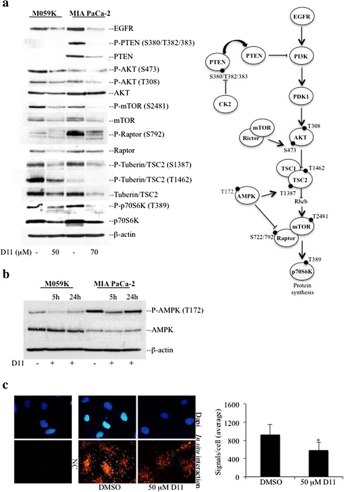
Background: Multi-drug resistance and predisposition to metastasize are major clinical problems in cancer treatment. Malignant primary brain tumor and pancreatic cancer are two well-known examples of malignant tumors resistant to conventional therapies where aberrant EGFR-mediated and NF-κB signal transduction pathways are likely to play an important role. We have recently identified 1,3-Dichloro-6-[(E)-((4-methoxyphenyl)imino)methyl] diben-zo(b,d) furan-2,7-diol (D11) as a potent and selective inhibitor of CK2 a serine/threonine protein kinase that modulates the aforementioned signaling cascades.

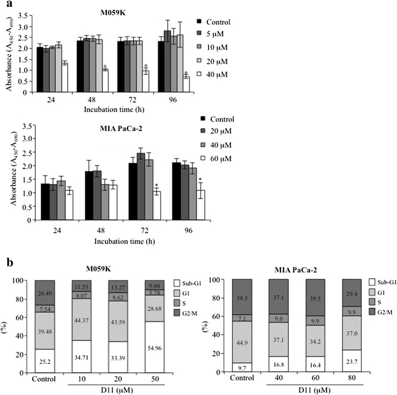
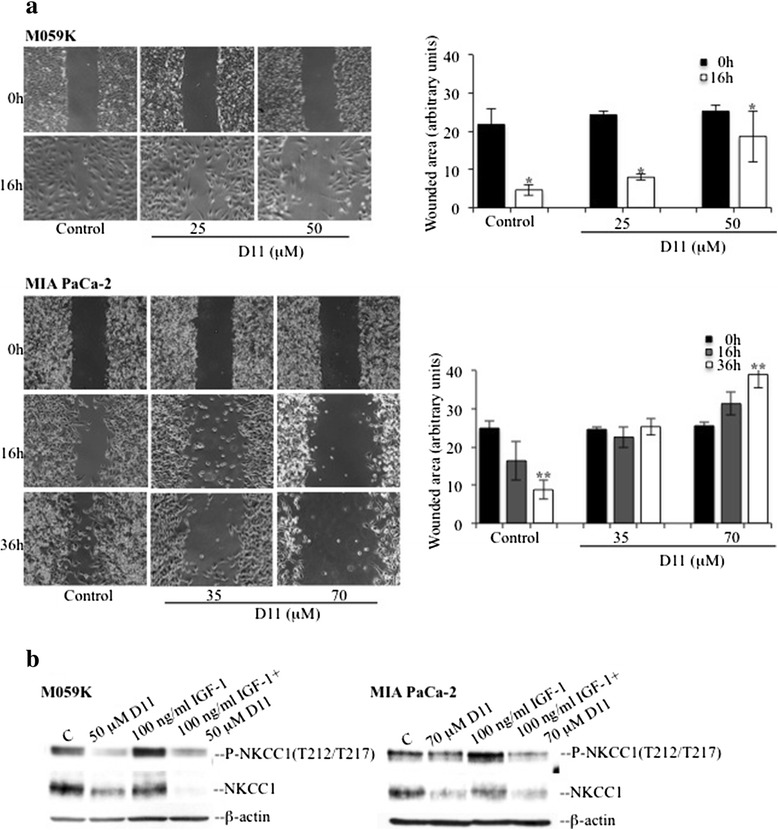
Methods: Human cancer cell lines (glioblastoma and pancreatic adenocarcinoma) resistant to conventional chemotherapeutic agents were incubated with increasing concentrations of D11 for variable amounts of time. Cell viability, cell death and effects on major signal transduction pathways deregulated in cancer cells were analyzed by ELISA, FACS and Western blot-based assays, respectively. Moreover, effects on cell migration and in cell protein-protein association were investigated by wound-healing and in situ proximity ligation assays, respectively.

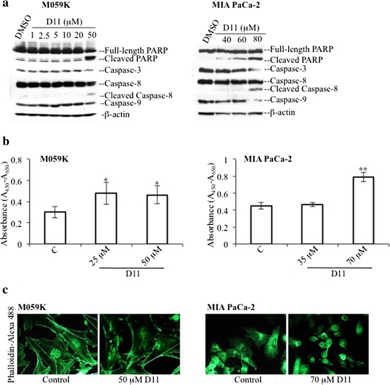
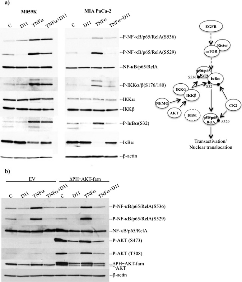
Results: We show here, that D11 treatment leads to i) significant caspase-mediated apoptotic cell death, ii) down-regulation of EGFR expression and iii) inhibition of NF-κB transcriptional activity. Furthermore, cell exposure to D11 results in impaired cell migration and correlates with reduced expression of the ion co-transporter and cell volume regulator Na(+)-K(+)-2Cl(-) (NKCC1).

Conclusions: Data reported here underline the therapeutic potential of D11 with respect to certain types of cancer that carry aberrant intracellular signaling cascades and/or exhibit sustained cell migration and suggest a new therapeutic strategy against chemotherapy resistance.


Figures

Fig. 1
Fig. 1
Anti-proliferative effects of D11 in human cancer cell lines. a M059K and MIA PaCa-2 cell lines were treated with increasing concentrations of D11 for a variable amount of time, respectively. Control experiment refers to cells treated with vehicle (0.1 % DMSO). The proportion of viable cells was determined by WST-1 assay and expressed in arbitrary units as a difference in absorbance measured at 450 nm and 690 nm (reference) wavelengths, respectively (mean +/− standard deviation, N = 6). Asterisks denote statistical significant differences between control and D11-treated cells for the corresponding time points, *, P <0.0001. b Flow cytometry analysis of cells treated with 0.1 % DMSO (Control) and increasing concentrations of D11 for 24 h (M059K) and 48 h (MIA PaCa-2), respectively. The amount of cells in the various phases of the cell cycle is indicated in percentage. Experiments were repeated three times obtaining similar results. Data from one representative experiment are shown
Fig. 2
Fig. 2
Exposure of cells to D11 leads to induction of apoptotic cell death. a Cells were treated with DMSO and increasing concentrations of D11 for 24 h (M059K) and 48 h (MIA PaCa-2), respectively. Whole cell lysate was subjected to Western blot analysis for apoptosis-associated proteins. b Whole cell lysate from cells treated for 24 h with the indicated concentrations were subjected to ROCK kinase assay in the presence of myosin phosphatase target subunit 1 (MYPT1). Data from one representative experiment (mean +/− standard deviation, N = 6, *, P <0.05; **, P <0.001) are shown. c Immunofluorescence analysis of control cells (0.1 % DMSO) or cells treated with the indicated concentrations of D11 for 24 h. Actin filaments were visualized by staining cells with phalloidin-Alexa 488. Representative images were acquired using fluorescence microscopy at 400x magnification
Fig. 3
Fig. 3
Analysis of the PI3K pathway in cells treated with D11. a Whole cell lysates from the indicated cell lines were subjected to Western blot analysis. Expression and/or phosphorylation of the indicated proteins were analyzed after 5 h incubation with D11. Control experiments (−) refer to cells incubated with vehicle (0.1 % DMSO). b Cells were treated with vehicle (−) or 50 μM (M059K) and 70 μM (MIA PaCa-2) D11, respectively, for the indicated times. Whole cell lysates were subjected to Western blot analysis of phosphorylated AMPK and AMPK protein. β-actin detection was used as a control for equal loading. c Association between HSP90 and CDC37 was revealed in M059K by in situ proximity ligation assay (PLA). NC refers to untreated cells subjected to PLA where one of the primary antibodies was omitted. Determination of the number of signals per cell as distinct red fluorescence spots indicative of HSP90-CDC37 proximity, was performed by computer-assisted image analysis as described in the Materials and methods. Nuclei were visualized by DAPI staining (blue fluorescence emission). Experiments were repeated at least three times obtaining similar results. The figure includes a schematic representation of the EGFR/PI3K signaling network in mammalian cells (*, P <0.05)
Fig. 4
Fig. 4
Cell incubation with D11 impairs activation of the NF-κB signaling pathway induced by TNFα. a Whole cell lysates from cells treated with 50 μM (M059K) and 70 μM D11 (MIA PaCa-2), respectively, for 5 h and stimulated with 20 ng/ml TNFα for 10 min prior harvesting, were analyzed by Western blot. The expression and phosphorylation levels of members of the NF-κB signaling pathway are shown. b MIA PaCa-2 cells transiently transfected with either empty vector (EV) or a construct containing farnesylated AKT devoid of the PH domain (ΔPH-AKT-farn) were treated as described in (a). Expression and activation of NF-κB as well as AKT were verified by Western blot analysis of whole cell lysates. β-actin detection was used as loading control. The figure shows a schematic model of the canonical NF-κB signaling pathway
Fig. 5
Fig. 5
D11-mediated effects on the transcriptional activity of NF-κB. a Cells were treated as described in Fig. 4 and Additional file 4: Figure S4. Subcellular localization of NF-κB was detected by immunofluorescence staining of cells with rabbit monoclonal anti-NF-κB antibody (green fluorescence, Alexa Fluor 488). Nuclei were visualized by DAPI staining. NC, negative control refers to cells stained with biotinylated secondary antibody and Alexa Fluor 488 streptavidin, only. Original magnification: 400x. b NF-κB transcriptional activity was determined as described in Materials and methods. The indicated cell lines were treated as described in Fig. 4. Asterisks denote statistical significant differences in TNFα-induced NF-κB activity in the absence or presence of D11 (N = 6, *, P <0.05). Experiments were repeated at least three times obtaining similar results
Fig. 6
Fig. 6
D11 treatment results in impaired cell migration and decreased expression of NKCC1. a M059K and MIA PaCa-2 cells were treated with vehicle or D11 as indicated in the figure and subjected to wound-healing assay. D11 was added to the cells 4 h before the wounds were made. Unwounded areas were measured as described in the Materials and methods (N = 6, *, P <0.05, **, P <0.0001). Original magnification: 50x. b Cells were treated with D11 essentially as described in Fig. 4. Stimulation with IGF-1 was performed for 15 min. β-actin detection was carried out as loading control. Experiments were repeated three times obtaining similar results

References

    1. Cancer Genome Atlas Research Network. Kandoth C, Schultz N, Cherniack AD, Akbani R, Liu Y, Shen H, Robertson AG, Pashtan I, Shen R, Benz CC, Yau C, Laird PW, Ding L, Zhang W, Mills GB, Kucherlapati R, Mardis ER, Levine DA. Integrated genomic characterization of endometrial carcinoma. Nature. 2013;497:67–73. doi: 10.1038/nature12113. - DOI - PMC - PubMed
    1. Garraway LA. Genomics-driven oncology: framework for an emerging paradigm. J Clin Oncol. 2013;31:1806–14. doi: 10.1200/JCO.2012.46.8934. - DOI - PubMed
    1. Herbst RS. Review of epidermal growth factor receptor biology. Int J Radiat Oncol Biol Phys. 2004;59:21–6. doi: 10.1016/j.ijrobp.2003.11.041. - DOI - PubMed
    1. Raychaudhuri B, Han Y, Lu T, Vogelbaum MA. Aberrant constitutive activation of nuclear factor kappaB in glioblastoma multiforme drives invasive phenotype. J Neurooncol. 2007;85:39–47. doi: 10.1007/s11060-007-9390-7. - DOI - PubMed
    1. Akimoto T, Hunter NR, Buchmiller L, Mason K, Ang KK, Milas L. Inverse relationship between epidermal growth factor receptor expression and radiocurability of murine carcinomas. Clin Cancer Res. 1999;5:2884–90. - PubMed

Publication types

MeSH terms