. 2015 Oct 19:6:201.
doi: 10.1186/s13287-015-0194-y.

Differential ability of MSCs isolated from placenta and cord as feeders for supporting ex vivo expansion of umbilical cord blood derived CD34(+) cells

Affiliations

Differential ability of MSCs isolated from placenta and cord as feeders for supporting ex vivo expansion of umbilical cord blood derived CD34(+) cells

Darshana Kadekar et al. Stem Cell Res Ther. .

Abstract

Introduction: Ex vivo expansion of umbilical cord blood (UCB) is attempted to increase cell numbers to overcome the limitation of cell dose. Presently, suspension cultures or feeder mediated co-cultures are performed for expansion of hematopoietic stem cells (HSCs). Mesenchymal stem cells (MSCs) have proved to be efficient feeders for the maintenance of HSCs. Here, we have established MSCs-HSCs co-culture system with MSCs isolated from less invasive and ethically acceptable sources like umbilical cord tissue (C-MSCs) and placenta (P-MSCs). MSCs derived from these tissues are often compared with bone marrow derived MSCs (BM-MSCs) which are considered as a gold standard. However, so far none of the studies have directly compared C-MSCs with P-MSCs as feeders for ex vivo expansion of HSCs. Thus, we for the first time performed a systematic comparison of hematopoietic supportive capability of C and P-MSCs using paired samples.

Methods: UCB-derived CD34(+) cells were isolated and co-cultured on irradiated C and P-MSCs for 10 days. C-MSCs and P-MSCs were isolated from the same donor. The cultures comprised of serum-free medium supplemented with 25 ng/ml each of SCF, TPO, Flt-3 L and IL-6. After 10 days cells were collected and analyzed for phenotype and functionality.

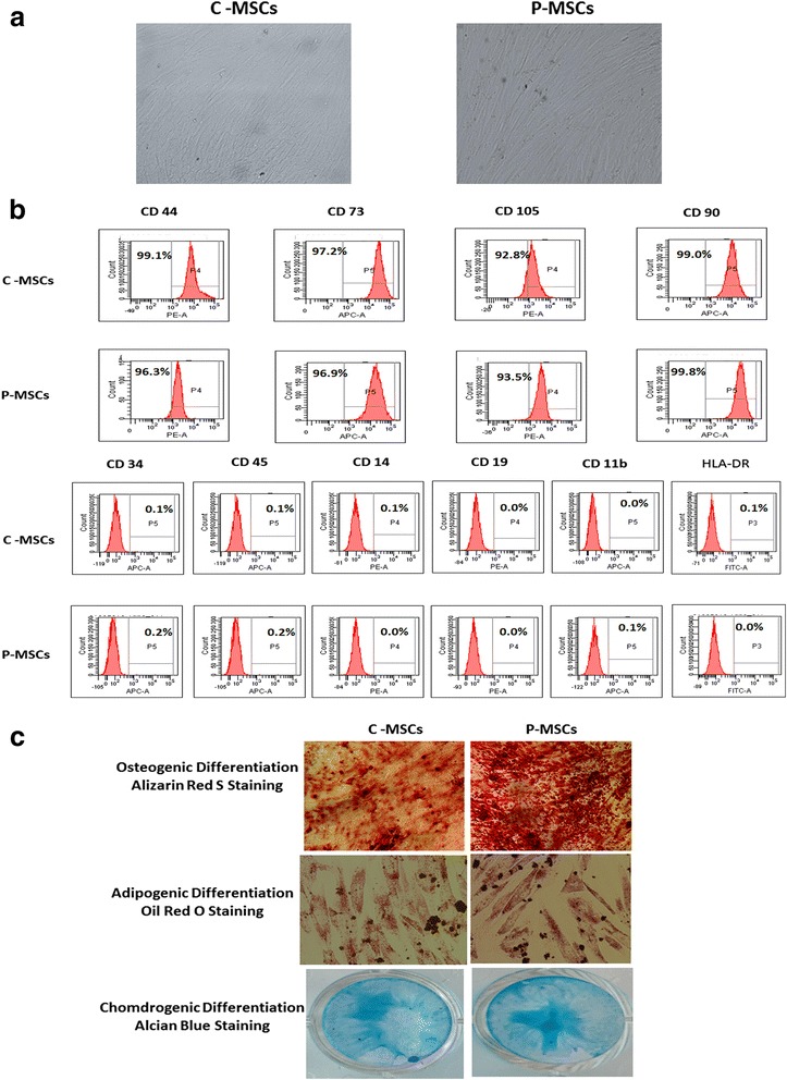
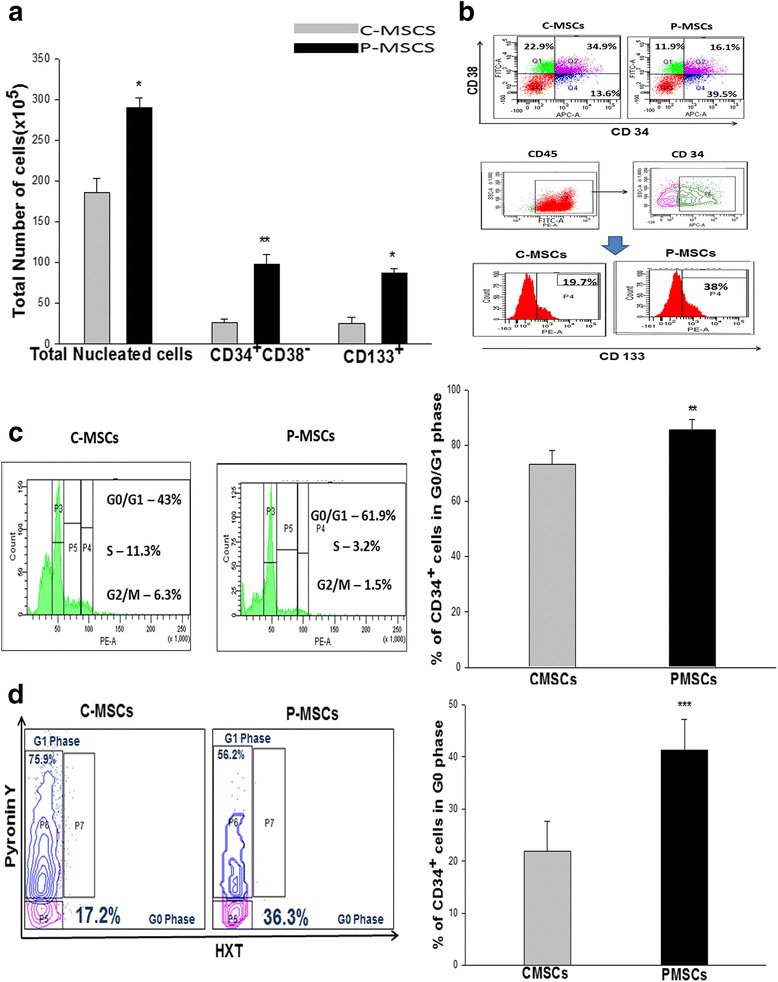
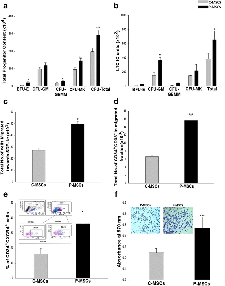
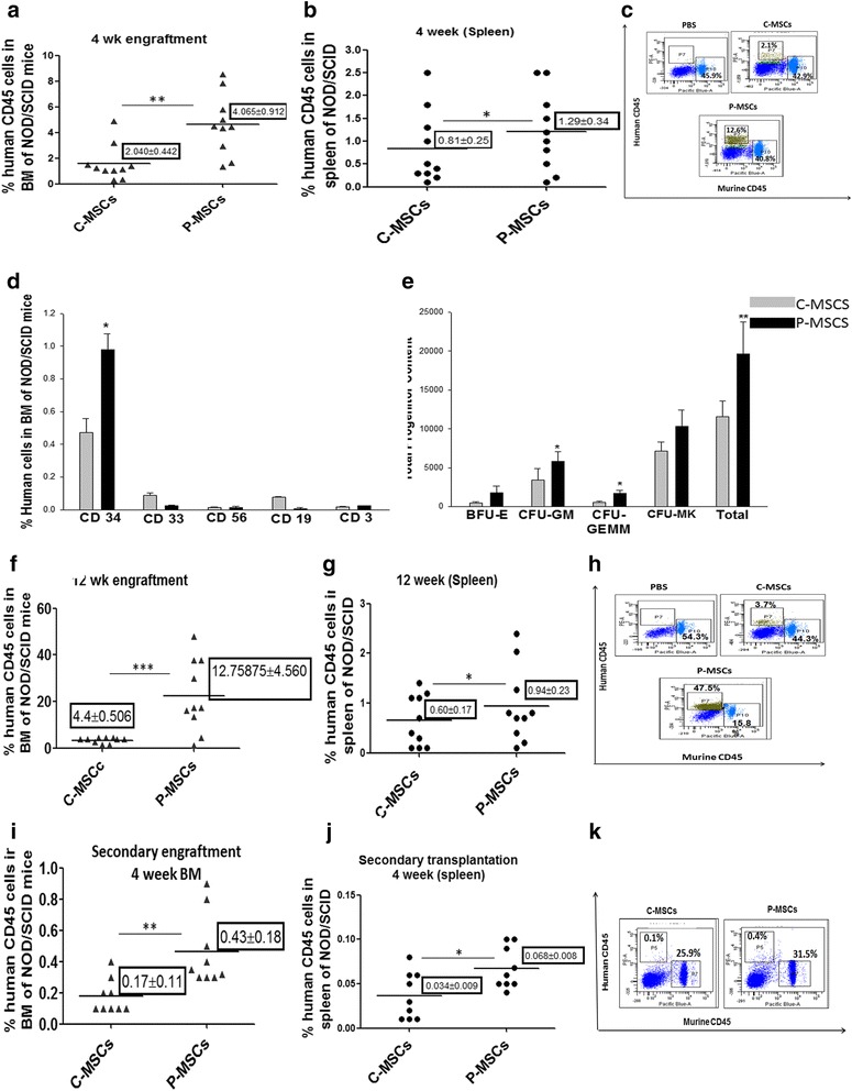
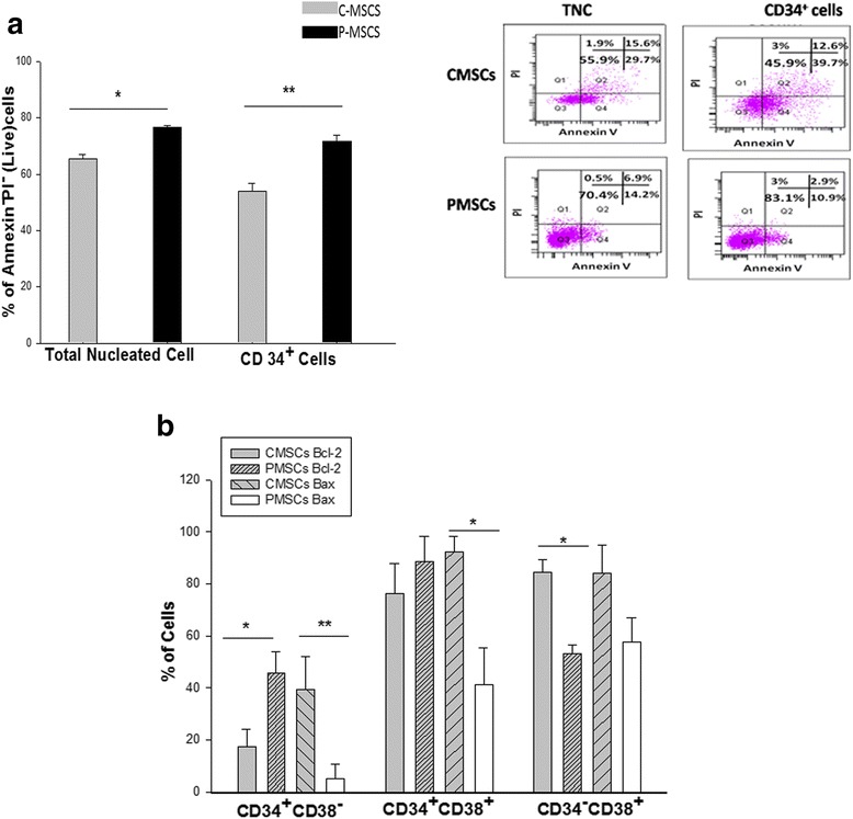
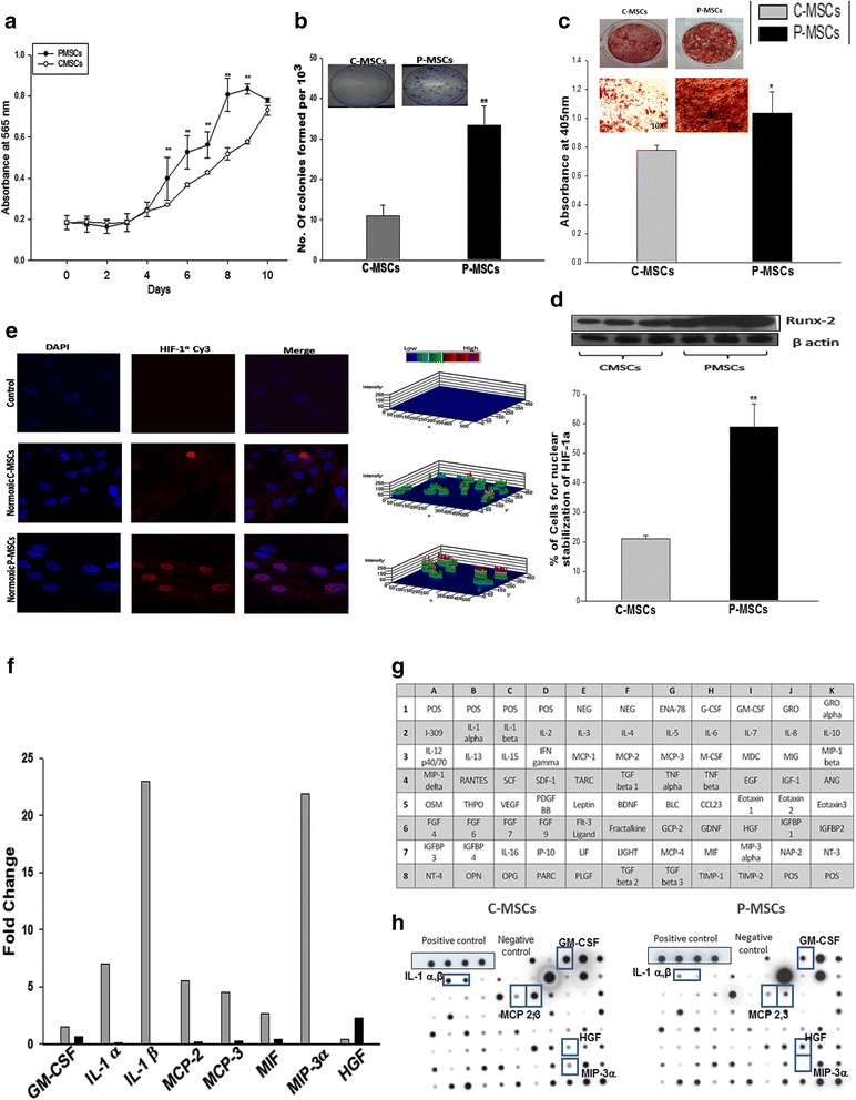
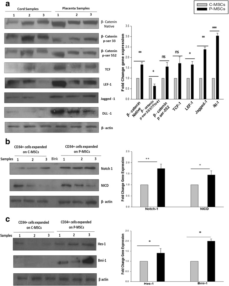
Results: C-MSCs and P-MSCs were found to be morphologically and phenotypically similar but exhibited differential ability to support ex vivo hematopoiesis. Cells expanded on P-MSCs showed higher percentage of primitive cells (CD34(+)CD38(-)), CFU (Colony forming unit) content and LTC-IC (Long term culture initiating cells) ability. CD34(+) cells expanded on P-MSCs also exhibited better in vitro adhesion to fibronectin and migration towards SDF-1α and enhanced NOD/SCID repopulation ability, as compared to those grown on C-MSCs. P-MSCs were found to be closer to BM-MSCs in their ability to expand HSCs. P-MSCs supported expansion of functionally superior HSCs by virtue of reduction in apoptosis of primitive HSCs, higher Wnt and Notch activity, HGF secretion and cell-cell contact. On the other hand, C-MSCs facilitated expansion of progenitors (CD34(+)CD38(+)) and differentiated (CD34(-)CD38(+)) cells by secretion of IL1-α, β, MCP-2, 3 and MIP-3α.

Conclusions: P-MSCs were found to be better feeders for ex vivo maintenance of primitive HSCs with higher engraftment potential than the cells expanded with C-MSCs as feeders.


Figures

Fig. 1
Fig. 1
C- and P-MSCs displayed similar morphology and immuno-phenotype. a Fibroblastic morphology exhibited by C- and P-MSCs as seen under phase contrast microscope (10X). b Histogram profile of representative samples of C- and P-MSCs exhibiting expression of markers such as CD44, CD90, CD73, and CD105. No expression was found for CD45, CD34, CD14, CD19, CD11b, and HLA-DR. c Upper panel represents osteogenic differentiation of MSCs by staining with alizarin red S. Middle panel is for adipogenic differentiation confirmed after lipid droplets stained by oil red o. Chondrogenic differentiation of C- and P-MSCs confirmed with Alcian blue stain. C-MSCs cord-derived mesenchymal stem cells, P-MSCs placenta-derived mesenchymal stem cells
Fig. 2
Fig. 2
P-MSCs supported robust expansion of CD34+ cells without deterring their quiescence state. a Co-culturing of CD34+ cells with P-MSCs harbored a significantly higher TNC yield, CD34+CD38 and CD133+ cells than C-MSCs as feeders. b Upper panel- A representative FACS dot plot showing higher CD34+CD38 cells with P-MSCs as feeders. Lower panel- A representative FACS histogram depicting total CD34+CD133+ cells in the co-culture C- and P MSCs as feeders. c Cell cycle analysis demonstrated an increase of CD34+ cells at G0/G1 phase with P-MSCs as feeders and higher numbers of cycling CD34+ cells in cultures comprised of C-MSCs as feeders. d FACS dot plot profile of Hoechst and PyroninY based cell cycle analysis showing P-MSCs as feeders had a higher percentage of CD34+ cells in G0 (quiescence) stage in the co-cultures with P-MSCs than C-MSCs. Data are represented as mean ± standard deviation from three independent sets of experiments. *p ≤ 0.05, **p ≤ 0.01, and ***p ≤ 0.001 P-MSCs placenta-derived mesenchymal stem cells, C-MSCs cord-derived mesenchymal stem cells, FACS fluorescence-activated cell sorting
Fig. 3
Fig. 3
CD34+ cells expanded on P-MSCs have augmented in vitro functionality of the expanded cells. a P-MSCs:CD34+ co-cultures displayed higher blast-forming unit erythroid (BFU-E), granulocyte–monocyte (GM), granulocyte-erythroid-monocyte megakaryocyte (GEMM), and megakaryocytes (MK) colonies when compared with C-MSCs as feeders. b P-MSCs as feeders maintained significantly higher LTC-IC units than C-MSCs. c Trans well migration assay displayed significantly augmented migration of the expanded cells towards SDF-1α in the P-MSCs set as compared to the C-MSCs set. d Migrated fraction from the co-culture with P-MSCs revealed significantly higher numbers of primitive CD34+CD38cells. e Superior migratory response might be attributed to the higher percentage of CD34+CXCR-4+ cells in co-cultures with P-MSCs as feeders. f Quantitation of the attachment to the fibronectin was carried out after lysing the adhered cells and measuring color intensity at 570 nm. The inset shows sort-purified CD34+ expanded on P-MSCs adhered to fibronectin in higher numbers. Data are represented as mean ± standard deviation from three independent sets of experiments. *p ≤ 0.05, **p ≤ 0.01, and ***p ≤ 0.001. P-MSCs placenta-derived mesenchymal stem cells, C-MSCs cord-derived mesenchymal stem cells, LTC-IC long term culture initiating cell
Fig. 4
Fig. 4
P-MSCS imparts significantly higher SCID repopulating ability on expanded CD34+ cells than C-MSCs. a,b At four weeks post transplantation (short term engraftment) the bone marrow(a) and spleen(b) of the mice receiving cells expanded on P-MSCs showed a significantly higher percentage of human CD45+ cells. Each dot in the graph represents an individual animal. c Representative FACS dot plots showing a higher percentage of human CD45+ cells in the BM of NOD/SCID mice infused with cells expanded on P-MSCs on the background of murine CD45+ cells. d Donor derived7 multi-lineage engraftment was seen in the BM of NOD/SCID mice for P- and C-MSCs expanded sets as determined by CD34 (stem cells), CD33 (myeloid cells), CD56(NK cells), CD19 (B cells), and CD3 (T cells). e Donor-derived committed progenitors were higher in the BM of mice receiving cells expanded on P-MSCs than on C-MSCs as evaluated by performing an in vitro colony formation assay on MBM using human specific growth factors. f, g Long term engraftment (12 weeks) indicated significantly higher chimerism (% human CD45) was detected in the BM (f) and spleen (g) of NOD/SCID mice receiving cells from P-MSCs co-cultures. h Representative FACS dot plots showing higher percentage of human CD45+ cells in BM of NOD/SCID mice infused with cells expanded on P-MSCs in the murine CD45+ background after 12 weeks. i, j Serial transplantation assay showed a significantly higher percentage of human CD45+ in BM (i) and spleen (j), the secondary recipient receiving cells from mice infused with cells expanded on P-MSCs. k Representative FACS dot plots showing a higher percentage of human CD45+ cells in the BM of NOD/SCID mice infused with cells from primary recipient. Data are represented as mean ± standard deviation from ten different mice. (n = 10) *p ≤ 0.05, **p ≤ 0.01, and ***p ≤ 0.001. P-MSCs placenta-derived mesenchymal stem cells, C-MSCs cord-derived mesenchymal stem cells, FACS fluorescence-activated cell sorting, BM bone marrow
Fig. 5
Fig. 5
Co-culturing of CD34+ cells with P-MSCs reduced the level of apoptosis and expansion of the primitive HSCs by preventing apoptosis via up regulation of BCL-2. a Higher percentage of live TNC as well as gated CD34+ cells were detected in P-MSCs:CD34+ co-cultures. Inset depicts representative FACS profile depicting the same P-MSCs:CD34+ co-cultures (Lower panel), C-MSCs:CD34+ co-cultures (Upper panel). b Significantly higher levels of Bcl-2 were detected in CD34+CD38 cells from the P-MSCs:CD34+ co-cultures. Co-cultures of C-MSCs displayed higher Bcl-2 levels in CD34+CD38+ and CD34CD38+ cells. Bax expression was significantly higher in all the subsets from C-MSCs: CD34+ co-cultures. Data are represented as mean ± standard deviation from three different independent experimental sets. *p ≤ 0.05, **p ≤ 0.01, and ***p ≤ 0.001. P-MSCs placenta-derived mesenchymal stem cells, TNC total nucleated cells, FACS fluorescence-activated cell sorting, C-MSCs cord-derived mesenchymal stem cells
Fig. 6
Fig. 6
C-MSCs and P-MSCs differ in the functional attributes relevant to expansion of HSCs. a P-MSCs had higher rate of proliferation than C-MSCs as determined by MTT assay. b P-MSCs exhibited higher clonogenecity than C-MSCs even at lowest cell concentration. Inset depicts the representative CFU-F colonies stained with crystal violet. c P-MSCs exhibits superior differentiation to osteoblasts depicted by the calcium deposits stained with alizarin red S (Left panel). Right panel shows quantitative analysis after extraction of the dye and its quantitation at 405 nm. d Superior osteoblastic differentiation may be due to the pre-osteoblastic nature of P-MSCs by virtue of higher RUNX-2 expression. e Higher normoxic stabilization of HIF-1α in the nucleus of the P-MSCs than C-MSCs. Left panel shows 3D representations of expression of the HIF-1α as 3-D histograms. Percentage of the positive nuclei from ten random fields/slide of three independent experiments is represented in the graph. f Fold change in the cytokines expression by C-MSCs and P-MSC, respectively. Data represented as mean value of two independent paired samples. g C-MSCs had a distinctive secretion profile mainly of pro-inflammatory cytokines as checked by membrane-based cytokine array. h Represents the hybridization results for a representative sample. Data are represented as mean ± standard deviation from three different independent experimental sets. *p ≤ 0.05, **p ≤ 0.01, and ***p ≤ 0.001. P-MSCs placenta-derived mesenchymal stem cells, C-MSCs cord-derived mesenchymal stem cells, HSCs hematopoietic stem cells, CFU-F colony forming unit-fibroblasts
Fig. 7
Fig. 7
P-MSCs displayed higher Wnt activity leading to collateral increase into Notch signaling in the expanded CD34+cells. a Higher expression of native β-catenin. Phosphorylated β-catenin (ser-552) was found in the P-MSCs. Higher expression of phosphorylated β-catenin (ser-33/34Thr41) in C-MSCs. The expression of downstream targets of Wnt, TCF, LEF-1, jagged-1 and δ-like one (dll-1) was higher in P-MSCs than C-MSCs. The graph represents the fold change in the expression after normalization to β-actin. b Sort purified CD34+ cells from P-MSCs. Co-cultures showed significant up regulation of Notch I and intracellular domain of Notch (NICD). The quantitation of the same is represented as a graph. c The CD34+ cells expanded on the P-MSCs exhibited higher expression of Hes-1 and Bmi-1, downstream target of Notch, quantitatively represented as a graph. Data are represented as mean ± standard deviation from three different samples. *p ≤ 0.05, **p ≤ 0.01, and ***p ≤ 0.001. P-MSCs placenta-derived mesenchymal stem cells, C-MSCs cord-derived mesenchymal stem cells

Similar articles

Cited by

References

    1. Hofmeister CC, Zhang J, Knight KL, Le P, Stiff PJ. Ex vivo expansion of umbilical cord blood stem cells for transplantation: growing knowledge from the hematopoietic niche. Bone Marrow Transplant. 2007;39:11–23. doi: 10.1038/sj.bmt.1705538. - DOI - PubMed
    1. Ponce DM, Gonzales A, Lubin M, Castro-Malaspina H, Giralt S, Goldberg JD, et al. Graft-versus-host disease after double-unit cord blood transplantation has unique features and an association with engrafting unit-to-recipient HLA match. Biol Blood Marrow Transplant. 2013;6:904–11. doi: 10.1016/j.bbmt.2013.02.008. - DOI - PMC - PubMed
    1. Haspel RL, Ballen KK. Double cord blood transplants: filling a niche? Stem Cell Rev. 2006;2:81–6. - PubMed
    1. Flores–Guzman P, Fernandez–Sanchezv V, Mayani H. Concise review: ex vivo expansion of cord blood-derived hematopoietic stem and progenitor cells: basic principles, experimental approaches, and impact in regenerative medicine. Stem Cells Trans Med. 2013;2:830–8. doi: 10.5966/sctm.2013-0071. - DOI - PMC - PubMed
    1. Kelly SS, Parmar S, De Lima M, Robinson S, Shpall E. Overcoming the barriers to umbilical cord blood transplantation. Cytotherapy. 2010;12:121–30. doi: 10.3109/14653240903440111. - DOI - PMC - PubMed

Publication types

MeSH terms