. 2015 Nov 9;28(5):610-622.
doi: 10.1016/j.ccell.2015.09.008. Epub 2015 Oct 17.

Erythropoietin Stimulates Tumor Growth via EphB4

Affiliations

Erythropoietin Stimulates Tumor Growth via EphB4

Sunila Pradeep et al. Cancer Cell. .

Abstract

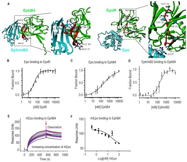
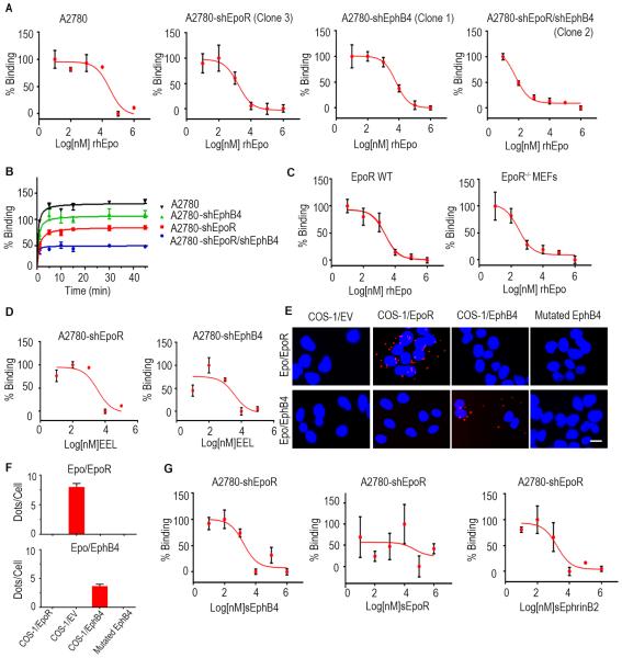
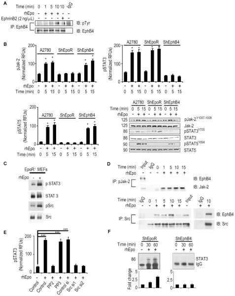
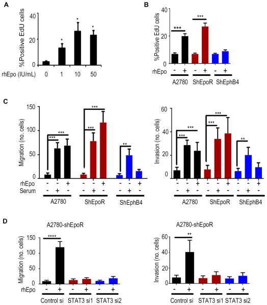
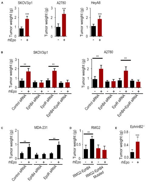
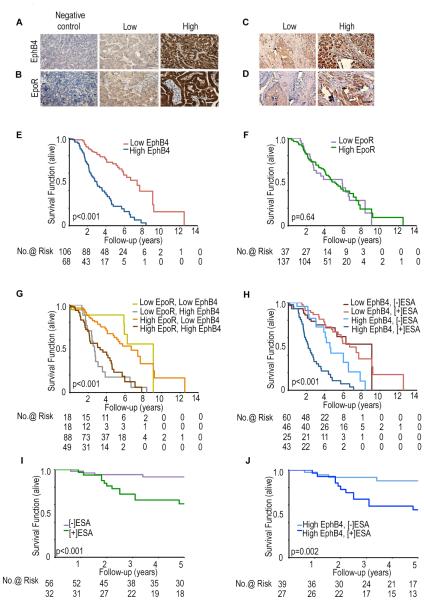
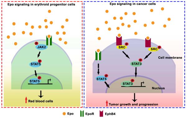
While recombinant human erythropoietin (rhEpo) has been widely used to treat anemia in cancer patients, concerns about its adverse effects on patient survival have emerged. A lack of correlation between expression of the canonical EpoR and rhEpo's effects on cancer cells prompted us to consider the existence of an alternative Epo receptor. Here, we identified EphB4 as an Epo receptor that triggers downstream signaling via STAT3 and promotes rhEpo-induced tumor growth and progression. In human ovarian and breast cancer samples, expression of EphB4 rather than the canonical EpoR correlated with decreased disease-specific survival in rhEpo-treated patients. These results identify EphB4 as a critical mediator of erythropoietin-induced tumor progression and further provide clinically significant dimension to the biology of erythropoietin.


Figures

Figure 1
Figure 1. Epo Binds to EphB4
(A) X-ray crystal structure of EphB4 (green), EphrinB2 (blue) and Epo (blue) -EpoR (green) complex. The C-D region from which the inhibitory peptide has been derived from is highlighted in red. E43, E44 and L45 are involved in EphrinB2 binding. Interactions between homology region with EpoR and ligand are shown in detail: E43, E44 and L45 of EphB4 interact with K60, T114 and K116 of EphrinB2. The acidic residues in the apical position of the loop (E50, E51) are shown as well. The A-B loop region homologous to the inhibitory peptide is highlighted in red. Interaction between K97 and R14 of Epo with acidic residue located on the apical loop of the homology region (E34). The region homologous to EphB4 ranges from E24, E25, L26 to F39. E24, E25 and L26 of EpoR are homologous to residues E43, E44 and L45 of EphB4 which are involved in Ephrin ligand binding. Acidic residues are located in the loop in an apical position similar to the location of acidic residues in the A-B loop region of Epo Receptor. (B) Fluorescence microscale thermophoresis analysis of Epo binding to EpoR or (C) EphB4. (D) EphrinB2 binding to labeled EphB4. (E) Surface competition assay (SCA) of Epo and EphrinB2 with coated EphB4 receptor using the BIAcore instrument for detection of bound protein. Serial dilutions of Epo were mixed with EphrinB2 and injected onto a CM5 chip to which EphB4 was bound. (F) Bound protein shown as response units (RU) at the end of association was plotted as a function of Epo concentration and fit with a three-parameter non-linear regression using Graphpad Prism 5.0. Samples and a buffer blank were injected in duplicate. Mean ± SEM values are shown. (n=3). (See also Figure S1).
Figure 2
Figure 2. Characterization of rhEpo binding to EphB4 cells
(A) Analysis of competitive binding of [I125]rhEpo to A2780-parental, -shEpoR, -shEphB4, and shEpoR/shEphB4 cells. (B) Kinetics of [I125]rhEpo binding to A2780-parental, -shEpoR, -shEphB4, and -shEpoR/shEphB4 cells. (C) Analysis of competitive binding of [I125]rhEpo to EpoR WT and EpoR−/− MEFs. (D) Competitive binding studies using the EEL peptide in A2780-shEpoR or -shEphB4 cells. (E) Proximity ligation assay using Cos-1EpoR, Cos-1EphB4, and mutated EphB4 cells. Scale bar represents 50 μm. (F) Bar graph represents the quantification of ligation assay. (G) Competitive binding studies using soluble EphB4, EpoR or EphrinB2 in A2780-shEpoR cells. CPM, counts per minute. Mean ± SEM values are shown. (n=3). (See also Figure S2 and Table S1-S2).
Figure 3
Figure 3. Epo binding to EphB4 activates the Src-STAT pathway
(A) Activation of EphB4 by rhEpo in A2780-parental cells. (B) Evaluation of pJak-2, pSTAT5, and pSTAT3 levels (ELISA and Western blot) following rhEpo treatment in A2780, -shEpoR, and -shEphB4 cells. (C) Evaluation of pSTAT3 levels following rhEpo treatment in EpoR−/− MEFs. (D) Effect of rhEpo on EphB4 binding to Jak-2 and Src in A2780-EpoR cells. (E) Effect of Src inhibitor (PP2, 10μM; inactive counterpart, PP3) or Src siRNA on pSTAT3 levels following treatment with rhEpo in A2780-shEpoR cells, (RFUs - relative fluorescence units), n=3. (F) Effect of rhEpo on STAT3 binding to a oligonucleotide containing consensus STAT3 binding site in A2780-shEpoR and -shEphB4 cells. Dose of rhEpo used for these experiments was 50 IU/mL. Mean ± SEM values are shown. *p < 0.05; ***p <0.001. (See also Figure S3).
Figure 4
Figure 4. An essential role of EphB4 in rhEpo-mediated effects on cancer cells
(A) Effect of rhEpo on proliferation at indicated dosage. (B) Proliferation, (C) migration, and invasion of A2780, -shEpoR, and -shEphB4 cells after rhEpo treatment. (D) Effect of STAT3 silencing on migration and invasion in A2780-shEpoR cells. Mean ± SEM values are shown. *p < 0.05; **p < 0.01; ***p <0.001; ****p <0.0001. n=3. (See also Figure S4).
Figure 5
Figure 5. EphB4 plays a key role in Epo-induced tumor growth in vivo
(A) Effect of rhEpo (50 IU given 3x/week i.p; n=10) on tumor growth (aggregate tumor weight after 3-5 weeks of rhEpo treatment) in orthotopic ovarian cancer models in vivo. (B) Effect of EphB4 and EpoR silencing on SKOV3ip1 and A2780 tumor growth in vivo with or without rhEpo treatment (n=10). Effect of EphB4 or EpoR silencing on the MDA-231 tumor growth in vivo with or without rhEpo treatment (n=10). (D) Effect of ectopically expressed EphB4 or EphB4 mutant on RMG2 tumor growth in vivo with or without rhEpo treatment (n=10). (E) Effect of rhEpo (50 IU given 3x/week i.p; n=10) on ID8-VEGF tumor growth (aggregate tumor weight after 3-5 weeks of rhEpo treatment; 1×10⁶ ID8-VEGF murine ovarian cancer cells were injected into EphrinB2−/− mice). Mean ± SEM values are shown. **p < 0.01; ***p <0.001. (See also Figure S5).
Figure 6
Figure 6. Clinical relevance of EphB4 and EpoR expression and ESA treatment in cancer patients
Representative immunohistochemical-peroxidase staining for (A) EphB4 and (B) EpoR expression in ovarian cancer, and (C) EphB4 and (D) EpoR expression in breast cancer samples. Kaplan-Meier curves of disease-specific mortality for ovarian (E-H) and breast (I-J) cancer patients stratified by tumoral expression of (E) EphB4, (F) EpoR, or (G) both EphB4 and EpoR. (H) Evaluation of disease-specific survival duration of ovarian cancer patients based on ESA-treatment and EphB4 expression; (I) Disease-specific survival analysis of breast cancer patients stratified by ESA treatment; (J) Disease-specific survival of breast cancer patients based on ESA treatment and EphB4 expression. Scale bar represents 50 μm. (See also Figure S6 and Table S3-S7).
Figure 7
Figure 7
Proposed model of Epo mediated EphB4 signaling in cancer cells.

References

    1. Aapro M, Jelkmann W, Constantinescu SN, Leyland-Jones B. Effects of erythropoietin receptors and erythropoiesis-stimulating agents on disease progression in cancer. British journal of cancer. 2012;106:1249–1258. - PMC - PubMed
    1. Ahmed AA, Lu Z, Jennings NB, Etemadmoghadam D, Capalbo L, Jacamo RO, Barbosa-Morais N, Le XF, Vivas-Mejia P, Lopez-Berestein G, et al. SIK2 is a centrosome kinase required for bipolar mitotic spindle formation that provides a potential target for therapy in ovarian cancer. Cancer cell. 2010;18:109–121. - PMC - PubMed
    1. Authier F, Desbuquois B. Glucagon receptors. Cellular and molecular life sciences : CMLS. 2008;65:1880–1899. - PMC - PubMed
    1. Belda-Iniesta C, Perona R, Carpeno Jde C, Cejas P, Casado E, Manguan-Garcia C, Ibanez de Caceres I, Sanchez-Perez I, Andreu FB, Ferreira JA, et al. Human recombinant erythropoietin does not promote cancer growth in presence of functional receptors expressed in cancer cells. Cancer biology & therapy. 2007;6:1600–1605. - PubMed
    1. Brines M, Cerami A. The receptor that tames the innate immune response. Molecular medicine (Cambridge, Mass) 2012;18:486–496. - PMC - PubMed

Publication types

MeSH terms