. 2015 Oct 20:5:15434.
doi: 10.1038/srep15434.

IL-4 Protects the Mitochondria Against TNFα and IFNγ Induced Insult During Clearance of Infection with Citrobacter rodentium and Escherichia coli

Affiliations

IL-4 Protects the Mitochondria Against TNFα and IFNγ Induced Insult During Clearance of Infection with Citrobacter rodentium and Escherichia coli

Arpan K Maiti et al. Sci Rep. .

Abstract

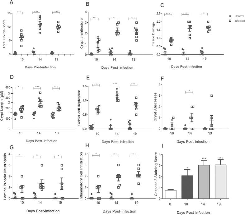
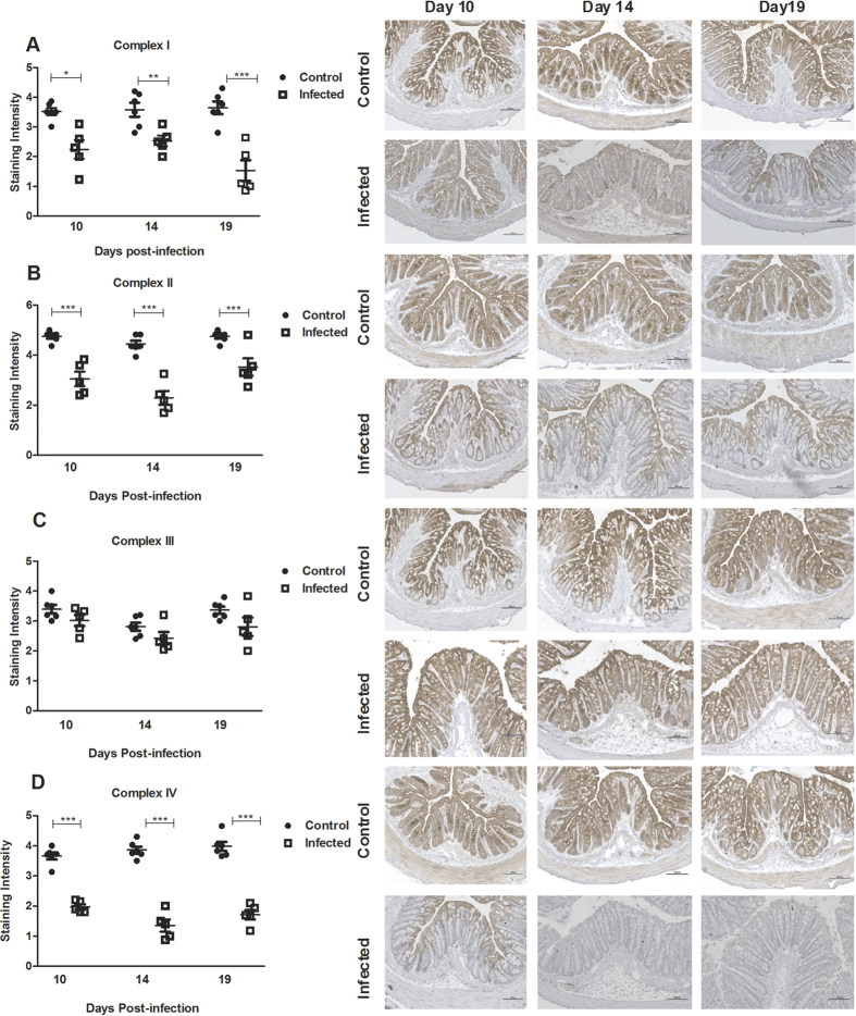
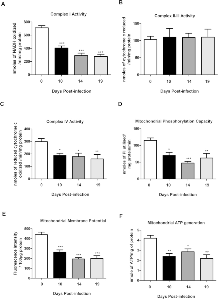
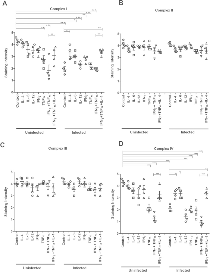
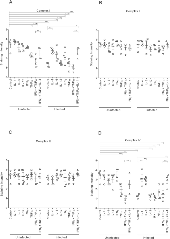
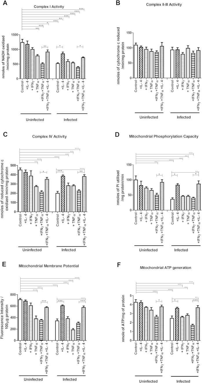
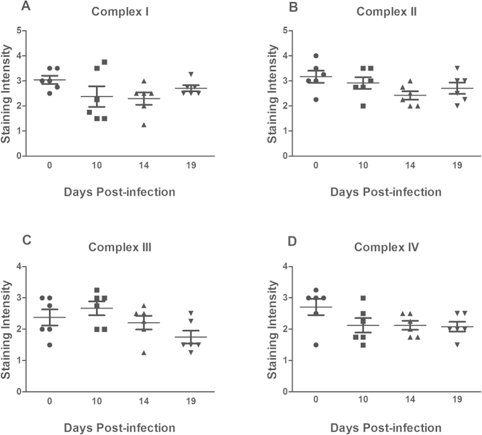
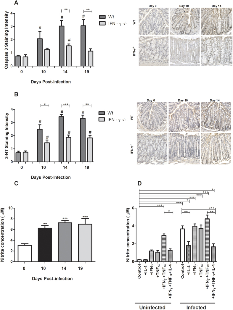
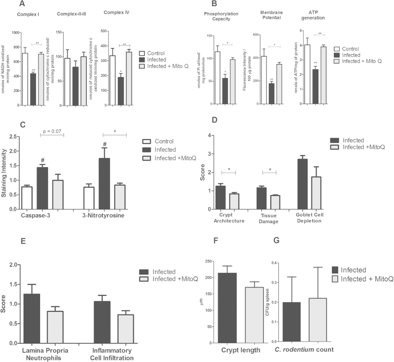
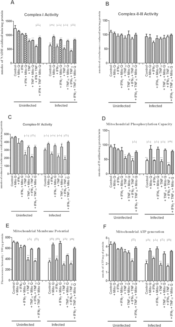
Citrobacter rodentium is a murine pathogen that serves as a model for enteropathogenic Escherichia coli. C. rodentium infection reduced the quantity and activity of mitochondrial respiratory complexes I and IV, as well as phosphorylation capacity, mitochondrial transmembrane potential and ATP generation at day 10, 14 and 19 post infection. Cytokine mRNA quantification showed increased levels of IFNγ, TNFα, IL-4, IL-6, and IL-12 during infection. The effects of adding these cytokines, C. rodentium and E. coli were hence elucidated using an in vitro colonic mucosa. Both infection and TNFα, individually and combined with IFNγ, decreased complex I and IV enzyme levels and mitochondrial function. However, IL-4 reversed these effects, and IL-6 protected against loss of complex IV. Both in vivo and in vitro, the dysfunction appeared caused by nitric oxide-generation, and was alleviated by an antioxidant targeting mitochondria. IFNγ -/- mice, containing a similar pathogen burden but higher IL-4 and IL-6, displayed no loss of any of the four complexes. Thus, the cytokine environment appears to be a more important determinant of mitochondrial function than direct actions of the pathogen. As IFNγ and TNFα levels increase during clearance of infection, the concomitant increase in IL-4 and IL-6 protects mitochondrial function.


Figures

Figure 1
Figure 1. Colitis and caspase-3 staining during C. rodentium infection in the distal colon of WT mice.
(A) The total colitis score represents the sum of the individual scores of the following parameters: (B) crypt architecture, (C) tissue damage, (D) crypt length (the numbers 1–3 on the y-axes indicates how the crypt lengths were translated to scores for the incorporation into the total colitis score), (E) goblet cell depletion, (F) crypt abscesses, (G) neutrophils in lamina propria and (H) inflammatory cell infiltration. Values are mean ± S.E.M (n = 6–7 mice). Statistics: unpaired t test, *P < 0.05, **P < 0.01, ***P < 0.001 compared to uninfected control. (I) Caspase-3 quantification. Statistics: ANOVA with Student Newman-Keuls Multiple Comparison post hoc test: *P < 0.05, **P < 0.01, ***P < 0.001. vs. control. The infection experiments were performed twice, and each time point contains results pooled from 4–9 mice.
Figure 2
Figure 2. Tissue localization and semi-quantification of the mitochondrial respiratory enzyme complexes in the murine distal colon during C. rodentium infection.
Immunohistochemical staining using antibodies for (A) MTND6 (complex I) (B) SDHA (complex-II) (C) CYC1 (complex-III) (D) CCO-VIc (complex-IV). Statistics: unpaired t test. *P < 0.05, **P < 0.01, ***P < 0.001 vs. control, n = 5–6 mice/group. The infection experiments were performed twice, and each time point contain results pooled from 5–6 mice. Scale bar 100 μm, magnification × 200.
Figure 3
Figure 3. Mitochondrial function in the murine distal colon after C. rodentium infection.
(A) complex I activity (B) complex II-III activity (C) complex IV activity (D) mitochondrial phosphorylation capacity (E) mitochondrial Membrane potential (F) mitochondrial ATP generation. Values are mean ± S.E.M. Statistics: ANOVA with Student Newman-Keuls Multiple Comparison post hoc test: *P < 0.05, **P < 0.01, ***P < 0.001 vs. control. (n = 4–9 mice/group). The infection experiments were performed twice, and each time point contains results pooled from 4–9 mice.
Figure 4
Figure 4. Semi-quantification of mitochondrial respiratory enzyme complexes of an in vitro intestinal model treated with cytokines and C. rodentium infection.
Immunohistochemical staining using antibodies for (A) MTND6 (complex I) (B) SDHA (complex-II) (C) CYC1 (complex-III) (D) CCO-VIc (complex-IV). Values are mean ± S.E.M. Statistics: ANOVA with Student Newman-Keuls Multiple Comparison post hoc test: *P < 0.05, **P < 0.01, ***P < 0.001.
Figure 5
Figure 5. Semi-quantification of mitochondrial respiratory enzyme complexes of an in vitro intestinal model treated with cytokines and E. coli infection.
Immunohistochemical staining using antibodies for (A) MTND6 (complex I) (B) SDHA (complex-II) (C) CYC1 (complex-III) (D) CCO-VIc (complex-IV). Values are mean ± S.E.M. Statistics: ANOVA with Student Newman-Keuls Multiple Comparison post hoc test: *P < 0.05, **P < 0.01, ***P < 0.001.
Figure 6
Figure 6. Mitochondrial function of an in vitro mucosal intestinal model treated with cytokines and C. rodentium infection.
(A) complex-I activity (B) complex-II-III activity (C) complex-IV activity (D) mitochondrial phosphorylation capacity (E) mitochondrial membrane potential (F) mitochondrial ATP generation. Values are mean ± S.E.M. Statistics: ANOVA Student Newman-Keuls Multiple Comparison post hoc test: *P < 0.05, **P < 0.01, ***P < 0.001.
Figure 7
Figure 7. Semi-quantitative analysis of the mitochondrial respiratory enzyme complexes in the distal colon of IFNγ−/− mice after C. rodentium infection.
Immunohistochemical staining using antibodies for (A) MTND6 (complex I) (B) SDHA (complex-II) (C) CYC1 (complex-III) (D) CCO-VIc (complex-IV). Statistics: ANOVA Student Newman-Keuls Multiple Comparison post hoc test: *P < 0.05, **P < 0.01, ***P < 0.001. vs. control.
Figure 8
Figure 8. Caspase-3, 3-NT staining scores and nitrite measurements after C. rodentium infection.
(A) The caspase-3 staining scores based on immunohistochemical staining and corresponding representative photos of the caspase-3 tissue localization in the distal colon of WT and IFNγ−/− mice. Values are mean ± S.E.M. Statistics: ANOVA with Newman-Keuls Multiple Comparison post hoc test, #P < 0.05 vs. corresponding non-infected genotype control, unpaired t test **P < 0.01 compared to WT of same infection period (n = 4–9 mice/group). Scale bar 50 μm, magnification × 400. (B) Immunohistochemical staining scores and photos representing the tissue localization of the 3-NT residues in the murine distal colons of WT and IFNγ−/− mice during infection. Scale bar 50 μm, magnification × 400. Statistics: ANOVA with Student Newman-Keuls Multiple Comparison post hoc test: #P < 0.05 vs. corresponding genotype control, unpaired t test: *P < 0.05, **P < 0.01, ***P < 0.001 vs. WT at the same time point of infection, n = 4–9 mice/group. (C) Nitrite concentration in the murine distal colon after C. rodentium infection. Statistics: ANOVA with Student Newman-Keuls Multiple Comparison post hoc test: *P < 0.05, **P < 0.01, ***P < 0.001. vs. control. (D) Nitrite concentration in the in vitro mucosal intestinal model treated with cytokines and C. rodentium infection. Statistics: ANOVA with Student Newman-Keuls Multiple Comparison post hoc test: *P < 0.05, **P < 0.01, ***P < 0.001.
Figure 9
Figure 9. Effects of MitoQ on mitochondrial functions, caspase-3, NO, colitis and C. rodentium translocation.
(A) complex I, II–III and IV activities and (B) mitochondrial phosphorylation capacity, membrane potential and mitochondrial ATP generation in infected and MitoQ treated mice. Values are mean ± S.E.M. Statistics: ANOVA with Student Newman-Keuls Multiple Comparison post hoc test: *P < 0.05, **P < 0.01, ***P < 0.001 (n = 4/group). (C) caspase-3 and 3-Nitrotyrosine immunostaining in infected and MitoQ treated mice. Values are mean ± S.E.M. Statistics: ANOVA with Student Newman-Keuls Multiple Comparison post hoc test: *P < 0.05 vs non-infected control, #P < 0.05 vs. infected control. (D) colonic scores of crypt architecture, tissue damage and goblet cell depletion, (E) Neutrophils in lamina propria and inflammatory cell infiltration, (F) crypt length and (G) C. rodentium counts in spleen of infected and MitoQ treated mice. Values are mean ± S.E.M. (n = 4 mice/group). Statistics: paired t test, *P < 0.05 compared to infected mice.
Figure 10
Figure 10. Effects of MitoQ on mitochondrial functions in the in vitro mucosal intestinal model treated cytokines with/without C. rodentium infection.
(A) complex-I activity, (B) complex-II–III activity, (C) complex-IV activity, (D) mitochondrial phosphorylation capacity, (E) mitochondrial membrane potential, and (F) mitochondrial ATP generation. Values are mean ± S.E.M. Statistics: ANOVA with Student Newman-Keuls Multiple Comparison post hoc test: *P < 0.05, **P < 0.01, ***P < 0.001.

Similar articles

Cited by

References

    1. Barnett F. D., Abul-Milh M., Huesca M. & Lingwood C. A. Enterohemorrhagic Escherichia coli induces apoptosis which augments bacterial binding and phosphatidylethanolamine exposure on the plasma membrane outer leaflet. Infect. Immun. 68(6), 3108–3115 (20l0). - PMC - PubMed
    1. Deng W., Li Y., Vallance B. A. & Finlay B. B. Locus of enterocyte effacement from Citrobacter rodentium: sequence analysis and evidence for horizontal transfer among attaching and effacing pathogens. Infect. Immun. 69(10), 6323–6335 (2001). - PMC - PubMed
    1. Frankel G. et al.. Intimin from enteropathogenic Escherichia coli restores murine virulence to a Citrobacter rodentium eaeA mutant: induction of an immunoglobulin A response to intimin and EspB. Infect. Immun. 64, 5315–5325 (1996). - PMC - PubMed
    1. Luperchio S. A. & Schauer D. B. Molecular pathogenesis of Citrobacter rodentium and transmissible murine colonic hyperplasia. Microbes Infect. 3, 333–340 (2001). - PubMed
    1. Vallance B. A., Deng W., Jacobson K. & Finlay B. B. Host susceptibility to the attaching and effacing bacterial pathogen Citrobacter rodentium. Infect. Immun. 71, 3443–3453 (2003). - PMC - PubMed

Publication types

MeSH terms