Review
. 2015 Nov;40(11):648-661.
doi: 10.1016/j.tibs.2015.09.001. Epub 2015 Oct 15.

Chemistry and Biology of Self-Cleaving Ribozymes

Affiliations
Review

Chemistry and Biology of Self-Cleaving Ribozymes

Randi M Jimenez et al. Trends Biochem Sci. 2015 Nov.

Abstract

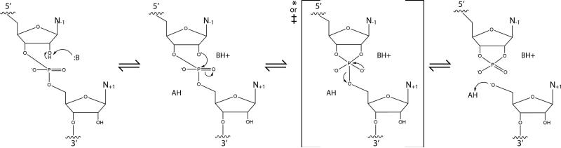
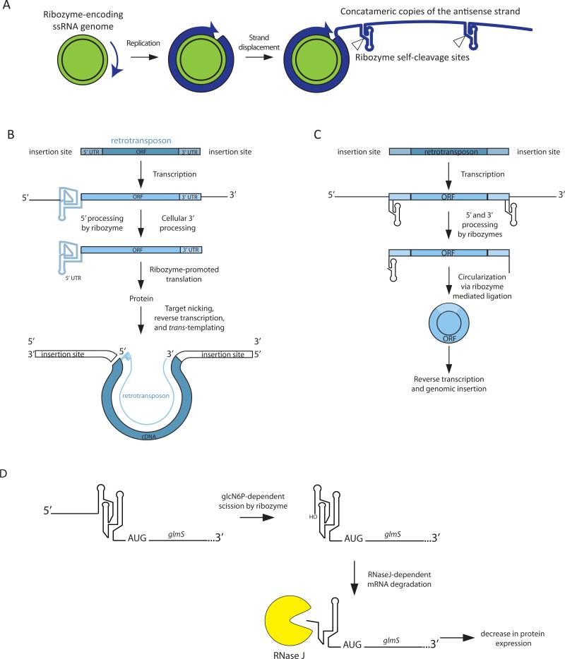
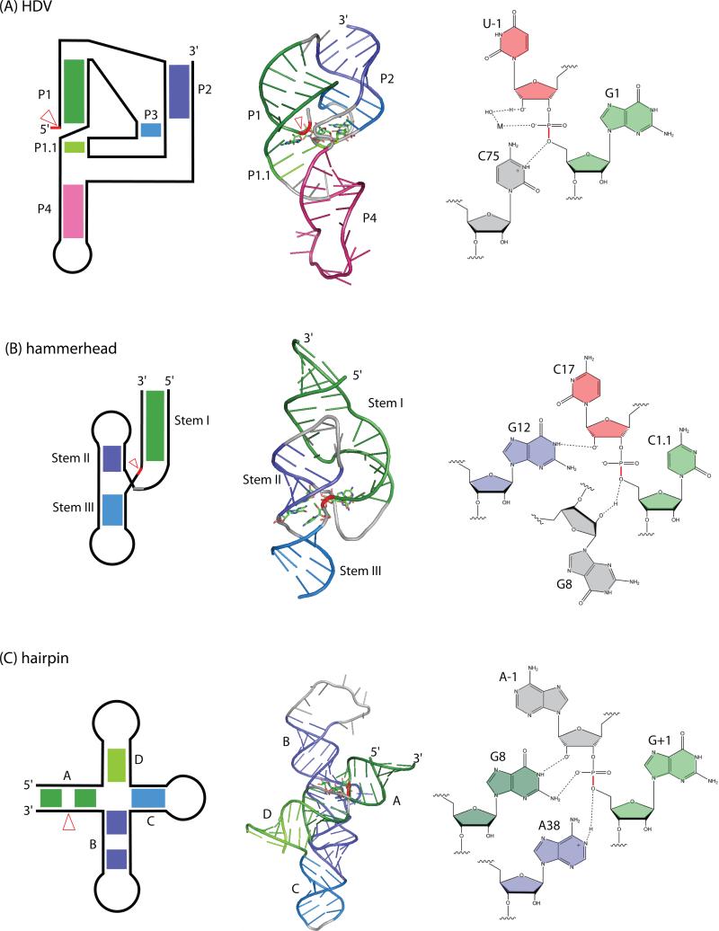
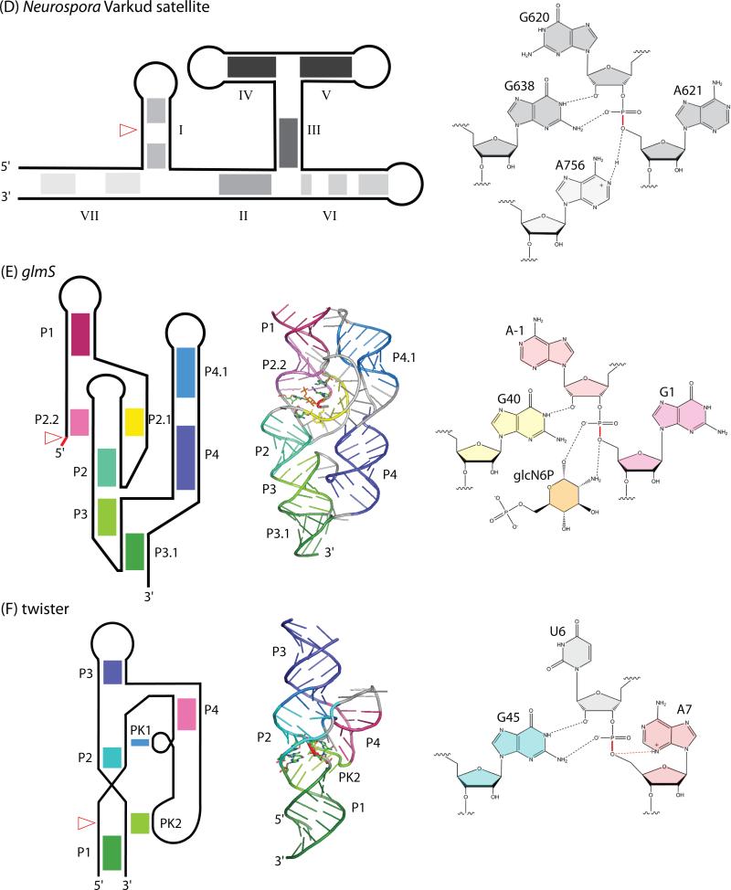
Self-cleaving ribozymes were discovered 30 years ago, but their biological distribution and catalytic mechanisms are only beginning to be defined. Each ribozyme family is defined by a distinct structure, with unique active sites accelerating the same transesterification reaction across the families. Biochemical studies show that general acid-base catalysis is the most common mechanism of self-cleavage, but metal ions and metabolites can be used as cofactors. Ribozymes have been discovered in highly diverse genomic contexts throughout nature, from viroids to vertebrates. Their biological roles include self-scission during rolling-circle replication of RNA genomes, co-transcriptional processing of retrotransposons, and metabolite-dependent gene expression regulation in bacteria. Other examples, including highly conserved mammalian ribozymes, suggest that many new biological roles are yet to be discovered.

Keywords: aptazymes; retrotransposon; riboregulation; riboswitch; ribozyme; self-scission.


Figures

Figure 1
Figure 1. Mechanism of RNA self-scission
A general acid-base catalysis involves a general base, which deprotonates the 2′ hydroxyl of the nucleophile, positioned in-line with the 5′ O leaving group. The transesterification proceeds via a phosphorane transition state or intermediate, depending on whether it is stabilized, yielding a 2′-3′ cyclic phosphate in the upstream nucleotide and an oxyanion on the downstream nucleotide. The leaving group is protonated by a general base. The small self-cleaving ribozymes described here use a variety of chemical groups to facilitate this process (Table 1).
Figure 2
Figure 2. Roles of self-cleaving ribozymes in different biological systems
(A) Most instances of self-cleaving ribozymes found in (sub-)viral genomes are involved in replication. During rolling-circle replication of a single-stranded RNA genome, concatameric copies of the opposite polarity are generated. The self-cleavage activity of ribozymes generates unit-length copies that must be circularized and replicated to complete the cycle. (B) Retrotransposon-associated hepatitis delta virus HDV-like ribozymes liberate the 5′ end of the retroelement from the full-length transcript. The ribozyme structure in the 5′ UTR promotes translation of the downstream ORF. The protein produced is necessary for target nicking, reverse transcription, and genomic insertion. Further, the HDV-like ribozyme on the 5′ end of the RNA promotes the template switching necessary to complete genomic insertion of the autonomous (LINE) element. (C) Hammerhead ribozymes mapping to non-autonomous retrotransposons (SINEs) serve to mobilize the retroelement. Complementarity of the 5′ and 3′ ends facilitates ribozyme self-ligation resulting in circularization of the RNA; this feature enhances the reverse transcription and genomic insertion processes that complete the retrotransposition cycle. (D) The glmS ribozyme is located in the 5′ UTR of the glmS gene, encoding the glutamine-fructose 6-phosphate aminotransferase enzyme responsible for generating glucosamine 6-phosphate (glcN6P), necessary for bacterial cell wall synthesis. The ribozyme requires glcN6P as a cofactor for catalysis. The resulting 5′-hydroxyl makes the processed transcript a substrate for the 5′-3′ exonuclease RNase J. Therefore, the glmS ribozyme is a riboswitch that turns off glcN6P synthase expression in response to glcN6P.
Figure 3
Figure 3. Overview of self-cleaving ribozymes: structural features and proposed cleavage mechanisms
The secondary structures of the (A) HDV-like, (B) hammerhead, (C) hairpin, (D) Neurospora Varkud satellite, (E) glmS, and (F) twister ribozymes are shown in the left column and labeled as they appear in current literature. Typically, the helical segments are numbered as “paired” elements (P1, P2, etc), by Roman numerals, or in alphabetical order in 5′ to 3′ direction as they appear in the first example of a given motif. Paired regions corresponding to pseudoknots are designated PK or simply as paired elements. The corresponding crystal structures for each ribozyme are shown in the middle column with colored helices to emphasize helical stacks and pseudoknots. The site of scission is pointed out by the red arrowheads in the secondary structures and highlighted in red in the respective crystal structures. A model of the cleavage site for each ribozyme is shown in the right column, which illustrates interactions that promote the cleavage event. Nucleobases flanking the scissile phosphate are splayed apart, which promotes the in-line orientation necessary to accomplish cleavage. The scissile bond is shown in red. Interactions proposed to stabilize the transition state of the reaction are shown for all ribozymes. Note that all cleavage sites, with the exception of the HDV-like ribozyme, which requires a hydrated metal (M), have a guanosine residue that participates as a proposed general base in the general acid-base reaction mechanism. The glmS ribozyme requires glucosamine 6-phosphate (glcN6P) as a cofactor necessary for catalysis. For the twister ribozyme, a hypothetical interaction (dashed red line) between N3 of A7 and the scissile phosphate suggests that A7 may act as the general acid during self-cleavage. PDBIDs: 3ZD5, ISJ3, IM5K, 2NZ4, 4QJH

Similar articles

Cited by

References

    1. Lai MM. The molecular biology of hepatitis delta virus. Annu Rev Biochem. 1995;64:259–286. - PubMed
    1. Taylor JM. Hepatitis delta virus. Intervirology. 1999;42:173–178. - PubMed
    1. Wu HN, Lin YJ, Lin FP, Makino S, Chang MF, Lai MMC. Human Hepatitis-Delta Virus-Rna Subfragments Contain an Autocleavage Activity. Proc Natl Acad Sci U S A. 1989;86:1831–1835. - PMC - PubMed
    1. Salehi-Ashtiani K, Luptak A, Litovchick A, Szostak JW. A genomewide search for ribozymes reveals an HDV-like sequence in the human CPEB3 gene. Science. 2006;313:1788–1792. - PubMed
    1. Webb CH, Riccitelli NJ, Ruminski DJ, Luptak A. Widespread occurrence of self-cleaving ribozymes. Science. 2009;326:953. - PMC - PubMed