

XingNaoJing, prescription of traditional Chinese medicine, prevents autophagy in experimental stroke by repressing p53-DRAM pathway
- PMID: 26481508
- PMCID: PMC4617486
- DOI: 10.1186/s12906-015-0882-2
XingNaoJing, prescription of traditional Chinese medicine, prevents autophagy in experimental stroke by repressing p53-DRAM pathway
Abstract
Background: Xingnaojing (XNJ), a well known prescription in traditional Chinese medicine, has been used for treatment of stroke in China. However, the effects and mechanisms of XNJ on autophagy are not clear. Here, we used the cell models of autophagy induced by serum-free condition and ischemia stroke in rats to further investigate whether the p53-DRAM pathway is involved in the effects of XNJ on autophagy.
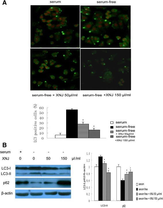
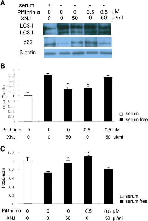
Methods: We used the cell model of autophagy induced by serum-free condition and the rat model of ischemia caused by a middle cerebral artery occlusion (MCAO). The effects of XNJ on p53 transcriptional activity of PC12 cells were evaluated by the luciferase activity assay. The mRNA levels and the expression of p53 and its target autophagy gene DRAM (damage-regulated autophagy modulator) were analyzed respectively by Quantitative-RTPCR and Western blot assay. The activation of autophagy was detected by the levels of autophagy markers, microtubule associated protein light chain 3 (LC3) and p62 by Immunofluorescence and Western blot. p53 inhibitor was used to determine whether p53 is responsible for the effects of XNJ on preventing autophagy.
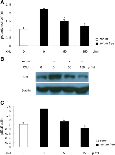
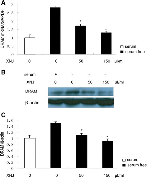
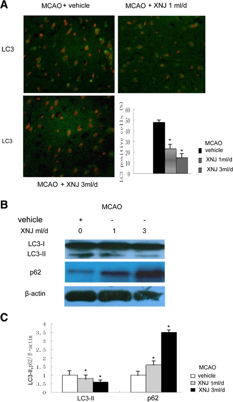
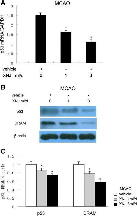
Results: The assay for luciferase activity of p53 promoter indicated that XNJ inhibited p53 transcriptional activity. XNJ reduced the expression of p53 and its target autophagy gene DRAM (damage-regulated autophagy modulator) in serum-free condition PC12 cells and the cortex in MCAO rats. XNJ reduced autophagy of PC12 cells induced by serum-free condition and the cortex in MCAO rats. Furthermore, suppression of p53 by p53 inhibitor significantly reduced the effects of XNJ on the autophagy of PC12 cells in serum-free condition.
Conclusion: XNJ prevents autophagy in experimental stroke by repressing p53/DRAM pathway. Our findings are therefore of considerable therapeutic significance and provide the novel and potential application of XNJ for the treatment of brain diseases.
Figures

Similar articles
-
XingNaoJing injection ameliorates cerebral ischaemia/reperfusion injury via SIRT1-mediated inflammatory response inhibition.Pharm Biol. 2020 Dec;58(1):16-24. doi: 10.1080/13880209.2019.1698619. Pharm Biol. 2020. PMID: 31854225 Free PMC article.
-
p53 Mediates Colistin-Induced Autophagy and Apoptosis in PC-12 Cells.Antimicrob Agents Chemother. 2016 Aug 22;60(9):5294-301. doi: 10.1128/AAC.00641-16. Print 2016 Sep. Antimicrob Agents Chemother. 2016. PMID: 27324771 Free PMC article.
-
Autophagy-mediated cytoplasmic accumulation of p53 leads to apoptosis through DRAM-BAX in cadmium-exposed human proximal tubular cells.Biochem Biophys Res Commun. 2021 Jan 1;534:128-133. doi: 10.1016/j.bbrc.2020.12.019. Epub 2020 Dec 13. Biochem Biophys Res Commun. 2021. PMID: 33321290
-
Evidence for the interplay between JNK and p53-DRAM signalling pathways in the regulation of autophagy.Autophagy. 2010 Jan;6(1):153-4. doi: 10.4161/auto.6.1.10537. Epub 2010 Jan 5. Autophagy. 2010. PMID: 19949306 Review.
-
DRAM links autophagy to p53 and programmed cell death.Autophagy. 2007 Jan-Feb;3(1):72-4. doi: 10.4161/auto.3438. Epub 2007 Jan 28. Autophagy. 2007. PMID: 17102582 Review.
Cited by
-
Therapeutic targets by traditional Chinese medicine for ischemia-reperfusion injury induced apoptosis on cardiovascular and cerebrovascular diseases.Front Pharmacol. 2022 Aug 19;13:934256. doi: 10.3389/fphar.2022.934256. eCollection 2022. Front Pharmacol. 2022. PMID: 36060007 Free PMC article. Review.
-
Network pharmacology to investigate the pharmacological mechanisms of muscone in Xingnaojing injections for the treatment of severe traumatic brain injury.PeerJ. 2021 Jul 20;9:e11696. doi: 10.7717/peerj.11696. eCollection 2021. PeerJ. 2021. PMID: 34322321 Free PMC article.
-
Uncovering the Mechanism of the Xingnaojing Injection against Ischemic Stroke Using a Combined Network Pharmacology Approach and Gut Microbiota Analysis.Evid Based Complement Alternat Med. 2022 May 20;2022:5886698. doi: 10.1155/2022/5886698. eCollection 2022. Evid Based Complement Alternat Med. 2022. PMID: 35646156 Free PMC article.
-
Challenges and Opportunities of Deferoxamine Delivery for Treatment of Alzheimer's Disease, Parkinson's Disease, and Intracerebral Hemorrhage.Mol Pharm. 2021 Feb 1;18(2):593-609. doi: 10.1021/acs.molpharmaceut.0c00474. Epub 2020 Oct 9. Mol Pharm. 2021. PMID: 32926630 Free PMC article. Review.
-
Poster Viewing Sessions PB01-B01 to PB03-V09.J Cereb Blood Flow Metab. 2019 Jul;39(1_suppl):167-523. doi: 10.1177/0271678X19851020. J Cereb Blood Flow Metab. 2019. PMID: 31265794 Free PMC article. No abstract available.
References
Publication types
MeSH terms
Substances
LinkOut - more resources
Full Text Sources
Other Literature Sources
Medical
Research Materials
Miscellaneous
