. 2015 Nov 16;212(12):2077-94.
doi: 10.1084/jem.20142162. Epub 2015 Oct 19.

TLR9 ligation in pancreatic stellate cells promotes tumorigenesis

Affiliations

TLR9 ligation in pancreatic stellate cells promotes tumorigenesis

Constantinos P Zambirinis et al. J Exp Med. .

Abstract

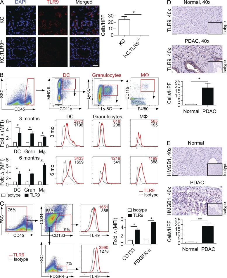
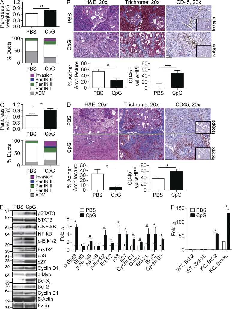
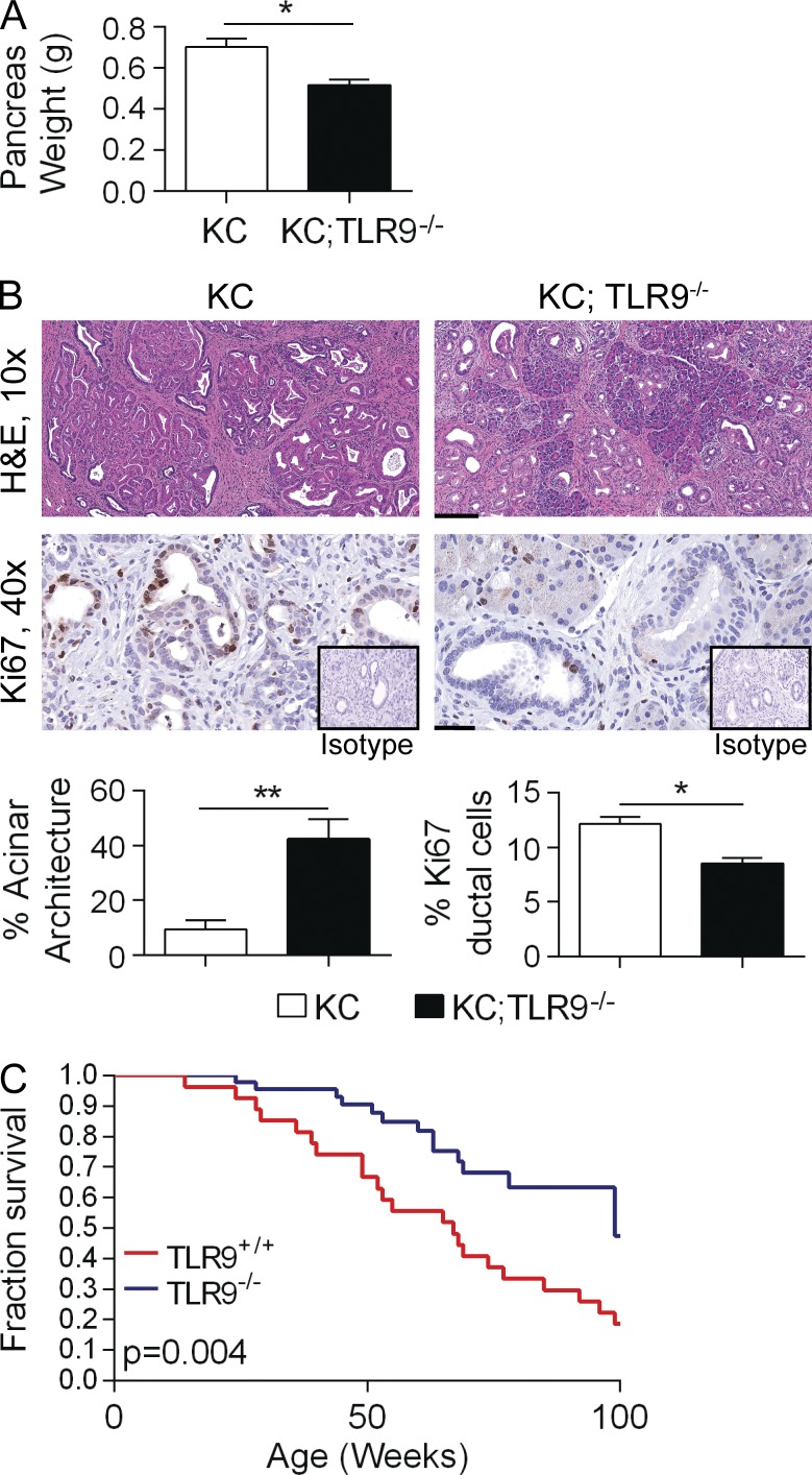
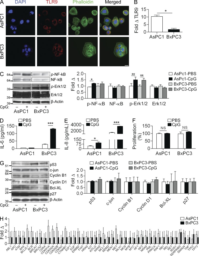
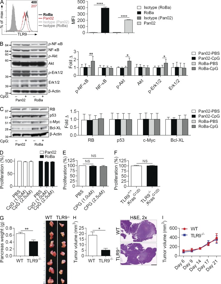
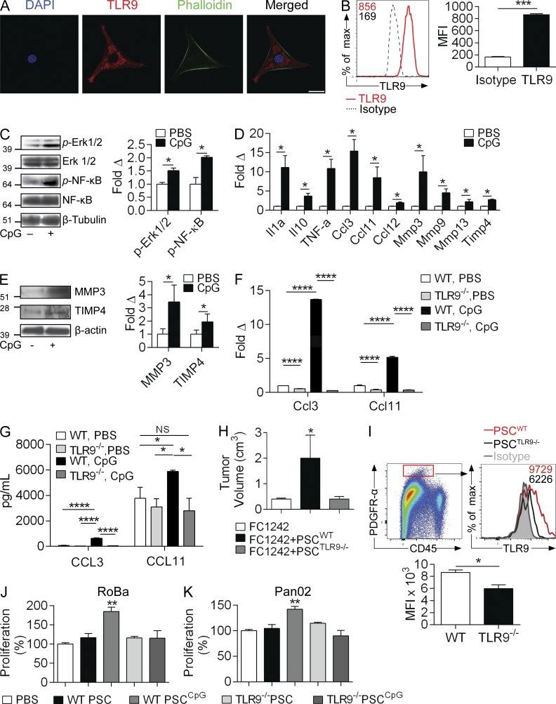
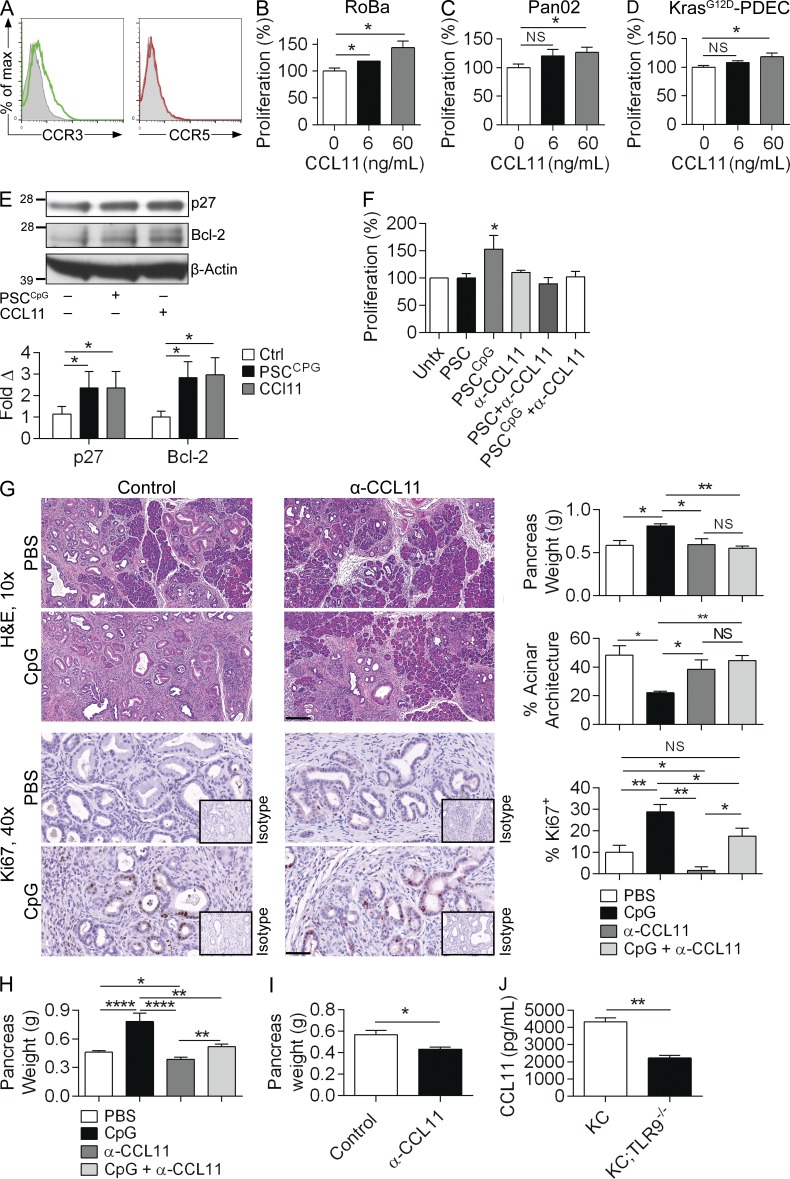
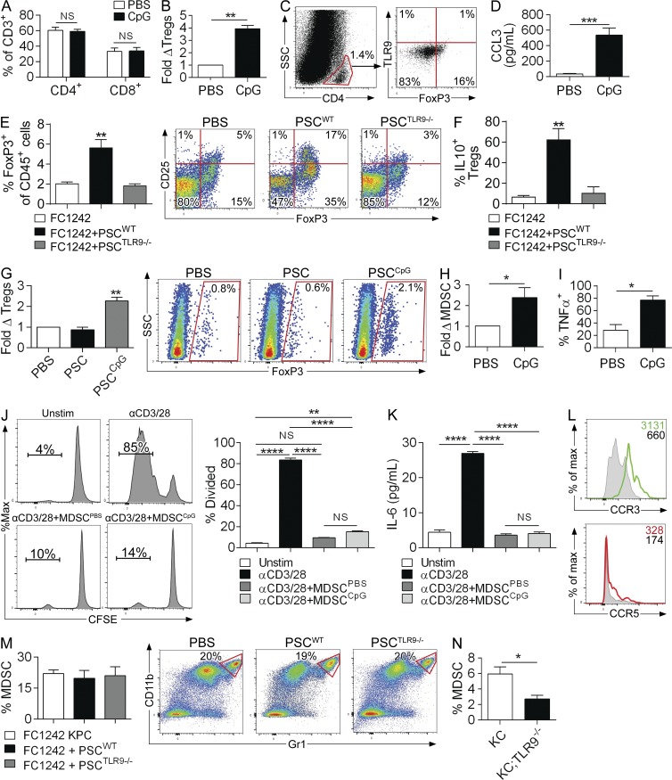
Modulation of Toll-like receptor (TLR) signaling can have protective or protumorigenic effects on oncogenesis depending on the cancer subtype and on specific inflammatory elements within the tumor milieu. We found that TLR9 is widely expressed early during the course of pancreatic transformation and that TLR9 ligands are ubiquitous within the tumor microenvironment. TLR9 ligation markedly accelerates oncogenesis, whereas TLR9 deletion is protective. We show that TLR9 activation has distinct effects on the epithelial, inflammatory, and fibrogenic cellular subsets in pancreatic carcinoma and plays a central role in cross talk between these compartments. Specifically, TLR9 activation can induce proinflammatory signaling in transformed epithelial cells, but does not elicit oncogene expression or cancer cell proliferation. Conversely, TLR9 ligation induces pancreatic stellate cells (PSCs) to become fibrogenic and secrete chemokines that promote epithelial cell proliferation. TLR9-activated PSCs mediate their protumorigenic effects on the epithelial compartment via CCL11. Additionally, TLR9 has immune-suppressive effects in the tumor microenvironment (TME) via induction of regulatory T cell recruitment and myeloid-derived suppressor cell proliferation. Collectively, our work shows that TLR9 has protumorigenic effects in pancreatic carcinoma which are distinct from its influence in extrapancreatic malignancies and from the mechanistic effects of other TLRs on pancreatic oncogenesis.


Figures

Figure 1.
Figure 1.
TLR9 is up-regulated during pancreatic oncogenesis in epithelial, inflammatory, and stromal cells. (A) Frozen sections from pancreata of 3-mo-old KC and KC;TLR9−/− mice were stained for DAPI and TLR9 and visualized on a confocal microscope (63×; bar = 30 µm). Results were quantified based on 10 HPFs per slide. (B) 3- and 6-mo-old KC mice were analyzed by flow cytometry for pancreatic TLR9 expression on DCs, granulocytes, and macrophages. Mean fluorescence intensity (MFI) is indicated compared with respective isotype controls. Representative data and summary statistics from three mice per data point are shown. (C) 3-mo-old KC mice were analyzed by flow cytometry for pancreatic TLR9 expression on epithelial cells (CD45CD34CD133+) and cancer-associated fibroblasts (CD45CD34CD133PDGFR-α+). Representative data and summary statistics from three mice per data point are shown. Mouse experiments were repeated more than five times with similar results. (D) TLR9 immunohistochemistry compared with isotype control was performed on normal human pancreata and pancreata from patients with PDAC (40×; bar = 60 µm). Representative images and quantitative data from four patients per group are shown. (E) Human PDAC and normal human pancreas specimens were stained using a mAb directed against HMGB1 (40×; bar = 60 µm). Representative images and quantitative data from four patients per group are shown (*, P < 0.05; **, P < 0.01 using the Student’s t test).
Figure 2.
Figure 2.
TLR9 activation accelerates pancreatic oncogenesis. (A) 10-wk-old KC mice were administered CpG or PBS thrice weekly by i.p. injection for 4 wk. Pancreata were harvested and weighed (n = 9/group). (B) Pancreatic sections were stained with H&E, Trichrome, and IHC was performed using an anti-CD45 mAb compared with isotype control. The fraction of pancreatic ducts exhibiting normal morphology, acinoductal metaplasia (ADM), various grades of PanIN, or foci of invasive carcinoma were quantified. Similarly, the fraction of preserved acinar area and the number of CD45+ cells per HPF were quantified (20×; bar = 100 µm). (C and D) Similarly, cohorts of 10-wk-old KC were serially administered CpG or PBS for 12 wk (n = 5/group). Pancreata were weighed, stained with H&E, trichrome, and CD45. Morphological transformation and inflammatory cell infiltration were quantified as above. (E) Pancreata from 10-wk-old KC mice treated with PBS or CpG for 4 wk were tested for expression of inflammatory signaling intermediates, cell cycle regulators, and oncogenic proteins by Western blotting. β-actin and Ezrin were used as loading controls. Representative data and averages of triplicates based on band intensity relative to respective loading controls are shown. (F) 10-wk-old WT and KC mice were administered CpG or PBS for 4 d and tested for expression of Bcl-2 and Bcl-XL by PCR. This experiment was performed in quadruplicate and repeated twice with similar results (n = 3/group; *, P < 0.05; ***, P < 0.001 using the Student’s t test).
Figure 3.
Figure 3.
Luminal TLR9 ligands can accelerate pancreatic oncogenesis. (A) 8-wk-old WT mice were administered CFSE-labeled S. mutans (108) by the i.p. or oral gavage route. Pancreata were harvested at 6 h and examined by flow cytometry for CFSE+ cells compared with PBS-treated control mice. Representative data are shown. The experiment was performed on three separate occasions using up to five mice per group. (B) Cerulein-treated WT mice were administered FITC-labeled CpG (25 µg) by oral gavage. Pancreata were harvested at 6 h, and MHCII+ cells were gated and tested for FITC. Representative and quantitative data are shown (n = 4/group). (C) 6-wk-old KC mice were administered CpG or PBS thrice weekly by oral gavage for 5 wk (n = 5/group). Pancreata were stained with H&E and the fraction of preserved acinar area was calculated (10×; bar = 200 µm; *, P < 0.05 using the Student’s t test).
Figure 4.
Figure 4.
TLR9 deletion is protective against pancreatic tumorigenesis. (A) 6-mo-old KC and KC;TLR9−/− mice were sacrificed and their pancreata were harvested and weighed (n = 5/group). (B) H&E stained sections were assessed histologically and the fraction of nontransformed acini was quantified (10×; bar = 200 µm). Tumor proliferation was assessed by Ki67 staining (40×; bar = 60 µm; *, P < 0.05; **, P < 0.01 using the Student’s t test). (C) Cohorts of KC (n = 29) and KC;TLR9−/− (n = 46) mice were observed for 2 yr in a Kaplan-Meier survival analysis (P = 0.004 using the Gehan-Breslow-Wilcoxon Test).
Figure 5.
Figure 5.
TLR9 ligation in human pancreatic cancer cells can induce inflammatory signaling but does not elicit oncogenic changes. (A) AsPC1 and BxPC3 cells were seeded on glass coverslips, stained with anti-TLR9 antibody, and imaged on a confocal microscope (40×; bar = 40 µm). (B) The relative levels of Tlr9 expression in AsPC1 and BxPC3 cells were quantified by qRT-PCR (normalized to β-actin). (C) AsPC1 and BxPC3 cells were either unstimulated or stimulated with CpG (1 µM) for 10 min and Western blotting was performed on whole-cell lysates for NF-κB and MAP kinase signaling intermediates. Results were quantified based on triplicates. (D and E) AsPC1 and BxPC3 cells were either unstimulated or stimulated with CpG (1 µM) for 24 h and levels of IL-6 (D) and IL-8 (E) in cell culture supernatants were quantified by cytometric bead array. (F) AsPC1 and BxPC3 cells were stimulated with CpG (1 µM) for 24 h and cellular proliferation was assessed using the XTT II assay (% proliferation was normalized to untreated control). (G) AsPC1 and BxPC3 cells were selectively stimulated with CpG (1 µM) for 24 h and Western blotting was performed on whole-cell lysates for select proteins involved in the regulation of pancreatic oncogenesis. Results were quantified based on triplicates. All experiments were reproduced two or three separate times. (H) Gene expression analysis using the preconfigured Human oncogene and tumor suppressor qPCR array was performed on AsPC1 and BxPC3 cells after 24-h treatment with CpG (1 µM). Fold-change compared with control untreated cells is shown (p = ns for all comparisons). PCR experiments were repeated twice with similar results (*, P < 0.05; **, P < 0.01; ***, P < 0.001 using the Student’s t test).
Figure 6.
Figure 6.
TLR9 ligation can be proinflammatory in murine pancreatic cancer cells but does not induce proliferative or oncogenic changes. (A) Pan02 cells and KPC-derived RoBa cells were tested for expression of TLR9 compared with isotype control. MFI is indicated in the upper right corner. Averages of triplicates based on MFI are shown. (B) Pan02 and RoBa cells were either unstimulated or stimulated with CpG (1 µM) for 10 min and tested for activation of inflammatory signaling pathways by Western blotting. (C) Similarly, Pan02 and RoBa cells were selectively stimulated with CpG (1 µM) for 24 h and Western blotting was performed on whole-cell lysates for select proteins involved in the regulation of pancreatic oncogenesis. Results were quantified based on triplicates. (D) Pan02 and RoBa cells were stimulated with increasing doses of CpG and cellular proliferation was assessed using the XTT assay (p = ns for all comparisons). (E) KrasG12D-PDEC were stimulated with increasing doses of CpG and proliferation measured as above (p = ns for all comparisons). (F) The comparative in vitro proliferation of TLR9+/+;KrasG12D-PDEC and TLR9−/−;KrasG12D-PDEC was tested using the XTT assay. In vitro experiments were performed in triplicate and repeated at least twice with similar results (*, P < 0.05; **, P < 0.01; ****, P < 0.0001). (G) WT and TLR9−/− mice were administered an orthotopic intrapancreatic injection of KPC-derived FC1242 cells. Tumors were harvested and weighed at 3 wk. Quantitative results and representative photographs of individual tumors are shown (n = 10/group; **, P < 0.01 using the Student’s t test). (H) WT and TLR9−/− mice were administered an orthotopic intrapancreatic injection of TLR9+/+;KrasG12D-PDEC. Tumors were harvested at 3 wk. Quantitative results and representative H&E stained sections from each group are shown (n = 7/group; *, P < 0.05 using the Student’s t test; 2; bar = 1 mm). (I) WT and TLR9−/− mice were administered a subcutaneous injection of FC1242 cells (n = 5/group). Tumor volume was calculated at serial intervals by measuring the long (D) and short (d) diameter of the tumor, and applying the formula V = 1/2 × (D × d2; p = ns at each time point).
Figure 7.
Figure 7.
TLR9 ligation activates pancreatic stellate cells into protumorigenic entities. (A) Cultured PSCs were stained with DAPI, anti-TLR9 mAb, and phalloidin and imaged by confocal microscopy (63×; bar = 40 µm). (B) PSCs were also tested for expression of TLR9 by flow cytometry compared with isotype control. MFIs are indicated. Averages of triplicates based on MFI are shown. (C) PSCs were treated with CpG (1 µM) for 10 min and whole-cell lysates were tested for MAP kinase and NF-κB pathway activation by Western blotting. Density analysis was performed based on triplicates. (D) Gene expression analysis using the preconfigured mouse fibrosis qPCR array was performed on PSCs treated with CpG for 24 h. This experiment was repeated twice in triplicates. (E) Similarly, PSCs were treated with CpG for 24 h and whole-cell lysates were tested for MMP3 and TIMP4 by Western blotting. Results were quantified based on triplicates. (F) WT and TLR9−/− PSCs were treated for 24 h with CpG (1 µM), and expression of Ccl3 and Ccl11 mRNA were quantified by qPCR in triplicate. (G) Similarly, CCL3 and CCL11 protein levels were measured in cell culture supernatant of WT and TLR9−/− PSCs treated CpG versus untreated controls. Averages of triplicates are shown. (H and I) WT mice were orthotopically transplanted with FC1242 pancreatic cancer cells alone, FC1242 + PSCs derived from WT mice, or FC1242 + PSCs derived from TLR9−/− mice (n = 4/group). (H) Tumor volume was recorded at 3 wk. This experiment was performed twice with similar results. (I) PDGFR-α+ CAFs from orthotopic FC1242 tumors that had been co-implanted with TLR9+/+ and TLR9−/− PSCs were gated on flow cytometry and tested for expression of TLR9. Averages of quadruplicates are shown. RoBa (J) and Pan02 (K) cells were cultured for 24 h in 96-well plates with conditioned media derived from WT or TLR9−/− PSCs that had been either unstimulated or pretreated with CpG (1 µM). Cellular proliferation was assessed using the XTT assay based on triplicates. All experiments were repeated two to three times with similar results (*, P < 0.05; **, P < 0.01; ***, P < 0.001; ****, P < 0.0001 using the Student’s t test or ANOVA).
Figure 8.
Figure 8.
PSC-derived CCL11 promotes tumor progression. (A) Pan02 cells were tested by flow cytometry for expression of CCR3 and CCR5 compared with isotype control. RoBa cells (B), Pan02 cells (C), and KrasG12D-PDECs (D) were stimulated with recombinant murine CCL11 at the designated concentrations for 24 h in 96-well plates, and proliferation was assessed with the XTT assay. Averages of triplicates are shown. (E) Pan02 cells stimulated with recombinant CCL11 (60 ng/ml) or conditioned media derived from CpG-treated PSCs and tumor cell lysates were tested for p27 and Bcl-2 expression by Western blotting. (F) Pan02 cells were cultured alone or with conditioned media derived from untreated or CpG-treated WT PSCs in the presence or absence of α-CCL11–neutralizing antibody. Proliferation was measured at 24 h. Averages of triplicates are shown. All cellular proliferation experiments were repeated at least twice. (G) 10-wk-old KC mice were administered CpG or PBS for 4 wk, with or without concurrent administration of an α-CCL11 neutralizing antibody. Pancreata were weighed and assessed by H&E (10×; bar = 200 µm) and Ki67 IHC (40×; bar = 50 µm). The extent of morphological transformation and epithelial cell proliferation, respectively, were quantified (n = 4/group). (H) 8-wk-old KPC mice without palpable tumor were treated with CpG or PBS for 6 wk, with concurrent administration of an α-CCL11 neutralizing antibody or isotype control. Pancreata were weighed (n = 6/group). (I) WT mice were challenged with orthotopic KPC-derived FC1242 tumor and treated with α-CCL11 or control. Pancreata were harvested at 3 wk and weighed (n = 5/group). (J) Lysates from pancreata of 3-mo-old KC and KC;TLR9−/− mice were tested for CCL11 levels by ELISA (n = 3/group; *, P < 0.05; **, P < 0.01; ****, P < 0.0001 using the Student’s t test or ANOVA).
Figure 9.
Figure 9.
TLR9 ligation recruits immune suppressive cellular subsets to the pancreatic tumor microenvironment. (A and B) 10-wk-old KC mice were administered CpG or PBS thrice weekly for 4 wk and the frequency of intrapancreatic CD4+ and CD8+ T cells determined by flow cytometry (n = 4/group). (B) Similarly, the relative number of CD45+CD3+CD4+FoxP3+CD25+ T reg cells was determined in PBS- and CpG-treated KC pancreata. (C) CD4+ pancreatic T lymphocytes in KC mice were gated and tested for coexpression of FoxP3 and TLR9. (D) 10-wk-old KC mice were treated with PBS or CpG for 4 wk and CCL3 levels in pancreatic lysates were quantified using a cytometric bead array (n = 3/group). (E and F) WT mice were treated with an intrapancreatic injection of FC1242 cells alone, FC1242 + WT PSCs, or FC1242 + TLR9−/− PSCs (n = 4/group). (E) The percentage of CD3+CD4+FoxP3+ pancreatic leukocytes among all CD45+ leukocytes was calculated. Representative dot plots showing CD25 and FoxP3 coexpression on CD4+ T cells are shown for each experimental group. (F) IL-10 expression was determined on CD4+CD25+FoxP3+ T reg cells by intracellular cytokine analysis. (G) CD4+ splenic T cells from WT mice were FACS-sorted and incubated alone or with conditioned media from PBS- or CpG-treated PSCs. On day 5, T cells were then tested for expression of FoxP3. Representative data and averages of quadruplicates are shown. (H and I) 10-wk-old KC mice were administered CpG or PBS thrice weekly for 4 wk (n = 4/group). (H) The percentage of CD45+CD11b+Gr1+ MDSCs in each cohort was determined by flow cytometry. (I) Similarly, the production of TNF by intrapancreatic MDSC was quantified by intracellular cytokine staining in CpG- and PBS-treated KC mice. (J and K) Splenic T cells labeled with CFSE were cultured without stimulation, stimulated with αCD3 + αCD28 mAbs alone, αCD3 + αCD28 mAbs + MDSC harvested from PBS-treated KC mice, or αCD3 + αCD28 mAbs + MDSC harvested from CpG-treated KC mice (5:1 T cell/MDSC ratio). (J) T cell proliferation was determined by dissolution of CFSE. (K) IL-6 was measured in cell culture supernatant by cytometric bead array. Representative data and averages of triplicates are shown. This experiment was performed twice. (L) CD45+CD11b+Gr1+ cells from 3-mo-old KC mice were tested for expression of CCR3 and CCR5 by flow cytometry. Shaded histograms correspond to isotype controls. (M) WT mice were administered an intrapancreatic injection of FC1242 cells alone, FC1242 + WT PSCs, or FC1242 + TLR9−/− PSCs (n = 4/group). 3 wk later, the percentage of CD11b+Gr1+ MDSC among intratumoral CD45+ cells was assessed by flow cytometry. Representative dot plots are shown for each experimental group (p = ns for all comparisons). (N) The percentage of CD11b+Gr1+ intrapancreatic MDSC among CD45+ cells were quantified by flow cytometry in 3-mo-old KC and KC;TLR9−/− mice (n = 4/group). All data were reproduced in three separate experiments (*, P < 0.05; **, P < 0.01; ***, P < 0.001; ****, P < 0.0001 using the Student’s t test or ANOVA).

Comment in

  • Pancreatic cancer takes its Toll.
    Mazur PK, Sage J. Mazur PK, et al. J Exp Med. 2015 Nov 16;212(12):1988. doi: 10.1084/jem.21212insight1. J Exp Med. 2015. PMID: 26573583 Free PMC article. No abstract available.

References

    1. Agarwal M., He C., Siddiqui J., Wei J.T., and Macoska J.A.. 2013. CCL11 (eotaxin-1): a new diagnostic serum marker for prostate cancer. Prostate. 73:573–581. 10.1002/pros.22597 - DOI - PMC - PubMed
    1. American Cancer Society 2013. Cancer Facts & Figures 2013. In American Cancer Society, Atlanta.
    1. Bartsch D.K., Gress T.M., and Langer P.. 2012. Familial pancreatic cancer--current knowledge. Nat. Rev. Gastroenterol. Hepatol. 9:445–453. 10.1038/nrgastro.2012.111 - DOI - PubMed
    1. Bera A., Zhao S., Cao L., Chiao P.J., and Freeman J.W.. 2013. Oncogenic K-Ras and loss of Smad4 mediate invasion by activating an EGFR/NF-κB Axis that induces expression of MMP9 and uPA in human pancreas progenitor cells. PLoS One. 8:e82282 10.1371/journal.pone.0082282 - DOI - PMC - PubMed
    1. Butts C., Murray N., Maksymiuk A., Goss G., Marshall E., Soulières D., Cormier Y., Ellis P., Price A., Sawhney R., et al. . 2005. Randomized phase IIB trial of BLP25 liposome vaccine in stage IIIB and IV non-small-cell lung cancer. J. Clin. Oncol. 23:6674–6681. 10.1200/JCO.2005.13.011 - DOI - PubMed

Publication types

MeSH terms