

Pretreatment with β-Boswellic Acid Improves Blood Stasis Induced Endothelial Dysfunction: Role of eNOS Activation
- PMID: 26482008
- PMCID: PMC4611516
- DOI: 10.1038/srep15357
Pretreatment with β-Boswellic Acid Improves Blood Stasis Induced Endothelial Dysfunction: Role of eNOS Activation
Abstract
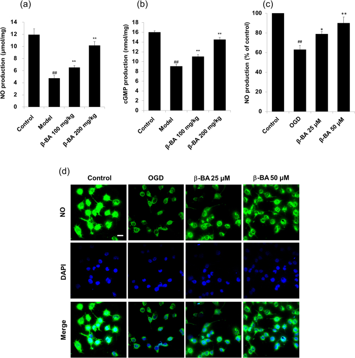
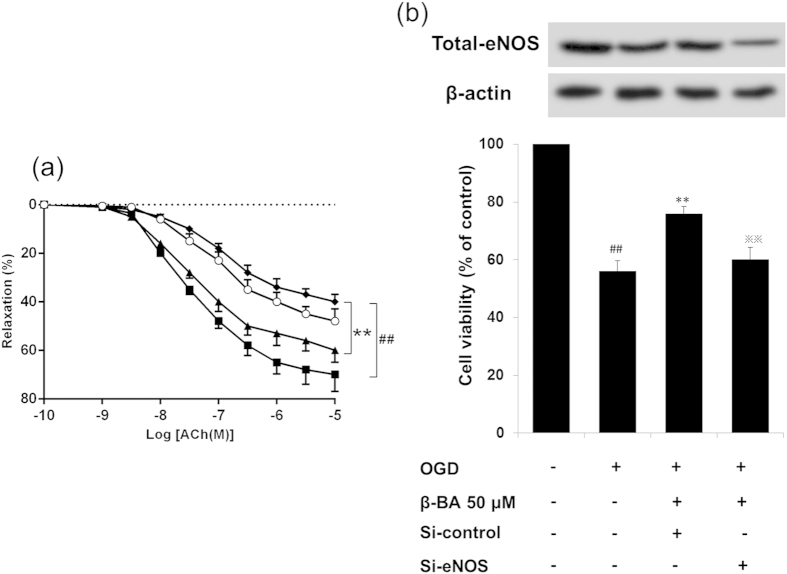
Vascular endothelial cells play an important role in modulating anti-thrombus and maintaining the natural function of vascular by secreting many active substances. β-boswellic acid (β-BA) is an active triterpenoid compound from the extract of boswellia serrate. In this study, it is demonstrated that β-BA ameliorates plasma coagulation parameters, protects endothelium from blood stasis induced injury and prevents blood stasis induced impairment of endothelium-dependent vasodilatation. Moreover, it is found that β-BA significantly increases nitric oxide (NO) and cyclic guanosine 3', 5'-monophosphate (cGMP) levels in carotid aortas of blood stasis rats. To stimulate blood stasis-like conditions in vitro, human umbilical vein endothelial cells (HUVECs) were exposed to transient oxygen and glucose deprivation (OGD). Treatment of β-BA significantly increased intracellular NO level. Western blot and immunofluorescence as well as immunohistochemistry reveal that β-BA increases phosphorylation of enzyme nitric oxide synthase (eNOS) at Ser1177. In addition, β-BA mediated endothelium-dependent vasodilatation can be markedly blocked by eNOS inhibitor L-NAME in blood stasis rats. In OGD treated HUEVCs, the protective effect of β-BA is attenuated by knockdown of eNOS. In conclusion, the above findings provide convincing evidence for the protective effects of β-BA on blood stasis induced endothelial dysfunction by eNOS signaling pathway.
Figures


 p < 0.01 versus β-BA group.
Similar articles
-
Rumex acetosa L. induces vasorelaxation in rat aorta via activation of PI3-kinase/Akt- AND Ca(2+)-eNOS-NO signaling in endothelial cells.J Physiol Pharmacol. 2015 Dec;66(6):907-15. J Physiol Pharmacol. 2015. PMID: 26769840
-
Propofol protects against high glucose-induced endothelial dysfunction in human umbilical vein endothelial cells.Anesth Analg. 2012 Feb;114(2):303-9. doi: 10.1213/ANE.0b013e31823f0c42. Epub 2011 Dec 9. Anesth Analg. 2012. PMID: 22156331
-
Arginase Inhibition Restores Peroxynitrite-Induced Endothelial Dysfunction via L-Arginine-Dependent Endothelial Nitric Oxide Synthase Phosphorylation.Yonsei Med J. 2016 Nov;57(6):1329-38. doi: 10.3349/ymj.2016.57.6.1329. Yonsei Med J. 2016. PMID: 27593859 Free PMC article.
-
Neurovascular Protective Function of Endothelial Nitric Oxide - Recent Advances.Circ J. 2016 Jun 24;80(7):1499-503. doi: 10.1253/circj.CJ-16-0423. Epub 2016 May 25. Circ J. 2016. PMID: 27238834 Review.
-
The use of human umbilical vein endothelial cells (HUVECs) as an in vitro model to assess the toxicity of nanoparticles to endothelium: a review.J Appl Toxicol. 2017 Dec;37(12):1359-1369. doi: 10.1002/jat.3470. Epub 2017 Apr 6. J Appl Toxicol. 2017. PMID: 28383141 Review.
Cited by
-
Synergistic neuroprotective effects of Danshensu and hydroxysafflor yellow A on cerebral ischemia-reperfusion injury in rats.Oncotarget. 2017 Dec 15;8(70):115434-115443. doi: 10.18632/oncotarget.23272. eCollection 2017 Dec 29. Oncotarget. 2017. PMID: 29383171 Free PMC article.
-
Role of Oxidative Stress in Peyronie's Disease: Biochemical Evidence and Experiences of Treatment with Antioxidants.Int J Mol Sci. 2022 Dec 15;23(24):15969. doi: 10.3390/ijms232415969. Int J Mol Sci. 2022. PMID: 36555611 Free PMC article. Review.
-
Anti-Inflammatory Activity of Boswellia serrata Extracts: An In Vitro Study on Porcine Aortic Endothelial Cells.Oxid Med Cell Longev. 2018 Jun 25;2018:2504305. doi: 10.1155/2018/2504305. eCollection 2018. Oxid Med Cell Longev. 2018. PMID: 30046370 Free PMC article.
-
Acetyl-11-Keto-β-Boswellic Acid Attenuates Prooxidant and Profibrotic Mechanisms Involving Transforming Growth Factor-β1, and Improves Vascular Remodeling in Spontaneously Hypertensive Rats.Sci Rep. 2016 Dec 23;6:39809. doi: 10.1038/srep39809. Sci Rep. 2016. PMID: 28009003 Free PMC article.
-
A Direct Relationship Between 'Blood Stasis' and Fibrinaloid Microclots in Chronic, Inflammatory, and Vascular Diseases, and Some Traditional Natural Products Approaches to Treatment.Pharmaceuticals (Basel). 2025 May 12;18(5):712. doi: 10.3390/ph18050712. Pharmaceuticals (Basel). 2025. PMID: 40430532 Free PMC article. Review.
References
-
- Chashoo G. et al. A propionyloxy derivative of 11-keto-β-boswellic acid induces apoptosis in HL-60 cells mediated through topoisomerase I & II inhibition. Chem-Biol Interact. 189, 60–71 (2011). - PubMed
-
- Kokkiripati P. K. et al. Gum resin of Boswellia serrata inhibited human monocytic (THP-1) cell activation and platelet aggregation. J Ethnopharmacol. 137, 893–901 (2011). - PubMed
-
- Han R. Highlight on the studies of anticancer drugs derived from plants in China. Stem Cells. 12, 53–63 (1994). - PubMed
-
- Elshazly S. M. Abd El Motteleb D. M. & Nassar N. N., The selective 5-LOX inhibitor 11-keto-β-boswellic acid protects against myocardial ischemia reperfusion injury in rats: involvement of redox and inflammatory cascades. Naunyn-Schmiedeberg’s Archives of Pharmacology. 386, 823–833 (2013). - PubMed
-
- Gerbeth K. et al. Determination of major boswellic acids in plasma by high-pressure liquid chromatography/mass spectrometry. J Pharmaceut Biomed. 56, 998–1005 (2011). - PubMed
Publication types
MeSH terms
Substances
LinkOut - more resources
Full Text Sources
Other Literature Sources
