. 2016 Mar 1;22(5):1185-96.
doi: 10.1158/1078-0432.CCR-15-1015. Epub 2015 Oct 19.

Preclinical Efficacy of the MDM2 Inhibitor RG7112 in MDM2-Amplified and TP53 Wild-type Glioblastomas

Affiliations

Preclinical Efficacy of the MDM2 Inhibitor RG7112 in MDM2-Amplified and TP53 Wild-type Glioblastomas

Maite Verreault et al. Clin Cancer Res. .

Erratum in

Abstract

Purpose: p53 pathway alterations are key molecular events in glioblastoma (GBM). MDM2 inhibitors increase expression and stability of p53 and are presumed to be most efficacious in patients with TP53 wild-type and MDM2-amplified cancers. However, this biomarker hypothesis has not been tested in patients or patient-derived models for GBM.

Experimental design: We performed a preclinical evaluation of RG7112 MDM2 inhibitor, across a panel of 36 patient-derived GBM cell lines (PDCL), each genetically characterized according to their P53 pathway status. We then performed a pharmacokinetic (PK) profiling of RG7112 distribution in mice and evaluated the therapeutic activity of RG7112 in orthotopic and subcutaneous GBM models.

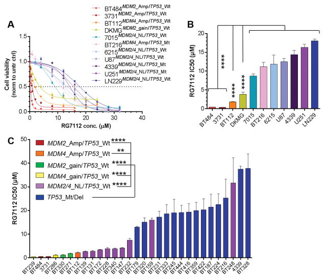
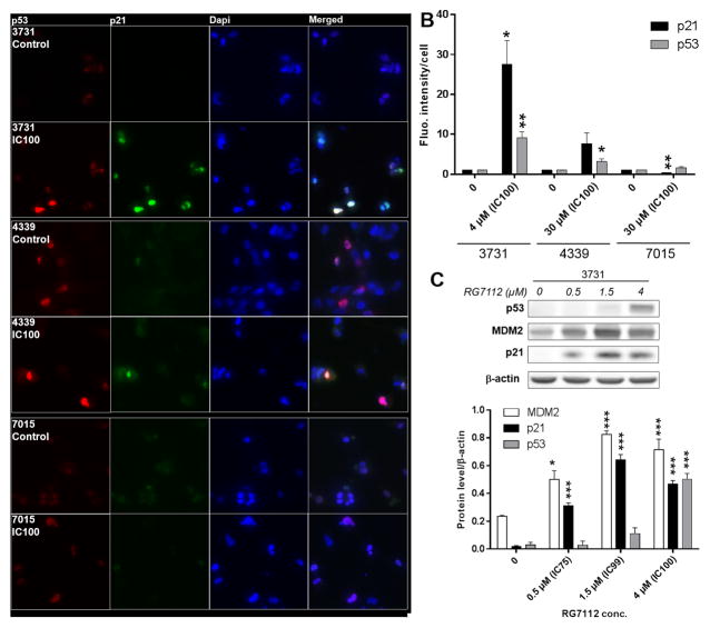
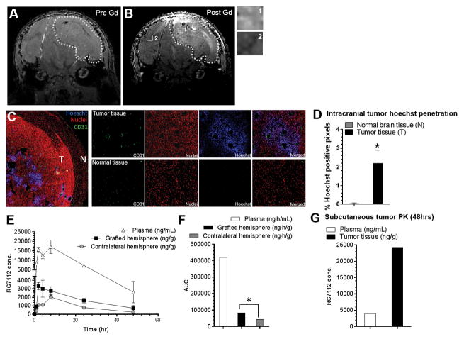
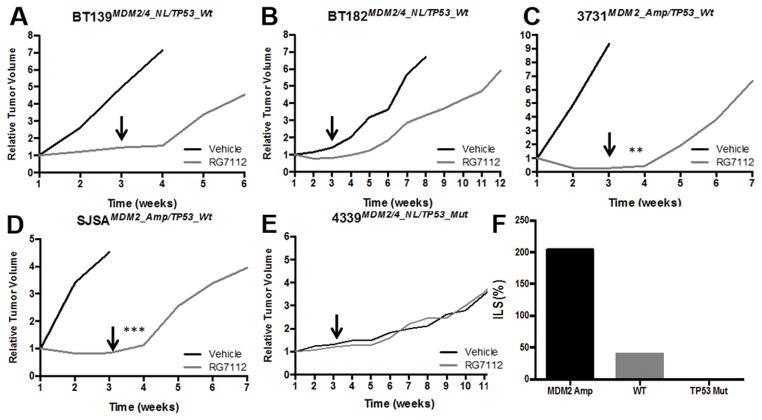
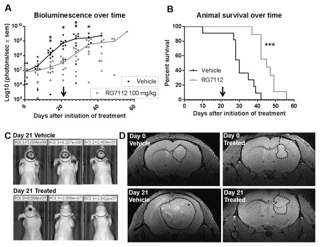
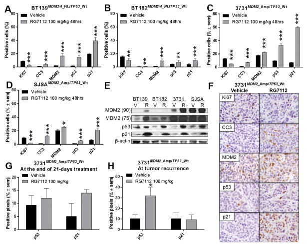
Results: MDM2-amplified PDCLs were 44 times more sensitive than TP53-mutated lines that showed complete resistance at therapeutically attainable concentrations (avg. IC50 of 0.52 μmol/L vs. 21.9 μmol/L). MDM4-amplified PDCLs were highly sensitive but showed intermediate response (avg. IC50 of 1.2 μmol/L), whereas response was heterogeneous in TP53 wild-type PDCLs with normal MDM2/4 levels (avg. IC50 of 7.7 μmol/L). In MDM2-amplified lines, RG7112 restored p53 activity inducing robust p21 expression and apoptosis. PK profiling of RG7112-treated PDCL intracranial xenografts demonstrated that the compound significantly crosses the blood-brain and the blood-tumor barriers. Most importantly, treatment of MDM2-amplified/TP53 wild-type PDCL-derived model (subcutaneous and orthotopic) reduced tumor growth, was cytotoxic, and significantly increased survival.

Conclusions: These data strongly support development of MDM2 inhibitors for clinical testing in MDM2-amplified GBM patients. Moreover, significant efficacy in a subset of non-MDM2-amplified models suggests that additional markers of response to MDM2 inhibitors must be identified.


Conflict of interest statement

Conflict of Interest: PW is a member of a Roche advisory board. AI was funded by Roche to attend a medical meeting.

Figures

Figure 1
Figure 1
Effects of RG7112 on cell proliferation and viability of GBM lines with various alterations of p53 pathway A) Cohort #1 (Amp: amplification, Mt: homozygous mutation, Wt: wild-type, NL: normal level): Proliferation and viability was 72 h after exposure to various concentrations of RG7112 on GBM cells carrying normal levels of MDM2 or MDM4 gene, and/or TP53 mutation (cold colors), or amplification of MDM2 or MDM4 gene, and a wild-type form of TP53 (warm colors). In this series, all lines with MDM2/4 amplification have Wt TP53, and all TP53 mutated lines have normal levels of MDM2/4 genes. B) Cohort #1: RG7112 IC50s (concentrations for which a 50% decrease in cell viability and proliferation was observed) in each cell line tested. C) Cohort #2: RG7112 IC50s in MDM2/4 amplified/gained cell lines, TP53 mutated/deleted cell lines, or cell lines with none of these alterations (MDM2/4_NL/TP53_Wt lines).
Figure 2
Figure 2
p53 and p21 protein levels in GBM lines with or without MDM2 gene amplification and treated with RG7112. A) Representative immunofluorescence images of 3731MDM2_Amp/TP53_Wt, 4339MDM2/4_NL/TP53_Mut, and 7015MDM2/4_NL/TP53_Wt treated or not with RG7112. p53 (red), p21 (green) and cell nuclei (blue) can be seen. B) Protein levels were quantified by three independent immunofluorescence assays. Each bar represents the fluorescence intensity per cell normalized over the value obtained in the corresponding untreated control cells. C) p53, p21 and MDM2 protein levels in MDM2-amplified GBM line treated with RG7112.
Figure 3
Figure 3
Assessment of the permeability of the blood-brain barrier in the MDM2-amplified orthotopic model and its impact on RG7112 pharmacokinetic distribution. MRI imaging of 3731MDM2_Amp/TP53_Wt xenograft. T1-weighted image A) before and B) after gadolinium injection are shown, with close-ups of tumor (1) and normal (2) tissue. Dotted line shows the outline of the tumor. B) Penetration of Hoechst 33342 in normal (N) and tumor tissue (T) after Hoechst intravenous injection. CD31 (green), nuclei (propidium iodide: red), Hoechst (blue) and a merged image can be seen. C) Hoechst staining was quantified in the tumor tissue compared to the normal brain tissue. D) Pharmacokinetic distribution of RG7112 in brain tissue of grafted and contralateral hemisphere and plasma after administration of a 100 mg/kg single dose. RG7112 was quantified by mass spectroscopy. E) Area-under-the-curves (AUCs) calculated from the pharmacokinetic data over a 48 hrs period. F) Drug distribution in subcutaneous tumor and plasma 48 h after RG7112 administration. *p-value < 0.05 between grafted hemisphere or tumor tissue compared to contralateral hemisphere or normal brain tissue.
Figure 4
Figure 4
Effect of RG7112 in GBM patient subcutaneous xenografts on tumor growth overtime of GBM lines with various alterations. A–B) MDM2/4 and TP53 wild-type cell line, C) MDM2 amplification and TP53 wild-type cell line and D) MDM2/4 normal level and TP53 mutant cell line. E) MDM2-amplified osteosarcoma model was included as a positive control. Once tumor was established, animals were treated with RG7112 or vehicle formulation for 3 weeks. Arrows show the end of treatment period. F) Effect of RG7112 on increased life span in MDM2-amplified tumor (3731), TP53 wild-type tumors (BT139 and BT182), and TP53 mutant tumor (4339).
Figure 5
Figure 5
Effect of RG7112 in 3731MDM2_Amp/TP53_Wt GBM orthotopic xenograft. A) Bioluminescence signal measured overtime. Each dot represents value for one animal. B) Animal survival. Once tumor was established, animals were treated with RG7112 or vehicle formulation for 3 weeks. Bioluminescence signal was measured weekly. Arrow shows the end of treatment period. C) Three representative animals from each group are shown after the end of treatment period (21 days) with bioluminescence signal acquired with a 0.5 second exposure time. D) MRI imaging (T2-weighted) of a vehicle and RG7112-treated animal prior to and at the end of the treatment period. Dotted line shows the outline of tumors.
Figure 6
Figure 6
Quantification of markers of response to RG7112. A–D) Quantification of Ki67, CC3, MDM2, p53 and p21 protein level by immunohistochemistry of: A–B) MDM2/4 and TP53 wild-type cell line, C) MDM2 amplification and TP53 wild-type cell line and D) MDM2-amplified osteosarcoma positive control, harvested 48 h after one dose of RG7112. E) Western immunoblot analysis of subcutaneous tumors showing MDM2 75 and 90 kDa forms, p53 and p21 proteins. F) Representative images showing marker staining in brown and nuclei in blue of vehicle and RG7112 treated 3731MDM2_Amp/TP53_Wt tumor. G) Quantification of p53 and p21 protein levels on the day after the 21 day treatment period, or H) at the moment of termination when animals showed signs of tumor-associated illness due to relapse after treatment.

References

    1. Ostrom QT, Bauchet L, Davis FG, Deltour I, Fisher JL, Langer CE, et al. The epidemiology of glioma in adults: a “state of the science” review. Neuro Oncol. 2014;16(7):896–913. - PMC - PubMed
    1. Stupp R, Hegi ME, Mason WP, van den Bent MJ, Taphoorn MJ, Janzer RC, et al. Effects of radiotherapy with concomitant and adjuvant temozolomide versus radiotherapy alone on survival in glioblastoma in a randomised phase III study: 5-year analysis of the EORTC-NCIC trial. Lancet Oncol. 2009;10(5):459–66. - PubMed
    1. TCGAP. Comprehensive genomic characterization defines human glioblastoma genes and core pathways. Nature. 2008;455(7216):1061–8. - PMC - PubMed
    1. Brennan CW, Verhaak RG, McKenna A, Campos B, Noushmehr H, Salama SR, et al. The somatic genomic landscape of glioblastoma. Cell. 2013;155(2):462–77. - PMC - PubMed
    1. Fridman JS, Lowe SW. Control of apoptosis by p53. Oncogene. 2003;22(56):9030–40. - PubMed

Publication types

MeSH terms