

Multifocal leukoencephalopathy in cocaine users: a report of two cases and review of the literature
- PMID: 26482228
- PMCID: PMC4615875
- DOI: 10.1186/s12883-015-0467-1
Multifocal leukoencephalopathy in cocaine users: a report of two cases and review of the literature
Abstract
Background: Cocaine abuse is associated with several mechanisms of brain injury including ischemic, hemorrhagic and metabolic. Recently two case reports of leukoencephalopathy in cocaine users implicated a commonly used cocaine adulterant, levamisole. One well-documented adverse effect of levamisole, when used alone as antihelminthic or immunomodulatory drug, is multifocal inflammatory leukoencephalopathy. Therefore, immune mechanisms may also contribute to cocaine-induced brain injury.
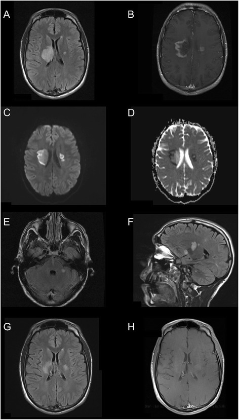
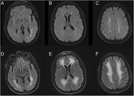
Case presentations: Two cocaine users with multifocal leukoencephalopathy, treated with steroids and plasmapheresis, are described. The first is a 25-year-old man who presented with unilateral motor and sensory impairment progressing to bilateral deficits, dysphagia, dysarthria and confusion over several days. Serial MRI showed increasing abnormal FLAIR signal lesions with patchy restricted diffusion and heterogenous enhancement deep in the right and left hemispheres, including periventricular white matter as well as in the pons and cerebellar peduncle. The second patient is a 41-year-old woman who presented with confusion and impaired balance. MRI showed bilateral periventricular FLAIR lesions with scattered restricted diffusion and subtle gadolinium enhancement of some of the lesions. She initially stabilized with supportive care only, but after further cocaine use was re-admitted six weeks later with marked neurological deterioration and MRI showed prominent worsening of the lesions. Both patients received steroid and plasma exchange and showed substantial improvement clinically and on imaging, which was sustained during out-patient follow-up.
Conclusion: Multifocal leukoencephalopathy associated with cocaine use may have an inflammatory/immune basis, possibly related to levamisole contamination, at least in some patients. Three cases, including the present two, have been described wherein good neurological improvement was seen in association with steroid treatment. However, in the absence of appropriate clinical trials, it remains unknown whether immunotherapy is truly beneficial for these patients.
Figures
References
Publication types
MeSH terms
Substances
LinkOut - more resources
Full Text Sources
Other Literature Sources
Medical
