. 2015 Dec;47(12):1482-8.
doi: 10.1038/ng.3423. Epub 2015 Oct 19.

Poly(A)-specific ribonuclease (PARN) mediates 3'-end maturation of the telomerase RNA component

Affiliations

Poly(A)-specific ribonuclease (PARN) mediates 3'-end maturation of the telomerase RNA component

Diane H Moon et al. Nat Genet. 2015 Dec.

Abstract

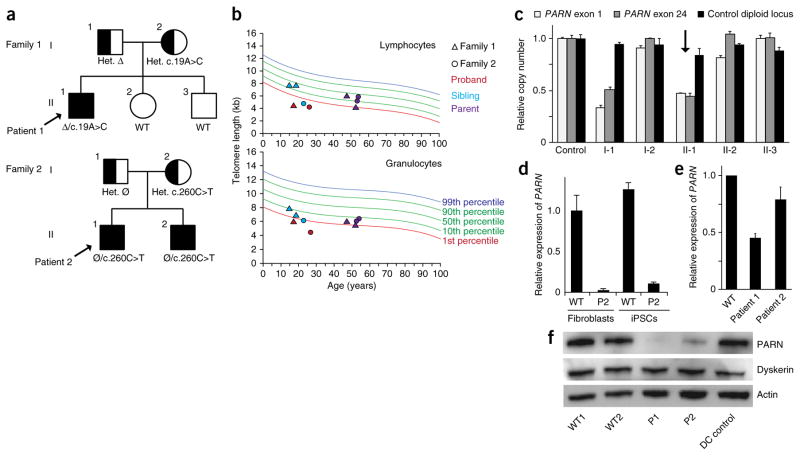
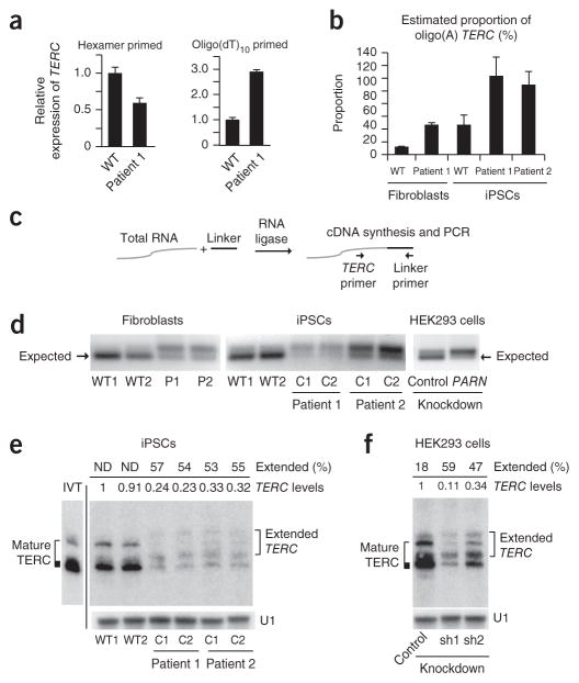
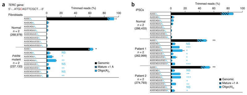
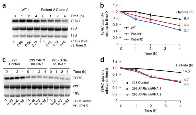
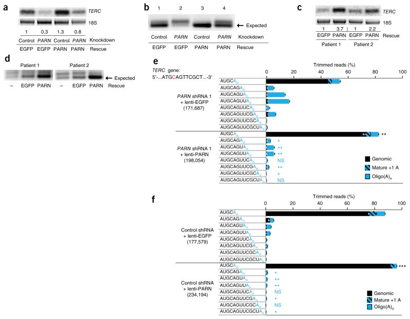
Mutations in the PARN gene (encoding poly(A)-specific ribonuclease) cause telomere diseases including familial idiopathic pulmonary fibrosis (IPF) and dyskeratosis congenita, but how PARN deficiency impairs telomere maintenance is unclear. Here, using somatic cells and induced pluripotent stem cells (iPSCs) from patients with dyskeratosis congenita with PARN mutations, we show that PARN is required for the 3'-end maturation of the telomerase RNA component (TERC). Patient-derived cells as well as immortalized cells in which PARN is disrupted show decreased levels of TERC. Deep sequencing of TERC RNA 3' termini shows that PARN is required for removal of post-transcriptionally acquired oligo(A) tails that target nuclear RNAs for degradation. Diminished TERC levels and the increased proportion of oligo(A) forms of TERC are normalized by restoring PARN, which is limiting for TERC maturation in cells. Our results demonstrate a new role for PARN in the biogenesis of TERC and provide a mechanism linking PARN mutations to telomere diseases.


Conflict of interest statement

COMPETING FINANCIAL INTERESTS

The authors declare no competing financial interests.

Figures

Figure 1
Figure 1
PARN mutations in two families with dyskeratosis congenita. (a) Segregation of compound heterozygous PARN mutations in families 1 and 2. Probands are indicated by arrows. Clinically affected individuals are shown as filled shapes, and carriers are shown as half-filled shapes. Nucleotide substitutions are indicated, as well as the inheritance of a large deletion (Δ) encompassing the PARN locus in family 1 and a noncoding defect affecting accumulation of PARN transcripts in family 2 (Ø). Het., heterozygous; WT, wild type. (b) Telomere length measurement by flow-FISH in lymphocytes (top) and granulocytes (bottom). (c) Quantitative PCR (qPCR) of relative PARN copy number in peripheral blood cell genomic DNA from a control and members of family 1, using primers from the first (exon 1) and last (exon 24) exons of the PARN locus, as compared to copy number for a control diploid locus (GPR15) (n = 2 technical replicates). Error bars, s.d. The proband is indicated by an arrow. (d) qPCR of cDNA using an allele-specific primer that distinguishes wild-type (c.C260) from mutant (c.T260) transcripts (n = 2 technical replicates). Error bars, s.d. Cells from patient 2 (P2) show deficits in transcript levels from the ‘normal’ allele (lacking a protein-coding mutation), which are expected to be 50% of those in wild-type cells. (e) qPCR of PARN transcripts from patient-derived and normal fibroblasts (n = 3 biological replicates). Error bars, s.d. (f) Immunoblot of PARN, dyskerin and actin protein levels in fibroblasts from normal individuals with wild-type PARN, patients 1 and 2 with PARN mutations, and a patient with dyskeratosis congenita without PARN mutations (DC control).
Figure 2
Figure 2
PARN deficiency results in decreased TERC levels, telomerase activity and telomere length. (a) qPCR of TERC transcripts in patient-derived and normal (wild-type) fibroblasts (n = 2 biological replicates). (b) qPCR of PARN transcript levels in iPSCs (n = 2 biological replicates). (c) Left, representative immunoblot of PARN, dyskerin and actin protein levels in iPSCs. Right, PARN and dyskerin protein levels normalized to actin levels (n = 4 biological replicates). (d) RNA blot of TERC following denaturing agarose gel electrophoresis of RNA from iPSCs. Ethidium bromide staining of 18S rRNA was used as a loading control. TERC levels normalized to those in WT1 cells are indicated. (e) Telomere repeat amplification protocol (TRAP) assay for telomerase activity in iPSCs, using fivefold dilutions of input cell extract. The internal control (IC) amplification standard is indicated. (f) Southern blot of telomere length by terminal restriction fragment length analysis in normal and patient-derived (clones C1 and C2) iPSCs, as well as control TERC-haploinsufficient (TR+/−) iPSCs. Passage numbers are indicated. MW, molecular weight marker. (g) qPCR of PARN, TERC and DKC1 transcripts from HEK293 cells transduced with lentivirus encoding shRNA directed against PARN versus luciferase (control) (n = 4 biological replicates). (h) Left, representative immunoblot of PARN, dyskerin and actin protein levels in HEK293 cells (from g) transduced with lentivirus encoding shRNA directed against PARN versus luciferase. Right, PARN and dyskerin protein levels normalized to actin levels (n = 4 biological replicates). For all panels, error bars represent s.d. Significance is indicated: *P ≤ 0.05, **P ≤ 0.01, ***P ≤ 0.001; NS, not significant.
Figure 3
Figure 3
PARN deficiency results in abnormal TERC 3′ ends. (a) qPCR of TERC transcripts in cDNA from normal and patient-derived fibroblasts generated using random hexamer versus oligo(dT)10 priming (n = 3 technical replicates). Error bars, s.d. (b) Estimated proportion of oligo(A)-containing TERC forms in fibroblasts and iPSCs (n = 2 technical replicates). Error bars, s.d. (c) The 3′ RACE strategy. A universal linker is ligated to the 3′ ends of total RNA. cDNA synthesis with a linker-specific primer followed by PCR using linker- and TERC-specific primers yields amplicons representing the diversity of TERC 3′ ends. (d) 3′ RACE products from normal and patient-derived fibroblasts (left) and iPSCs (middle) and from HEK293 cells subjected to shRNA-mediated knockdown of PARN versus luciferase (control) (right). Amplicons were separated by agarose gel electrophoresis; the expected size (184 bp) of mature TERC is indicated (arrows). (e) RNA blot of TERC using RNA from iPSCs, separated by denaturing 5% PAGE. U1 small nuclear RNA (snRNA) represents loading control. The migration pattern of in vitro–transcribed full-length TERC RNA is shown (IVT) and is considered to represent mature TERC. Extended TERC species found in patient-derived iPSCs are indicated on the right. Levels of total TERC species normalized to the levels in WT1 are shown. The percentage of extended forms in comparison to total TERC levels within each sample is indicated. Discrete, extended TERC species could not be distinguished in wild-type iPSCs (ND). (f) RNA blot (urea 5% PAGE) of TERC using RNA from HEK293 cells subjected to shRNA-mediated knockdown of PARN (sh) versus luciferase. Analysis was as described in e.
Figure 4
Figure 4
Decreased proportion of mature TERC and increased proportion of 3′-extended TERC species in PARN-deficient patient-derived cells. (a) Fibroblasts. 3′ RACE PCR products from normal and PARN-mutant patient-derived fibroblasts were subjected to deep sequencing, and reads were aligned to the TERC gene. The canonical TERC 3′ terminus is shown in red in the context of the TERC gene sequence. The relative proportions of mature TERC and TERC RNA species extending up to eight bases into the genomic sequence, with or without post-transcriptionally added oligo(A) tails, are depicted. Species with genomically encoded termini are in black, mature TERC with a single adenosine (which may be genomically encoded) is hatched and species with oligo(A) additions of any length (n) are in solid blue. Proportions are averaged for normal fibroblasts and patient-derived fibroblasts (n = 2 for each group). The total number of trimmed reads for each group is shown in parentheses. Error bars, s.d. For statistical evaluations, mature TERC forms and all genomically extended TERC species with oligo(A)n ends were compared between normal and patient-derived cells in a two-tailed t test. Significance is indicated: *P ≤ 0.05, **P ≤ 0.01, ***P ≤ 0.001; NS, not significant (black, mature TERC; blue, extended, oligo(A)n forms of TERC). (b) iPSCs. 3′ RACE PCR products from normal and PARN-mutant patient-derived iPSCs were subjected to deep sequencing, and reads were aligned to the TERC gene. Analyses and statistical comparisons were performed as in a. Proportions are averaged for normal iPSCs and patient-derived iPSCs (n = 2 for each group).
Figure 5
Figure 5
Decreased stability of TERC in PARN-deficient cells. (a) Representative RNA blot of TERC RNA levels in normal versus patient-derived iPSCs, at 0, 1, 2 and 4 h after the addition of actinomycin D. Results for WT1 and patient 2 clone 2 iPSCs are shown. Ethidium bromide staining of 28S and 18S rRNAs is shown; 18S rRNA was used as a loading control. TERC levels normalized to those at time 0 for each sample are indicated. (b) Graph of the TERC decay rate in normal and patient-derived iPSCs, calculated from RNA blot analysis as shown in a (n = 2 for each group; error bars, s.d.). The dotted lines reflect the slope determined by simple linear regression, and half-life is the calculated x intercept at y = 0.5. (c, d) RNA blot (c) and decay rates (d) of TERC RNA in HEK293 cells subjected to shRNA-mediated knockdown of PARN versus control. Quantification was performed as in a and b.
Figure 6
Figure 6
Ectopic expression of PARN rescues TERC maturation in PARN-deficient cells and shows that PARN is limiting for TERC biogenesis. (a) RNA blot of TERC RNA from HEK293 cells transduced with lentivirus encoding shRNA directed against PARN versus luciferase (control) and then rescued with lentivirus expressing PARN versus EGFP as a control. Ethidium bromide staining of 18S rRNA is used as a loading control. TERC levels normalized to those in cells with control knockdown and EGFP expression are shown. (b) 3′ RACE products from HEK293 cells subjected to shRNA-mediated knockdown of PARN f versus luciferase and rescued with PARN versus EGFP, separated by agarose gel electrophoresis. (c) RNA blot of TERC RNA from patient-derived fibroblasts transduced with lentivirus encoding PARN versus EGFP control. Normalized TERC levels are indicated. (d) 3′ RACE products from PARN-mutant patient-derived fibroblasts rescued with lentivirus expressing PARN versus EGFP. (e) Deep sequencing of 3′ RACE PCR products from HEK293 cells in which PARN is disrupted by shRNA and rescued by lentivirus expressing PARN or EGFP. Analysis was performed as in Figure 4. Statistical comparisons were between cells ectopically expressing PARN and those expressing EGFP. Significance is indicated: *P ≤ 0.05, **P ≤ 0.01, ***P ≤ 0.001; NS, not significant (black, mature TERC; blue, extended, oligo(A)n TERC forms). Error bars, s.d. (f) Deep sequencing of 3′ RACE PCR products from HEK293 control cells transduced with lentivirus overexpressing PARN versus EGFP. Statistical comparisons were performed as in e. Error bars, s.d.

References

    1. Stuart BD, et al. Exome sequencing links mutations in PARN and RTEL1 with familial pulmonary fibrosis and telomere shortening. Nat Genet. 2015;47:512–517. - PMC - PubMed
    1. Tummala H, et al. Poly(A)-specific ribonuclease deficiency impacts telomere biology and causes dyskeratosis congenita. J Clin Invest. 2015;125:2151–2160. - PMC - PubMed
    1. Dehlin E, Wormington M, Korner CG, Wahle E. Cap-dependent deadenylation of mRNA. EMBO J. 2000;19:1079–1086. - PMC - PubMed
    1. Körner CG, Wahle E. Poly(A) tail shortening by a mammalian poly(A)-specific 3′-exoribonuclease. J Biol Chem. 1997;272:10448–10456. - PubMed
    1. Körner CG, et al. The deadenylating nuclease (DAN) is involved in poly(A) tail removal during the meiotic maturation of Xenopus oocytes. EMBO J. 1998;17:5427–5437. - PMC - PubMed

Publication types

Associated data