Review
. 2015 Dec;12(12):689-710.
doi: 10.1038/nrcardio.2015.161. Epub 2015 Oct 20.

Mitral valve disease--morphology and mechanisms

Affiliations
Review

Mitral valve disease--morphology and mechanisms

Robert A Levine et al. Nat Rev Cardiol. 2015 Dec.

Abstract

Mitral valve disease is a frequent cause of heart failure and death. Emerging evidence indicates that the mitral valve is not a passive structure, but--even in adult life--remains dynamic and accessible for treatment. This concept motivates efforts to reduce the clinical progression of mitral valve disease through early detection and modification of underlying mechanisms. Discoveries of genetic mutations causing mitral valve elongation and prolapse have revealed that growth factor signalling and cell migration pathways are regulated by structural molecules in ways that can be modified to limit progression from developmental defects to valve degeneration with clinical complications. Mitral valve enlargement can determine left ventricular outflow tract obstruction in hypertrophic cardiomyopathy, and might be stimulated by potentially modifiable biological valvular-ventricular interactions. Mitral valve plasticity also allows adaptive growth in response to ventricular remodelling. However, adverse cellular and mechanobiological processes create relative leaflet deficiency in the ischaemic setting, leading to mitral regurgitation with increased heart failure and mortality. Our approach, which bridges clinicians and basic scientists, enables the correlation of observed disease with cellular and molecular mechanisms, leading to the discovery of new opportunities for improving the natural history of mitral valve disease.


Conflict of interest statement

Competing interests

The authors declare no competing interests.

Figures

Figure 1
Figure 1
Models of mitral valve disease. a | Previous conceptual model of mitral valve disease caused by mutations in structural proteins. In this model, the mutations impair valve biomechanical integrity and thereby cause mitral regurgitation and clinical disease. Therapeutic options are limited to surgical valve repair or replacement, as the underlying mechanisms are inaccessible. b | Our conceptual model of mitral valve disease caused by mutations in structural proteins. This model is amenable to molecular therapy. In addition to impairing valve biomechanical integrity, mutations in structural proteins can also alter valve regulatory and signalling pathways, leading to abnormal cellular responses and extracellular matrix remodelling. Paracrine factors provide further therapeutic targets.
Figure 2
Figure 2
Mitral valve structure. a | The mitral valve has two leaflets—the anterior leaflet below the aortic valve, and the posterior leaflet composed of three scallops: P1, P2, and P3. At the leaflet edges, the chordae tendineae link the leaflets to the PM anchors on the left ventricular wall. b | The leaflet structure is well organized, with a dense fibrous layer of collagen, known as the fibrosa, which arises from the mitral annulus and faces the left ventricle. The fibrosa is thicker close to the annulus and thinner at the edge of the leaflet. The fibrosa is covered by the spongiosa, a loose connective tissue layer rich in GAGs. The spongiosa is thicker at the leaflet edge and thinner at the annulus. The atrialis is made up of lamellar collagen and elastin sheets, which extend from the left atrial endocardium into the leaflet. Endothelial cells cover the valve. The deep subendothelial layers contain quiescent VICs—noncontractile, fibroblast-like cells that originate from endocardial endothelial cells. Abbreviations: GAG, glycosaminoglycan; PM, papillary muscle; VIC, valvular interstitial cell.
Figure 3
Figure 3
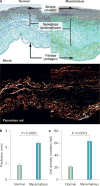
Morphological features of normal and myxomatous mitral valves. a | Normal mitral valves (left) and valves with myxomatous degeneration (right). Myxomatous valves have an abnormal layered architecture: loose collagen in fibrosa, expanded spongiosa strongly positive for proteoglycans, and disrupted elastin in atrialis. Top: Movat pentachrome stain (collagen stains yellow; proteoglycans, blue-green; and elastin, black). Bottom: Picrosirius red staining viewed under polarized light detected disruption and lower birefringence of collagen fibres in myxomatous leaflets. Magnification ×100. b | Quantitative analysis of valve thickness, demonstrating thickening of myxomatous valves. c | Increased density of interstitial cells in myxomatous spongiosa. Bars indicate SEM. Abbreviation: hpf, high-power field. Reprinted from Rabkin, E. et al. Activated interstitial myofibroblasts express catabolic enzymes and mediate matrix remodeling in myxomatous heart valves. Circulation 104 (21), 2525–2532 (2001).
Figure 4
Figure 4
Cell lineages that contribute to valve formation. The myocardial and endocardial cells are segregated at the onset or just after gastrulation in the E7.5 mouse embryo. This process occurs before, or at the onset of, gastrulation within the cardiac mesoderm. Endocardial progenitor cells emerge highly expressing VEGFR2. These cells undergo EMT to give rise to different cell types that participate in the valve leaflets. Some of these prospective valve cells are also likely to contribute to the endocardium. Myocardial progenitor cells, expressing only low levels of VEGFR2, give rise to both the FHL and SHL. SHL cells increase their expression of VEGFR2 and become endocardial progenitor cells. Epicardial progenitors also give rise to cells found within the valves. Abbreviations: EMT, endothelial-to-mesenchymal transition; FHL, first heart lineage; SHL, second heart lineage; VEGFR2, vascular endothelial growth factor receptor 2; VIC, valve interstitial cell.
Figure 5
Figure 5
Mitral valve growth and development. a | Mitral valve development from an endocardial cushion. The cells undergo a process of EMT to generate mesenchyme (brown cells) that invades the underlying matrix. These cells undergo proliferative expansion and differentiate into collagen-secreting VICs (green) that become progressively aligned along the proximal–distal tissue axis. Finally, the valve elongates and thins resulting in a mature valve comprised of stratified and aligned cells and matrix. b | Valve growth and response to stress. In a growing or stressed valve, or a valve stimulated by TGF-β, endothelial cells undergo EMT, increasing the number of matrix-producing interstitial cells. Abbreviations: EMT, endothelial-to-mesenchymal transition; GAG, glycosaminoglycan; TGF-β, transforming growth factor β; VIC, valve interstitial cell.
Figure 6
Figure 6
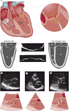
Echocardiographic diagnosis of mitral valve prolapse. a | Diagnosis of mitral valve prolapse must take into account the normal saddle shape of the valve and annulus, which produces opposite leaflet–annular relationships in perpendicular views. Mitral valve prolapse is most specifically diagnosed by leaflet displacement above the annular high points, imaged in long-axis views; and by leaflet misalignment at their point of coaptation. b | Parasternal long-axis echocardiographic view of posterior leaflet prolapse (arrows) beyond the annular hinge points (dashed line). c | Anterior leaflet prolapse and partial flail (partial eversion of the leaflet tip into the dilated LA; arrows) relative to the posterior leaflet, which is restricted, tethered by the dilated LV. These opposite leaflet displacements increase the regurgitant gap between the leaflets. d | Patient with extensive leaflet thickening and anterior leaflet flail (arrows). Abbreviations: Ao, aorta; LA, left atrium; LV, left ventricle; RA, right atrium; RV, right ventricle.
Figure 7
Figure 7
Anatomy of mitral valve prolapse. a | A resected mitral valve with prominent leaflet thickening and opacity and a prolapsing and domed central scallop (P2) of the posterior leaflet (arrow). b | Mitral valve removed from a patient with mitral valve prolapse. Note that the lesions, reflected by leaflet bulging towards the observer, are present throughout and most prominent in the medial scallop (P1) of the posterior leaflet.
Figure 8
Figure 8
Mechanisms of mitral valve prolapse. a | A mitral valve stained with haemotoxylin and eosin to define the lesion of mitral valve prolapse as disruption of the fibrosa by myxoid extracellular matrix (*), which also infiltrates the collagen core of the chordae tendineae, one of which was ruptured (arrow). The elastin lamina beneath the atrialis is also disrupted. b | A schematic showing the mechanism of myxomatous degeneration, with activation of valve interstitial cells to myofibroblasts that increase matrix production and turnover, secrete MMPs that drive collagen and elastin fragmentation, and release TGF-β that in turn promotes further cell proliferation and myofibroblast differentiation. Abbreviations: GAG, glycosaminoglycan; MMP, matrix metalloprotease; TGF-β, transforming growth factor β.
Figure 9
Figure 9
Proposed mechanism of mitral valve disease in Marfan syndrome. A mutation in fibrillin-1 decreases local binding of the TGF-β LLC, consisting of LAP, LTBP, and TGF-β ligands. TGF-β binds to its receptor (TGFBR1/2), activating canonical (Smad2/3, Smad4) and noncanonical (ERK1, JNK1, MAPK, MEK1, p38, Ras-GTP, ShcA, TAK1, TRAF6) signalling. Activation of these pathways leads to increased nuclear transcription of several downstream products, including CTGF, IL6, MCP1, MMPs, PAI-1, TSP1, and TGF-β itself. Filamin A inhibits activation of ERK1 and MEK1, and its deficiency increases these pathways and might contribute to mitral valve proliferation through this mechanism. Cross-talk between the Ang II receptors (AT1R and AT2R) and TGF-β receptors provides a therapeutic benefit of selective AT1R antagonism for aortic aneurysms in mice with fibrillin deficiency, and this approach is under investigation for treatment of myxomatous mitral valve disease. Abbreviations: Ang-II, angiotensin II; AT1R, angiotensin II type 1 receptor; AT2R, angiotensin II type 2 receptor; CTGF, connective tissue growth factor; ERK1, mitogen-activated protein kinase 3; JNK, mitogen-activated protein kinase 8; LAP, latency-associated peptide; LLC, large latent complex; LTBP, latent TGF-β-binding protein; MAPK, mitogen-activated protein kinase; MCP-1, C-C motif chemokine 2 (also known as monocyte chemoattractant protein 1); MEK1, dual specificity mitogen-activated protein kinase kinase 1; MMP, matrix metalloproteinase; PAI-1, plasminogen activator inhibitor 1; ShcA, SHC-transforming protein 1; TGF-β, transforming growth factor β; TGF-βR, transforming growth factor β receptor; TAK1, TGF-β-activated kinase 1; TRAF6, TNF receptor-associated factor 6; TSP-1, thrombospondin-1.
Figure 10
Figure 10
Mitral valve enlargement in SAM. a | SAM relates to a combination of a susceptible mitral valve, which is anteriorly positioned and elongated, encountering a posteriorly shifted left ventricular outflow stream, diverted by hypertrophy of the upper IVS. Anterior PM displacement decreases the posterior restraining force on the leaflets. Leaflet elongation allows greater leaflet excursion to create obstructive SAM. b | SAM of elongated leaflets (arrows) approaching the IVS and obstructing left ventricular outflow. Abbreviations: AML, anterior mitral leaflet; Ao, aorta; IVS, interventricular septum; LA, left atrium; LV, left ventricle; PM, papillary muscle; PML, posterior mitral leaflet; SAM, systolic anterior motion.
Figure 11
Figure 11
3D echocardiography showing increase in systolic anterior motion and LVOTO with increased mitral leaflet area. a,b | Component view from a healthy control individual with diastolic leaflet traces for 3D reconstruction. ce | Representative open mitral leaflet area measurements in green and purple for anterior and posterior leaflets viewed from the side. Top row: lateral commissure in foreground. Bottom row: left ventricular outflow tract aspect. Smallest values are seen in healthy controls (c), intermediate values with ASH and no resting LVOTO (d), and greatest leaflet areas in patients with ASH and resting LVOTO (e). Abbreviations: Ao, aorta; ASH, asymmetrical septal hypertrophy; LA, left atrium; LV, left ventricle; LVOTO, left ventricular outflow tract obstruction. Reprinted from Kim, D. H. et al. In vivo measurement of mitral leaflet surface area and subvalvular geometry in patients with asymmetrical septal hypertrophy: insights into the mechanism of outflow tract obstruction. Circulation 122 (13), 1298–1307 (2010).
Figure 12
Figure 12
Mechanism of ischaemic mitral regurgitation. a | Mechanism of ischaemic mitral regurgitation caused by increased mitral leaflet tethering owing to left ventricular remodelling after MI (shaded wall) with outward bulging (three arrows on the outer surface of the heart). Leaflet closure is restricted by increased tethering forces on the leaflets exerted via the chordae (arrows within the heart, exceeding normal on the left). b | Echocardiogram from a patient with inferior wall infarction and tethered mitral leaflets (arrows in left panel) with characteristic anterior leaflet bend and concavity towards the LA indicating chordal tethering, mitral regurgitation orifice (small arrows in right panel), and mitral regurgitant flow (large arrow in right panel). Abbreviations: Ao, aorta; LA, left atrium; LV, left ventricle; MI, myocardial infarction; MR, mitral regurgitation; PM, papillary muscle.
Figure 13
Figure 13
HE and Masson staining in the normal (left) and stretched (right) MV demonstrating increased leaflet thickness and spongiosa matrix after 2 months of stretching in a sheep model. Blue indicates collagen. Abbreviations: HE, hematoxylin and eosin; MV, mitral valve; S, spongiosa. Reprinted from Dal-Bianco, J. P. et al. Active adaptation of the tethered mitral valve: insights into a compensatory mechanism for functional mitral regurgitation. Circulation 120 (4), 334–342 (2009).
Figure 14
Figure 14
Evidence for reactivated EMT owing to leaflet stretch. a | Staining for the endothelial marker CD31 is reduced in the atrialis, and staining for smooth muscle α-actin is detected, which is normally absent, with positive cells (*) migrating into the interstitium, all features of EMT. Left panels: unstretched MV showing no staining for α-SMA along the CD31+ endothelium. Right panels: staining for α-SMA in the atrial endothelium (also CD31+) of a stretched MV, with nests of cells positive for α-SMA seeming to penetrate the interstitium (*). Upper panels: arrowheads indicate CD31+ endothelial cells. Lower panels: arrowheads indicate α-SMA+ endothelial cells, indicative of EMT. b | Schematic of active MV adaptation by EMT. Abbreviations: α-SMA, smooth muscle α-actin; EMT, endothelial-to-mesenchymal transition; MV, mitral valve. Reprinted from Dal-Bianco, J. P. et al. Active adaptation of the tethered mitral valve: insights into a compensatory mechanism for functional mitral regurgitation. Circulation 120 (4), 334–342 (2009). Bottom panel modified from Armstrong, E. J. & Bischoff, J. Heart valve development: endothelial cell signaling and differentiation. Circ. Res. 95 (5), 459–470 (2004).

References

    1. Nkomo VT, et al. Burden of valvular heart diseases: a population-based study. Lancet. 2006;368:1005–1011. - PubMed
    1. Enriquez-Sarano M, et al. Quantitative determinants of the outcome of asymptomatic mitral regurgitation. N Engl J Med. 2005;352:875–883. - PubMed
    1. Judge DP, Markwald RR, Hagege AA, Levine RA. Translational research on the mitral valve: from developmental mechanisms to new therapies. J Cardiovasc Transl Res. 2011;4:699–701. - PMC - PubMed
    1. Roberts R. Another chromosomal locus for mitral valve prolapse: close but no cigar. Circulation. 2005;112:1924–1926. - PubMed
    1. Williams TH, Jew JY. Is the mitral valve passive flap theory overstated? An active valve is hypothesized. Med Hypotheses. 2004;62:605–611. - PubMed

Publication types