. 2015 Dec 1;142(23):4038-48.
doi: 10.1242/dev.125674. Epub 2015 Oct 19.

Dach2-Hdac9 signaling regulates reinnervation of muscle endplates

Affiliations

Dach2-Hdac9 signaling regulates reinnervation of muscle endplates

Peter C D Macpherson et al. Development. .

Abstract

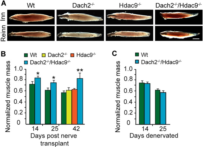
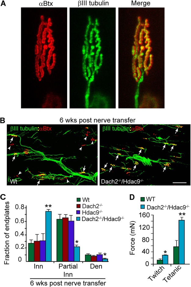
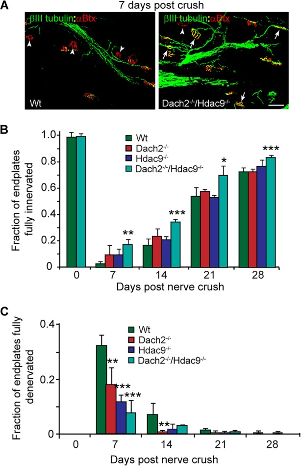
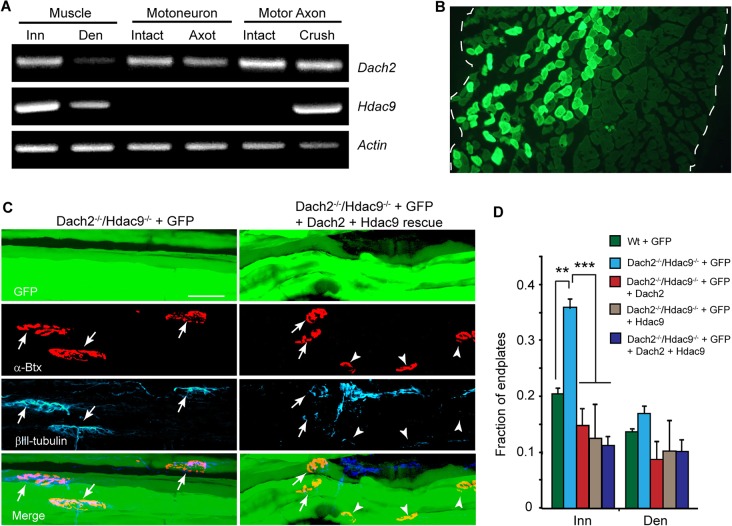
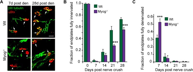
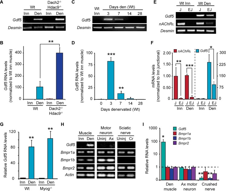
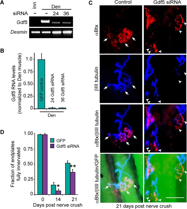
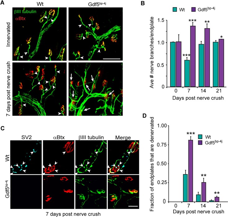
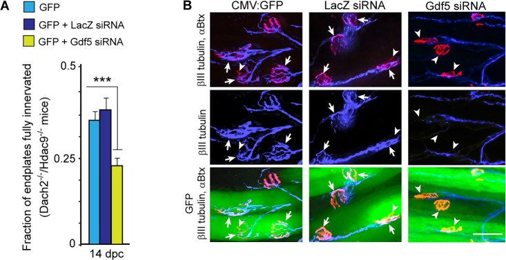
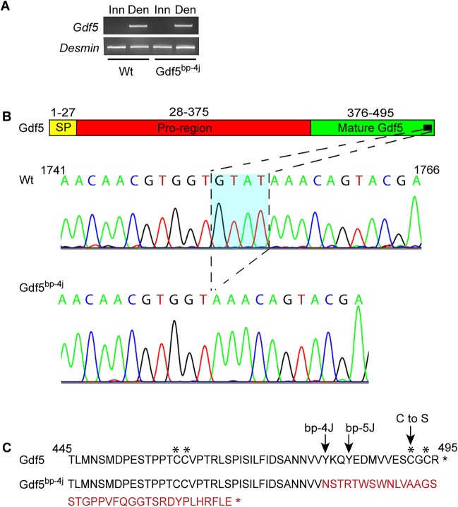
Muscle denervation resulting from injury, disease or aging results in impaired motor function. Restoring neuromuscular communication requires axonal regrowth and endplate reinnervation. Muscle activity inhibits the reinnervation of denervated muscle. The mechanism by which muscle activity regulates muscle reinnervation is poorly understood. Dach2 and Hdac9 are activity-regulated transcriptional co-repressors that are highly expressed in innervated muscle and suppressed following muscle denervation. Dach2 and Hdac9 control the expression of endplate-associated genes such as those encoding nicotinic acetylcholine receptors (nAChRs). Here we tested the idea that Dach2 and Hdac9 mediate the effects of muscle activity on muscle reinnervation. Dach2 and Hdac9 were found to act in a collaborative fashion to inhibit reinnervation of denervated mouse skeletal muscle and appear to act, at least in part, by inhibiting denervation-dependent induction of Myog and Gdf5 gene expression. Although Dach2 and Hdac9 inhibit Myog and Gdf5 mRNA expression, Myog does not regulate Gdf5 transcription. Thus, Myog and Gdf5 appear to stimulate muscle reinnervation through parallel pathways. These studies suggest that manipulating the Dach2-Hdac9 signaling system, and Gdf5 in particular, might be a good approach for enhancing motor function in instances where neuromuscular communication has been disrupted.

Keywords: Brachypodia; Dach2; Denervation; Gdf5; Hdac9; Motor nerve; Muscle; Myogenin; Nerve; Neuromuscular junction; Regeneration.


Conflict of interest statement

Competing interests

The authors declare no competing or financial interests.

Figures

Fig. 1.
Fig. 1.
Recovery of soleus muscle mass and function reflects muscle reinnervation in Dach2−/−/Hdac9−/− mice. (A) Representative images of dissected soleus muscles from Wt, Dach2−/−, Hdac9−/− and Dach2−/−/Hdac9−/− mice at 6 weeks post muscle denervation and nerve transplant. Scale bar: 2 mm. (B) Quantification of reinnervated soleus muscle mass normalized to the contralateral innervated control. Error bars are s.d.; n=3 for 14 and 25 days and n=5 for 42 days post nerve transplant. *P<0.05; **P<0.01 relative to Wt. (C) Quantification of denervated soleus muscle mass normalized to the contralateral innervated control. Error bars are s.e.m.; n=3.
Fig. 2.
Fig. 2.
Dach2 and Hdac9 inhibit reinnervation of endplates in denervated soleus muscle following nerve transfer. (A) βIII tubulin staining of motor nerve terminals overlaps precisely with αBtx stained endplates. (B) Representative images and (C) quantification of reinnervated endplates at 6 weeks post nerve transfer. βIII tubulin+ regenerating tibial nerve branches are green and αBtx+ endplates are red. Arrows point to fully innervated endplates. Arrowheads point to denervated endplates. Scale bar: 50 µm. Error bars are s.e.m.; n=4 for Wt and Dach2−/−; n=3 for Hdac9−/− and Dach2−/−/Hdac9−/−. *P<0.05; **P<0.01 relative to Wt. (D) Soleus force measurements following indirect stimulation through the transplanted tibial nerve. Error bars are s.e.m.; n=6 for Wt and n=5 for Dach2−/−/Hdac9−/−. *P<0.05; **P<0.01 relative to Wt. Inn, innervated; Den, denervated.
Fig. 3.
Fig. 3.
Dach2 and Hdac9 inhibit reinnervation of endplates in denervated soleus muscle following nerve crush. (A) Representative images and (B,C) quantification of innervated and denervated NMJs at various times post nerve crush. βIII tubulin+ regenerating tibial nerve branches are green and αBtx+ endplates are red. Arrows point to fully innervated endplates. Arrowheads point to denervated endplates. Scale bar: 50 µm. Error bars are s.e.m.; n=3. *P<0.05; **P<0.01; ***P<0.001 relative to Wt.
Fig. 4.
Fig. 4.
Dach2/Hdac9 inhibits muscle reinnervation. (A) RT-PCR analysis of Dach2, Hdac9 and γ-Actin mRNA in innervated (Inn) and denervated (Den) muscle; intact or axotomized (Axot) motor neurons and intact or crushed motor nerves and associated cells (Crush). (B) Representative image of soleus muscle cross section electroporated with CMV:GFP expression vector and stained for GFP immunofluorescence. Dashed line marks the muscle borders. (C) Representative images and (D) quantification of reinnervated endplates at 12 days post nerve crush in Wt and Dach2−/−/Hdac9−/− soleus muscles that were electroporated with GFP or Dach2, Hdac9 and GFP expression vectors. GFP marks electroporated muscle fibers green, αBtx marks endplates red and βIII tubulin marks regenerating axons cyan. Arrows point to fully and partially innervated endplates, whereas arrowheads point to denervated endplates. Error bars are s.d.; n=3 for Wt+GFP; n=4 for Dach2−/−/Hdac9−/−+GFP, n=3 for Dach2−/−/Hdac9−/− + GFP + Hdac9 and Dach2−/−/Hdac9−/− + GFP + Dach2 + Hdac9. **P<0.01; ***P<0.001 relative to Dach2−/−/Hdac9−/− + GFP. Scale bar: 50 µm.
Fig. 5.
Fig. 5.
Myog stimulates endplate reinnervation. (A) Representative images and (B,C) quantification of innervated and denervated endplates in Wt and Myog−/− soleus muscle at different times post nerve crush. βIII tubulin+ regenerating motor nerve branches are green and αBtx+ endplates are red. Arrows point to partially or fully innervated endplates and arrowheads point to denervated endplates. Scale bar: 50 µm. Error bars are s.d.; n=3 for 7 days and n=4 for 14, 21 and 28 days post nerve crush. ***P<0.001 relative to Wt.
Fig. 6.
Fig. 6.
Gdf5 gene expression is regulated by muscle activity in a Dach2/Hdac9-dependent manner. (A) RT-PCR and (B) qPCR show Gdf5 mRNA levels in innervated (Inn) and denervated (Den) soleus muscle from Wt and Dach2−/−/Hdac9−/− mice. Error bars are s.d.; n=3. **P<0.01. (C) RT-PCR and (D) qPCR shows changes in Gdf5 mRNA at various times post muscle denervation. Error bars are s.d.; n=3. **P<0.01; ***P<0.001. (E) RT-PCR and (F) qPCR show Gdf5 and nAChRε mRNA expression in junctional and extrajunctional regions of innervated and denervated soleus muscle. mRNA values are relative to innervated nAChRε mRNA (Y-axis on the left) and to innervated Gdf5 mRNA (Y-axis on the right). Error bars are s.d.; n=4. *P<0.05; **P<0.01; ***P<0.001. (G) qPCR show Gdf5 mRNA levels in innervated (Inn) and denervated (Den) soleus muscle from Wt and Myog−/− mice. Error bars are s.d.; n=3 for Wt and n=2 for Myog−/−. (H) RT-PCR and (I) qPCR shows expression levels of the indicated mRNAs in innervated and denervated soleus muscle, uninjured and axotomized (Ax) motor neurons and cells associated with uninjured or crushed (Cr) sciatic nerve. For qPCR values were normalized to γ-Actin for muscle and Gapdh for motor neuron and sciatic nerve. Error bars are s.d.; n=3. *P<0.05; **P<0.01 relative to uninjured for each tissue.
Fig. 7.
Fig. 7.
Gdf5 knockdown inhibits muscle reinnervation. (A) RT-PCR and (B) qPCR shows siRNAs 24 and 36 suppress Gdf5 mRNA levels. Error bars are s.d.; n=3 for control, n=1 for siRNA 24 and n=2 for siRNA 36. (C) Representative images of reinnervated endplates at 21 days post nerve crush in Wt soleus muscle electroporated with sCMV:GFP or a combination of sCMV:GFP and Gdf5-targeting siRNA24. αBtx marks endplates red; βIII tubulin marks regenerating axons cyan; and GFP identifies electroporated fibers. Arrows point to innervated endplates; arrowheads point to denervated endplates. Scale bar: 50 µm. (D) Quantification of regenerated neuromuscular synapses with and without Gdf5 knockdown. Error bars are s.d.; n=4 for GFP-treated 14 days post nerve crush and n=3 for 21 days post nerve crush. *P<0.05; **P<0.01 relative to GFP control.
Fig. 8.
Fig. 8.
Gdf5 stimulates muscle reinnervation. (A) Representative images showing increased axonal branching over the soleus muscle in Gdf5bp-4j mice at 7 days post nerve crush. βIII tubulin+ regenerating motor nerve branches are green and αBtx+ endplates are red. Arrowheads point to a single axonal branch innervating endplates, whereas arrows point to multiple axonal branches coursing over the endplate. Scale bar: 100 µm. (B) Quantification of the number of regenerating axonal branches/endplate in Wt and Gdf5bp-4j mice. Error bars are s.d.; n=3. *P<0.05; **P<0.01; ***P<0.001 relative to Wt. (C) Representative images showing reduced differentiation of axon terminals at endplates in Gdf5bp-4j mice at 7 days post nerve crush compared with Wt mice. SV2+ differentiated axon terminals are cyan, βIII tubulin+ regenerating motor nerves are green and αBtx+ endplates are red. Arrowheads point to SV2+ differentiated axon terminals that are also βIII tubulin+ and innervating αBtx+ endplates. (D) Quantification of denervated endplates in Wt and Gdf5bp-4j mice. Error bars are s.d.; n=3 for 7 and 14 days post nerve crush; n=4 for 21 days post nerve crush. **P<0.01; ***P<0.001 relative to Wt.
Fig. 9.
Fig. 9.
Gdf5 knockdown inhibits endplate reinnervation in Dach2−/−/Hdac9−/− mice. (A) Quantification of the percentage of fully innervated endplates at 14 days post nerve crush (dpc) in Dach2−/−/Hdac9−/− mice whose denervated soleus muscles were electroporated with a CMV:GFP expression vector and a siRNA targeting either E. coli lacZ mRNA or mouse Gdf5 mRNA. Error bars are s.d.; n=3. ***P<0.001. (B) Representative images of CMV:GFP and siRNA electroporated Dach2−/−/Hdac9−/− muscle fibers. GFP (green) identifies electroporated fibers, αBtx (red) identifies muscle endplates and βIII tubulin (cyan) identifies motor nerves. Arrowheads point to partially innervated and denervated endplates, whereas arrows point to fully innervated endplates. Scale bar: 50 µm.
Fig. 10.
Fig. 10.
Identification of Gdf5 gene mutation in Gdf5bp-4j mice. (A) RT-PCR shows normal denervation-dependent induction of Gdf5 mRNA in the Gdf5bp-4j mouse soleus muscle. (B) Diagram of Gdf5 protein sequence and DNA sequence of the Gdf5 carboxy-terminal region cDNA prepared from denervated Wt and Gdf5bp-4j muscle. A four nucleotide deletion is identified that alters the Gdf5 reading frame (C) in Gdf5bp-4j mice (amino acids in red). Mutations known to cause brachypodism in this region of the Gdf5 protein are indicated with arrows above the protein sequence. Cysteines that contribute to intrachain disulfide bonds are indicated with asterisks above the protein sequence.

Similar articles

Cited by

References

    1. Akaaboune M., Ma J., Festoff B. W., Greenberg B. D. and Hantaï D. (1993). The influence of denervation on beta-amyloid protein precursor and alpha 1-antichymotrypsin in mouse skeletal muscle. Neuromuscul. Disord. 3, 477-481. 10.1016/0960-8966(93)90100-X - DOI - PubMed
    1. Akaaboune M., Ma J., Festoff B. W., Greenberg B. D. and Hantaï D. (1994). Neurotrophic regulation of mouse muscle beta-amyloid protein precursor and alpha 1-antichymotrypsin as revealed by axotomy. J. Neurobiol. 25, 503-514. 10.1002/neu.480250505 - DOI - PubMed
    1. Berghella L., De Angelis L., De Buysscher T., Mortazavi A., Biressi S., Forcales S. V., Sirabella D., Cossu G. and Wold B. J. (2008). A highly conserved molecular switch binds MSY-3 to regulate myogenin repression in postnatal muscle. Genes Dev. 22, 2125-2138. 10.1101/gad.468508 - DOI - PMC - PubMed
    1. Berke B., Wittnam J., McNeill E., Van Vactor D. L. and Keshishian H. (2013). Retrograde BMP signaling at the synapse: a permissive signal for synapse maturation and activity-dependent plasticity. J. Neurosci. 33, 17937-17950. 10.1523/JNEUROSCI.6075-11.2013 - DOI - PMC - PubMed
    1. Buonanno A., Apone L., Morasso M. I., Beers R., Brenner H. R. and Eftimie R. (1992). The MyoD family of myogenic factors is regulated by electrical activity: isolation and characterization of a mouse Myf-5 cDNA. Nucleic Acids Res. 20, 539-544. 10.1093/nar/20.3.539 - DOI - PMC - PubMed

Publication types

MeSH terms

Associated data