. 2015 Dec 1;128(23):4341-52.
doi: 10.1242/jcs.172742. Epub 2015 Oct 19.

SLAT promotes TCR-mediated, Rap1-dependent LFA-1 activation and adhesion through interaction of its PH domain with Rap1

Affiliations

SLAT promotes TCR-mediated, Rap1-dependent LFA-1 activation and adhesion through interaction of its PH domain with Rap1

Marjorie Côte et al. J Cell Sci. .

Abstract

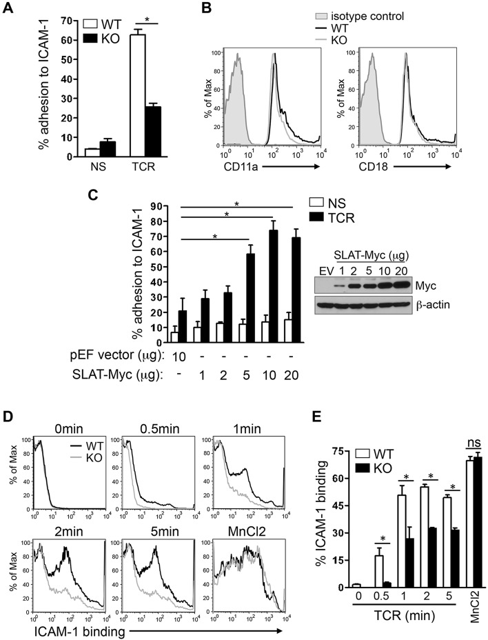
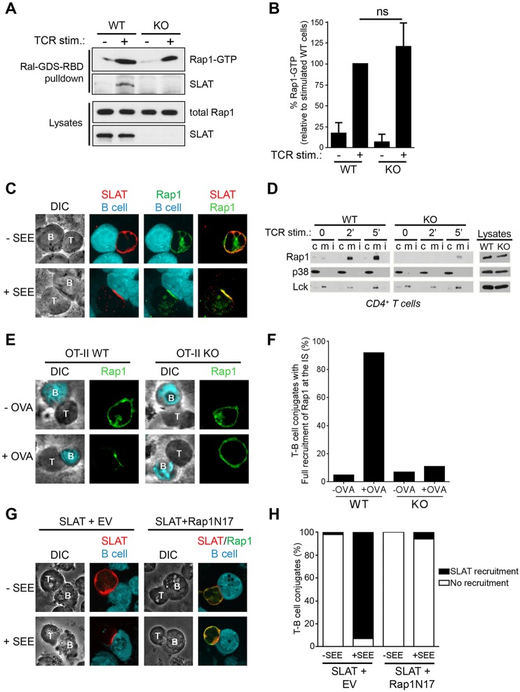
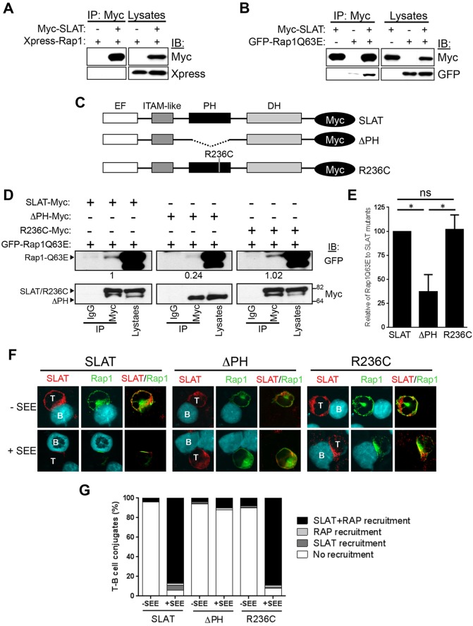
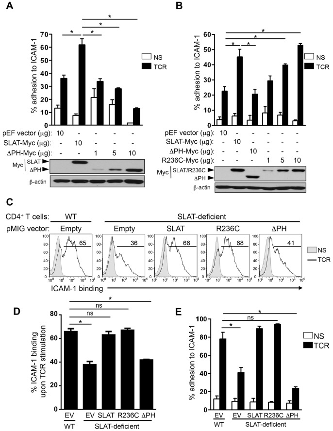
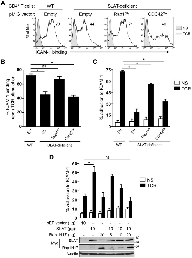
SLAT (also known as DEF6) promotes T cell activation and differentiation by regulating NFAT-Ca(2+) signaling. However, its role in TCR-mediated inside-out signaling, which induces integrin activation and T cell adhesion, a central process in T cell immunity and inflammation, has not been explored. Here, we show that SLAT is crucial for TCR-induced adhesion to ICAM-1 and affinity maturation of LFA-1 in CD4(+) T cells. Mechanistic studies revealed that SLAT interacts, through its PH domain, with a key component of inside-out signaling, namely the active form of the small GTPase Rap1 (which has two isoforms, Rap1A and Rap1B). This interaction has been further shown to facilitate the interdependent recruitment of Rap1 and SLAT to the T cell immunological synapse upon TCR engagement. Furthermore, a SLAT mutant lacking its PH domain drastically inhibited LFA-1 activation and CD4(+) T cell adhesion. Finally, we established that a constitutively active form of Rap1, which is present at the plasma membrane, rescues the defective LFA-1 activation and ICAM-1 adhesion in SLAT-deficient (Def6(-/-)) T cells. These findings ascribe a new function to SLAT, and identify Rap1 as a target of SLAT function in TCR-mediated inside-out signaling.

Keywords: Def6; Immunological synapse; Inside-out signaling; Integrin activation; Rap1; SLAT; T cell-adhesion.


Conflict of interest statement

Competing interests

The authors declare no competing or financial interests.

Figures

Fig. 1.
Fig. 1.
SLAT is required for TCR-induced adhesion to ICAM-1 and LFA-1 affinity maturation in CD4+ T cells. (A) Purified splenic WT and Def6−/− (KO) CD4+ T cells were left untreated (NS) or stimulated with 10 µg/ml anti-CD3 mAb (TCR) and subsequently analyzed for their ability to bind plate-bound Fc-ICAM-1 (mean±s.d., n=4). (B) WT (black line) and KO (gray line) CD4+ T cells were analyzed for the surface expression of CD11a and CD18 by flow cytometry, with normal IgG used as an isotype control (shaded). (C) Jurkat T cells transfected with 10 µg empty pEF vector (EV) or with pEF vector encoding Myc-tagged SLAT (1–20 µg) were either left unstimulated (NS) or stimulated with 10 µg/ml anti-CD3 mAb OKT3 (TCR), and subsequently analyzed for adhesion to plate-bound Fc-ICAM-1. SLAT expression was analyzed by anti-Myc immunoblotting and β-actin expression served as a loading control. Adhesion data represents the means±s.d. of four independent experiments. (D,E) WT or KO CD4+ T cells were analyzed by flow cytometry for their ability to bind soluble Fc-ICAM-1 (as a measurement of LFA-1 affinity) in response to anti-CD3 (10 µg/ml) mAb stimulation for the indicated times or 1 mM MnCl2 (positive control) treatment for 5 min. Quantitative analysis of the results shown in D, representing the mean±s.d. percentage of ICAM-1 binding (determined in triplicates) is shown in E. One out of five representative experiments is shown. *P<0.01; ns, not significant (Student's t-test).
Fig. 2.
Fig. 2.
Interdependent co-recruitment of SLAT and Rap1 to the plasma membrane and immunological synapse upon TCR stimulation. (A) WT or Def6−/− (KO) CD4+ T cells were either left untreated or stimulated with 10 µg/ml anti-CD3 mAb for 3 min. Active (GTP-loaded) Rap1 was precipitated using a GST–RalGDS-RBD fusion protein. Precipitates (Ral-GDS-RBD pulldown) and whole-cell lysates were analyzed by immunoblotting with the indicated antibodies. One out of four representative experiments is shown. (B) Bar graphs representing mean±s.d. densitometry data pooled from four independent pull-down experiments as shown in A. (C) Jurkat JA16 cells were co-transfected with Myc-tagged SLAT and Xpress-tagged Rap1. After 24 h, the cells were stimulated or not with SEE-pulsed CMAC-labeled Raji B cells and conjugates were bound to poly-L-lysine-coated coverslips, fixed, and stained with anti-Myc and anti-Xpress plus secondary Alexa-Fluor-555-coupled anti-rabbit-Ig or Alexa-Fluor-488-coupled anti-mouse-Ig antibodies, respectively. Individual and overlay of the green (SLAT), red (Rap1) and blue (B, B cell) images along with differential interference contrast (DIC) images are shown. T, T cell. Data shown are representative of three independent experiments. (D) Purified splenic CD4+ T cells from WT or Def6−/− (KO) mice were stimulated for the indicated times with an anti-CD3 mAb. Membrane (m), cytosolic (c) and detergent insoluble (i) fractions were prepared, resolved by SDS-PAGE, and immunoblotted with the indicated antibodies (left panels). Expression of p38 MAPK (detected using an antibody recognizing all isoforms) and Lck in the cytosolic and membrane fractions, respectively, confirmed proper separation of the respective fractions. Analysis of protein expression in whole-cell lysates (right panels) revealed no difference of expression of Rap1, Lck or p38 MAPK in WT versus KO cells. (E) WT or Def6−/− OT-II CD4+ T cells were pulsed with OVA323–339 peptide (+OVA) or not pulsed (−OVA) dendritic cells for 20 min. T-cell–dendritic cell conjugates were then stained for Rap1 expression and localization. Corresponding DIC images are shown. (F) Quantitative analysis of the results shown in E. Rap1 and/or SLAT localization in the immunological synapse was analyzed in 100 T-dendritic cell conjugates. The graph represents the mean percentage of imaged cells scored in each group (recruitment versus no recruitment to the immunological synapse) and is representative of three experiments. (G) Jurkat JA16 cells were co-transfected with Xpress-tagged SLAT along with empty vector (EV) or Myc-tagged dominant-negative Rap1 (Rap1N17). After 24 h, the cells were stimulated or not with SEE-pulsed CMAC-labeled Raji B cells and conjugates were bound to poly-L-lysine-coated coverslips, fixed, and stained with anti-Xpress and anti-Myc plus secondary Alexa-Fluor-555-coupled anti-mouse-Ig or Alexa-Fluor-488-coupled anti-rabbit-Ig antibodies, respectively. Individual and overlay of the red (SLAT), green (Rap1N17) and blue (B cell) images along with DIC images are shown. (H) Quantitative analysis of the results shown in G, representing approximately 100 T-cell–B-cell conjugates analyzed.
Fig. 3.
Fig. 3.
The PH domain of SLAT interacts with active Rap1 and is required to facilitate SLAT and Rap1 co-recruitment to the immunological synapse. (A,B) 293 T cells were co-transfected with Myc–SLAT and the indicated plasmids encoding Xpress–Rap1 (A) or GFP–Rap1Q63E (B). SLAT immunoprecipitation (IP) and cell lysates were analyzed by immunoblotting (IB). The results shown are representative of four independent experiments. (C) Schematic representation of Myc-tagged SLAT mutants. The EF hand, ITAM-like, PH and DH domains are shown. (D) 293 T cells were co-transfected with the indicated Myc-tagged SLAT plasmids together with GFP–Rap1Q63E. Cell lysates were immunoprecipitated with an anti-Myc antibody or normal IgG (as an immunoprecipitation control) and analyzed by immunoblotting with anti-Myc and anti-GFP antibodies. Cell lysates were also blotted with the same antibodies. Numbers under the Myc blots indicate the relative binding of Rap1Q63E to the SLAT mutants, as determined by densitometry. (E) Bar graphs representing the mean±s.d. relative binding of Rap1Q63E to SLAT mutants as determined by densitometry from three independent experiments as shown in D. (F) JA16 cells were co-transfected and stimulated as in Fig. 2C. Overlays of the red (SLAT) and green (Rap1) images are shown. B, B cell; T, T cell. The data are representative of three independent experiments. (G) Quantitative analysis of SLAT and Rap1 localization in the immunological synapse shown in F, representing approximately 100 T-cell–B-cell conjugates analyzed.
Fig. 4.
Fig. 4.
TCR-induced LFA-1 dependent T cell adhesion depends on the SLAT PH domain. (A,B) Jurkat JA16T cells were transfected with empty pEF vector or pEF-Myc-SLAT (10 µg each) plus the indicated amounts of Myc-tagged SLAT-ΔPH mutant (A,B) or SLAT-R236C mutant (B). Cells were either left unstimulated (NS) or stimulated with OKT3 mAb (TCR) for 45 min, and subsequently analyzed for adhesion to plate-bound Fc-ICAM-1. Adhesion data represents the mean±s.d. of four independent experiments. Lower panels, expression of transfected proteins was detected by anti-Myc immunoblotting. An anti-β-actin immunoblot serves as a loading control. (C–E) Primary WT and Def6−/− (KO) CD4+ T cells were activated with anti-CD3 plus anti-CD28 mAbs and IL-2 and transduced with retroviral pMIG vectors expressing either GFP alone (Empty) or GFP plus the indicated SLAT cDNAs. Sorted GFP+ CD4+ T cells were left unstimulated (NS) or restimulated with anti-CD3 mAb (TCR) for 3 min (C,D) or 45 min (E), and analyzed either for their ability to bind soluble Fc-ICAM-1 by flow cytometry (C,D) or for their ability to adhere to plate-bound Fc-ICAM-1 (E). Numbers shown in FACS histograms (C) indicate the percentage of GFP+ CD4+ T cells able to bind soluble Fc-ICAM in a representative experiment. Data shown are representative of four independent experiments. (D,E) ICAM-1 binding and adhesion data represent the mean±s.d. of the percentage of four independent experiments. *P<0.01, WT versus Def6−/− cells; ns, not significant (two-tailed Student's t-test).
Fig. 5.
Fig. 5.
SLAT regulates TCR-mediated LFA-1 activation and T cell adhesion in a Rap1-dependent manner. (A–C) Primary WT and Def6−/− (KO) CD4+ T cells were activated with anti-CD3 and anti-CD28 mAbs plus IL-2, and transduced with retroviral pMIG vectors expressing either GFP alone (Empty) or GFP plus Rap1V12 (Rap1CA), or Cdc42Q61L (CDC42CA). Sorted GFP+ CD4+ T cells were left unstimulated (NS) or restimulated with anti-CD3 mAb (TCR) for 3 (A,B) or 45 (C) min, and analyzed for their ability to bind soluble Fc-ICAM-1 by flow cytometry (A,B) or adhere to plate-bound Fc-ICAM-1 (C). Numbers shown in FACS histograms (A) indicate the percentage of GFP+ CD4+ T cells that bound soluble Fc-ICAM-1 in a representative experiment, and the bar graphs shown in B and C represent the mean±s.d. of ICAM-1 binding of three independent experiments. (D) Jurkat JA16T cells were transfected with empty pEF vector or Myc–SLAT plasmids (10 µg each) along with dominant-negative Rap1 (Rap1N17) plasmid (5-20 µg). Cells were then either left unstimulated or stimulated for 45 min and subsequently analyzed for their ability to bind plate-bound Fc-ICAM-1. Lower panel, whole-cell lysates were immunoblotted with anti-Myc (SLAT and Rap1N17) and anti-β-actin antibodies to assess the expression of the transfected proteins and to control loading, respectively. Data are representative of three (A–C) and four (D) independent experiments. *P<0.01, WT versus Def6−/− cells; ns, not significant (two-tailed Student's t-test).

Similar articles

Cited by

References

    1. Abram C. L. and Lowell C. A. (2009). The ins and outs of leukocyte integrin signaling. Annu. Rev. Immunol. 27, 339-362. 10.1146/annurev.immunol.021908.132554 - DOI - PMC - PubMed
    1. Alon R. and Dustin M. L. (2007). Force as a facilitator of integrin conformational changes during leukocyte arrest on blood vessels and antigen-presenting cells. Immunity 26, 17-27. 10.1016/j.immuni.2007.01.002 - DOI - PubMed
    1. Balla T. (2005). Inositol-lipid binding motifs: signal integrators through protein-lipid and protein-protein interactions. J. Cell Sci. 118, 2093-2104. 10.1242/jcs.02387 - DOI - PubMed
    1. Baumeister M. A., Martinu L., Rossman K. L., Sondek J., Lemmon M. A. and Chou M. M. (2003). Loss of phosphatidylinositol 3-phosphate binding by the C-terminal Tiam-1 pleckstrin homology domain prevents in vivo Rac1 activation without affecting membrane targeting. J. Biol. Chem. 278, 11457-11464. 10.1074/jbc.M211901200 - DOI - PubMed
    1. Bécart S. and Altman A. (2009). SWAP-70-like adapter of T cells: a novel Lck-regulated guanine nucleotide exchange factor coordinating actin cytoskeleton reorganization and Ca2+ signaling in T cells. Immunol. Rev. 232, 319-333. 10.1111/j.1600-065X.2009.00839.x - DOI - PMC - PubMed

Publication types

MeSH terms